Automated method for detecting cancers and high grade hyperplasias

a high-grade hyperplasia and automatic detection technology, applied in the field of automatic detection of cancer and dysplasia, can solve the problems of difficult analysis, long process, human error, and inability to accurately detect abnormal cells, and achieve the effect of quick detection of chromosomal regions and abnormal cells

Inactive Publication Date: 2009-08-20
IKONISYS INC
View PDF3 Cites 54 Cited by
  • Summary
  • Abstract
  • Description
  • Claims
  • Application Information

AI Technical Summary

Benefits of technology

[0062]Automatic scanning in such embodiments may be performed, for example, by an automated microscope wherein the biological specimen is placed on slides which are manually or automatically loaded onto the microscope stage, and the slide automatically scanned. Automated microscopes that may find employment in such system are such as described in other of applicant's patent applications (see below). Scanning may also be made of other substrates onto / into which the biological sample is placed. Scanning may comprise scanning the biological sample in one plane, or in more than one plane, such as, for example, two, three or more planes. By scanning in multiple planes, detection of abnormal cells, which may be rare in terms of total number of cells in the sample, may be significantly improved. The probes may make use of FISH probes in which the fluorescent signal is picked up by the detector. The probes may produce a signal with or without another input signal, e.g. they may be radioactive, or fluoresce when impinged by an activating signal (such as an appropriate wavelength of light or other electromagnetic radiation). The probes may be directed to different replication associated cancel loci, and may comprise different fluorescent tags so as to produce different signals. The detector may be selected in accordance with signal(s) which are to be produced by the tags, e.g. a fluorescence detector for detecting fluorescent tags, with the detector operatively configured to permit detection of the particular fluorescent signals produced by the fluorescent tags. Reporting may include a simple report of the particular tags associated with the particular chromosome and / or may comprise an automatic diagnostic (indicating the type of cancer associated with the particular hybridization pattern of the chromosomes). The vector value as compared to normal specimens may be selected to be less than a particular threshold, such as less than about 60, less than about 40, less than about 30, less than about 20, less than about 10, or less than about 0.500. A useful system may comprise automatic scanning and detection in multiple signal channels at once, or in a relatively short period of time (e.g., less than 1 minute) from one another. The system may be operatively configured to process each of the multiple signals in real time, simultaneously or concurrently (or a mix of the same), to allow for quick detection of chromosomal regions, and / or regional replications, which are indicative of one or more particular cancer / hyperplasia.

Problems solved by technology

The analysis is made more difficult by the time constraints imposed by the phenomena.
Even under the best conditions, the process has proven to be tedious, lengthily and subject to human error.
While such tests may indicate the need for interventional therapy, such as chemotherapy, they are not designed to lead one in the direction of the most appropriate interventional therapy.
As such snap-shot methods may entail considerable economic costs, as well as risks in themselves (e.g., radiation exposure), such snap-shots may be taken at considerably longer intervals than might be desired given the need for rapid intervention into resolving the disease state.

Method used


Image

  • Automated method for detecting cancers and high grade hyperplasias
  • Automated method for detecting cancers and high grade hyperplasias

Examples


Embodiment Construction

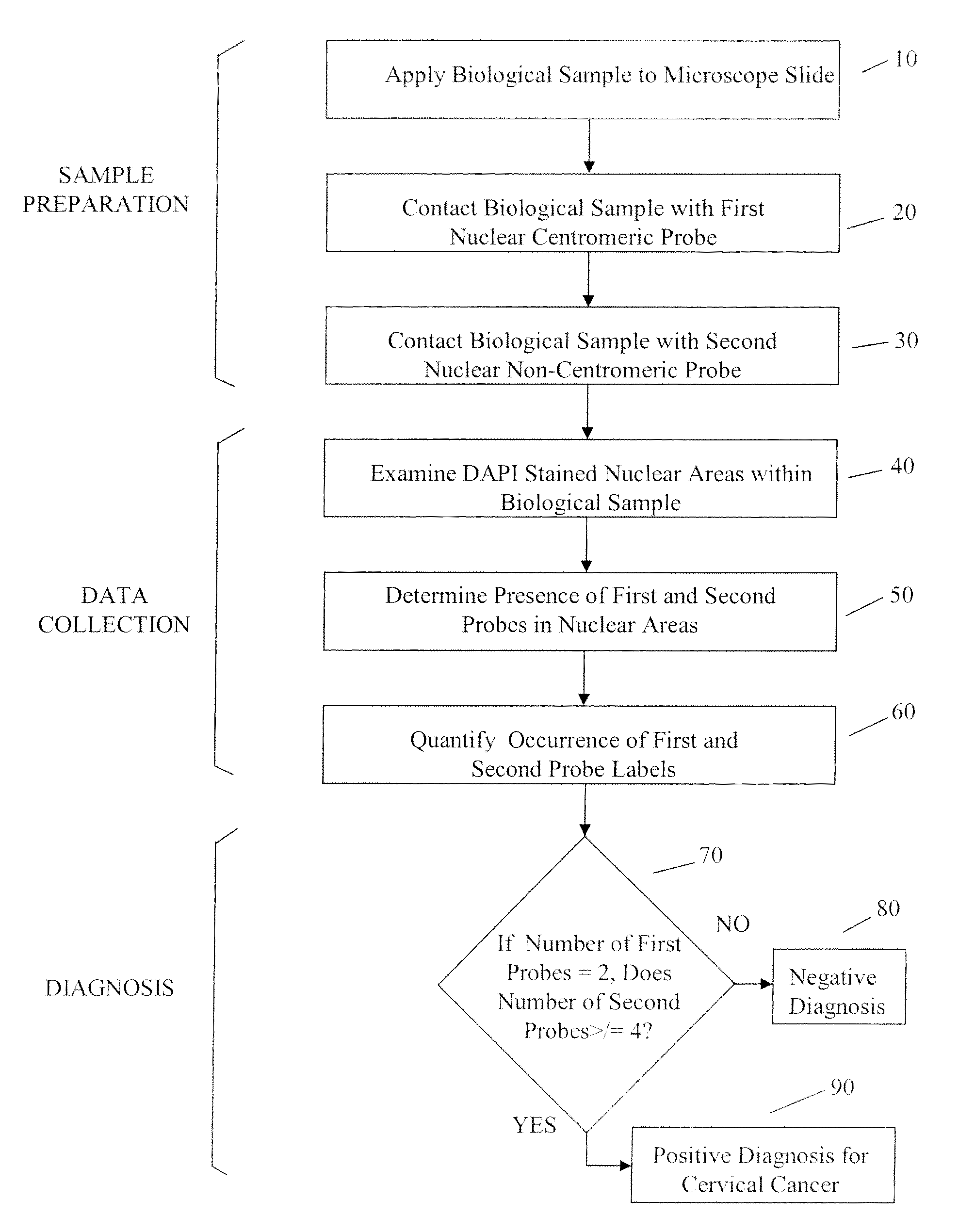
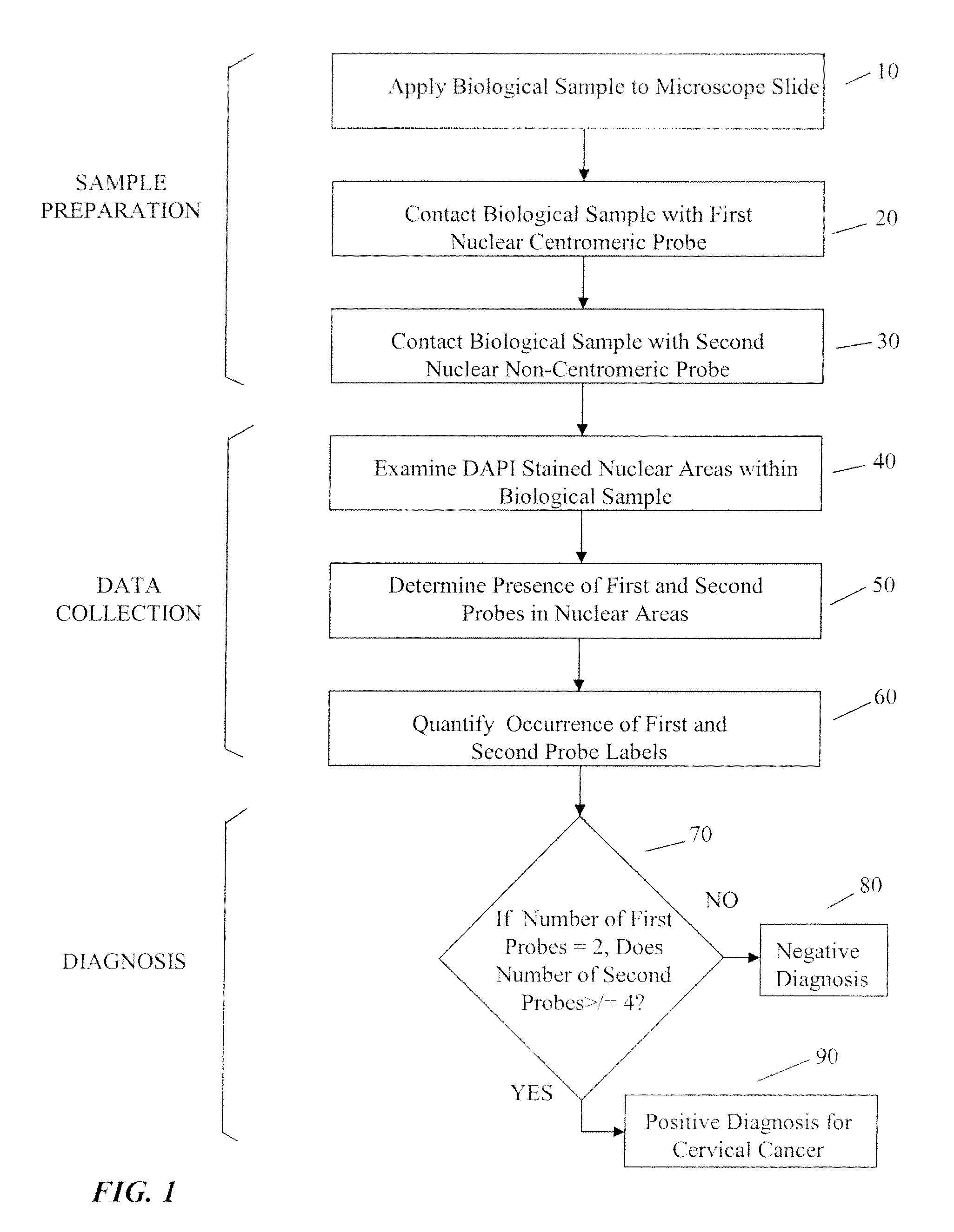
[0064]As used herein, “tag” and “label” relate synonymously to a moiety conjugated to a probe to render the probe detectable by a particular detection method and modality.

[0065]As used herein “probe” relates generally to a substance specifically designed to bind to a cellular target, and not to bind significantly to cellular moieties or structures not intended to be a target. In several embodiments a probe may be a nucleic acid, polynucleotide or oligonucleotide whose sequence is sufficiently complementary to a target sequence in a cellular chromosome or other nucleic acid to hybridize to the latter structure under appropriate conditions. In various additional embodiments a probe may be an antibody or a portion thereof bearing a specificity determining binding site that specifically targets a cellular structure.

[0066]As used herein “representation” relates generally to any visual, graphical, numerical. or similar assembly of information that characterizes a result obtained using a p...


PUM

PropertyMeasurementUnit
areasaaaaaaaaaa
fluorescentaaaaaaaaaa
microscopeaaaaaaaaaa

Abstract

Automated methods for detecting cancer and related hyperplasias in biological samples.

Description

CROSS REFERENCE TO RELATED APPLICATIONS[0001]This application is a continuation-in-part application of U.S. patent application Ser. No. 11 925,123, filed Oct. 26, 2007 which is a continuation application of U.S. patent application Ser. No.11 / 924,293, filed Oct. 25, 2007, which claims the benefit of priority of U. S. Provisional Application No. 60 / 862,974, filed Oct. 25, 2006. This reference and all additional references cited in this specification, and their references, are incorporated by reference herein where appropriate for teachings of additional or alternative details, features, and / or technical background.BACKGROUND OF THE INVENTION[0002]1. Field of the Invention[0003]The present invention generally relates to an automated method for detecting cancer, and dysplasias, particularly high grade dysplasias, in an individual. There is presented in one aspect a method for monitoring the effectiveness of treatment protocols in the treatment of one or more cancers.[0004]2. Description...

Claims


Application Information

Patent Timeline
Patent Type & Authority Applications(United States)
IPC IPC(8): C12Q1/68
CPCC12Q1/6841
Inventor TAFAS, TRIANTAFYLLOS P.TSIPOURAS, PETROSKILPATRICK, MICHAELSARKIS, GARY
Owner IKONISYS INC
Why Eureka
Try Eureka