Method for Analysis of Interaction Between Small Molecules and Cells and Apparatus thereof

a small molecule and interaction technology, applied in the field of interaction analysis between small molecules and cells, can solve the problems of difficult operation and time consumption, affecting the quality of cells, and separating cells by density gradient, and achieves the effect of reducing the cost involved and simplifying and rapid operation procedures

Inactive Publication Date: 2009-08-20
CHIH SHIN BIOMEDICAL TECH
View PDF11 Cites 1 Cited by
  • Summary
  • Abstract
  • Description
  • Claims
  • Application Information

AI Technical Summary

Benefits of technology

[0021]The invention provides an apparatus and a method for analysis of the interaction between small molecules and cells without using density gradient and antibodies, characterized in that, in addition to provide simpler and rapid operation procedure, as well as need not use antibody, a more economic apparatus and method can be realized so as to lower the cost involved in research and development.

Problems solved by technology

Separation of cells by means of density gradient, however, is a difficult operation and time-consumed.
Furthermore, the cell separation medium could be toxic on cells to be separated as well as affect the quality of cells.
However, since these apparatuses screen particular cells using their respective antibody, in addition to the high cost of reagents, the development of antibodies might be a limiting factor and at the same time, the utilization of an antibody may render the cell separated being incapable of being used directly in clinical application.
However, much higher or lower pH value (basic or acidic condition) could not be employed in the column for elution of alive cell.
Further, it could not be used to analyze the interaction between small molecules and cell surface.
In view of the forgoing, there are many disadvantages associated with the above-described conventional methods.
Moreover, the operation time of these conventional methods usually take a time period of 2 to 9 hours, which not only is time-consumed, but also might affect the quality of the cell during separation.
Therefore, these conventional methods do not have perfect design and need further improved.

Method used


Image

  • Method for Analysis of Interaction Between Small Molecules and Cells and Apparatus thereof
  • Method for Analysis of Interaction Between Small Molecules and Cells and Apparatus thereof
  • Method for Analysis of Interaction Between Small Molecules and Cells and Apparatus thereof

Examples


example 1

Interaction of Cells and Small Molecules

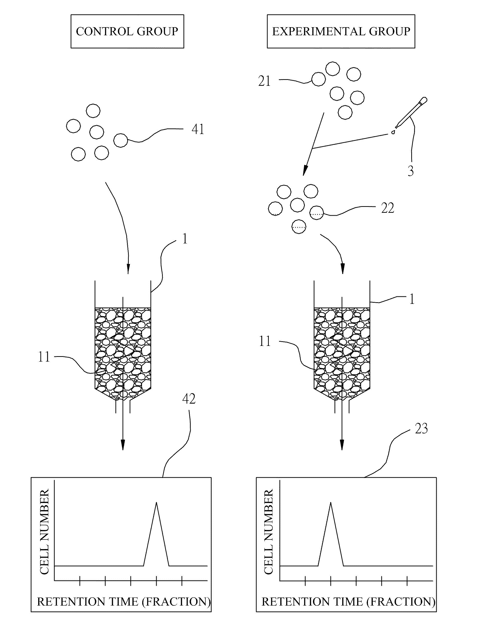
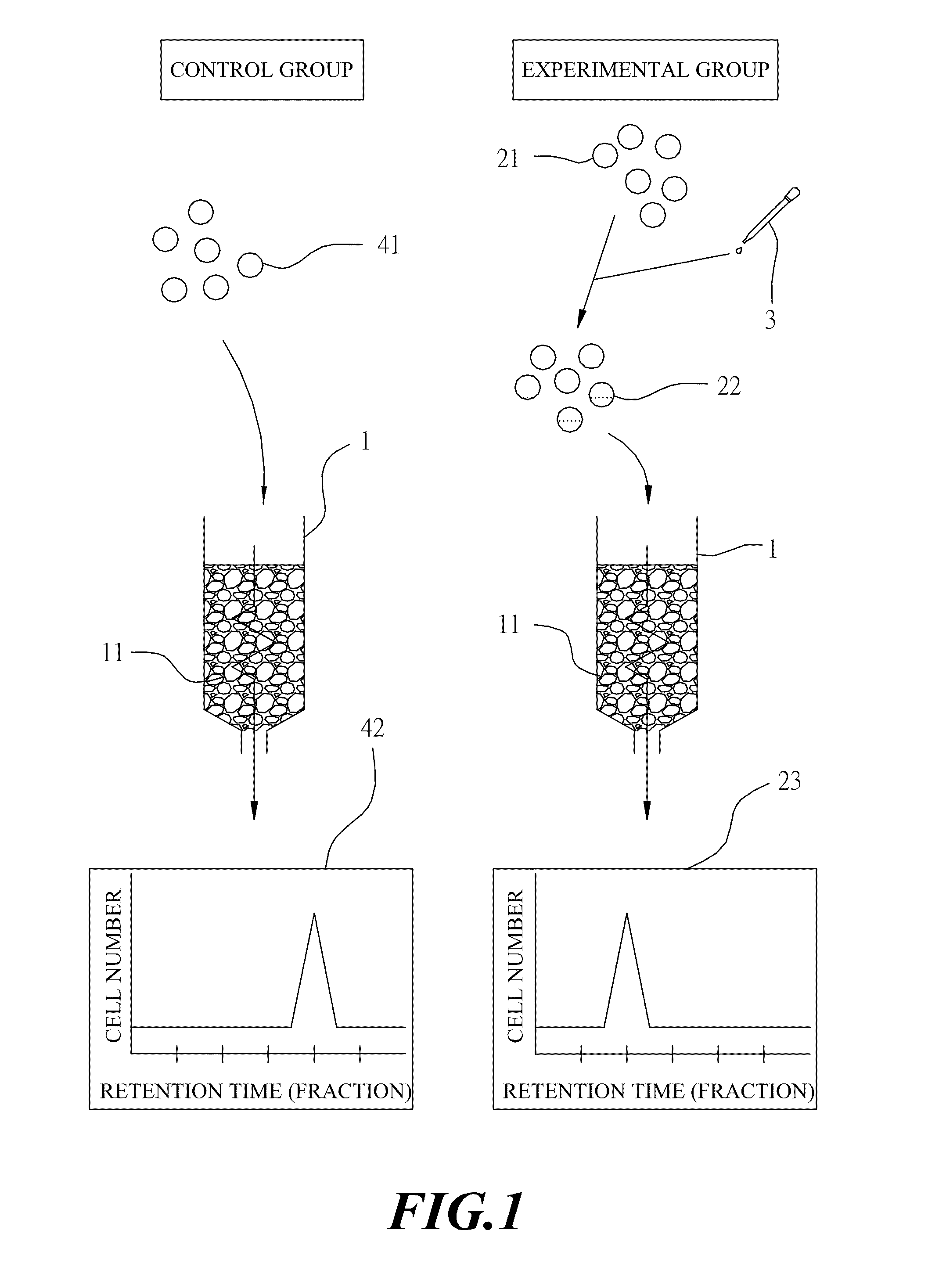
[0038]The invention provides a method and apparatus to screen and analyze the interaction between small molecules and cells. The interactions between cell surface and resin particles resulting in the different retention time of cells treating by different small molecules in the column contributed to examining whether the small molecule could interact with cell or not. In this example, using lectin and shikonin as an analyzed drug (small molecule). Different small molecules may cause different interactions with the same cell, such as binding to the different receptors, or interacting with different cell surface molecules or altering the cell membrane. Hence, this will cause the retention time of the drug-treated cell (or un-treated cell) with the small molecules in the column to be different. According, the method and apparatus can be used to screen the interaction exist or not between small molecules and cell surface.

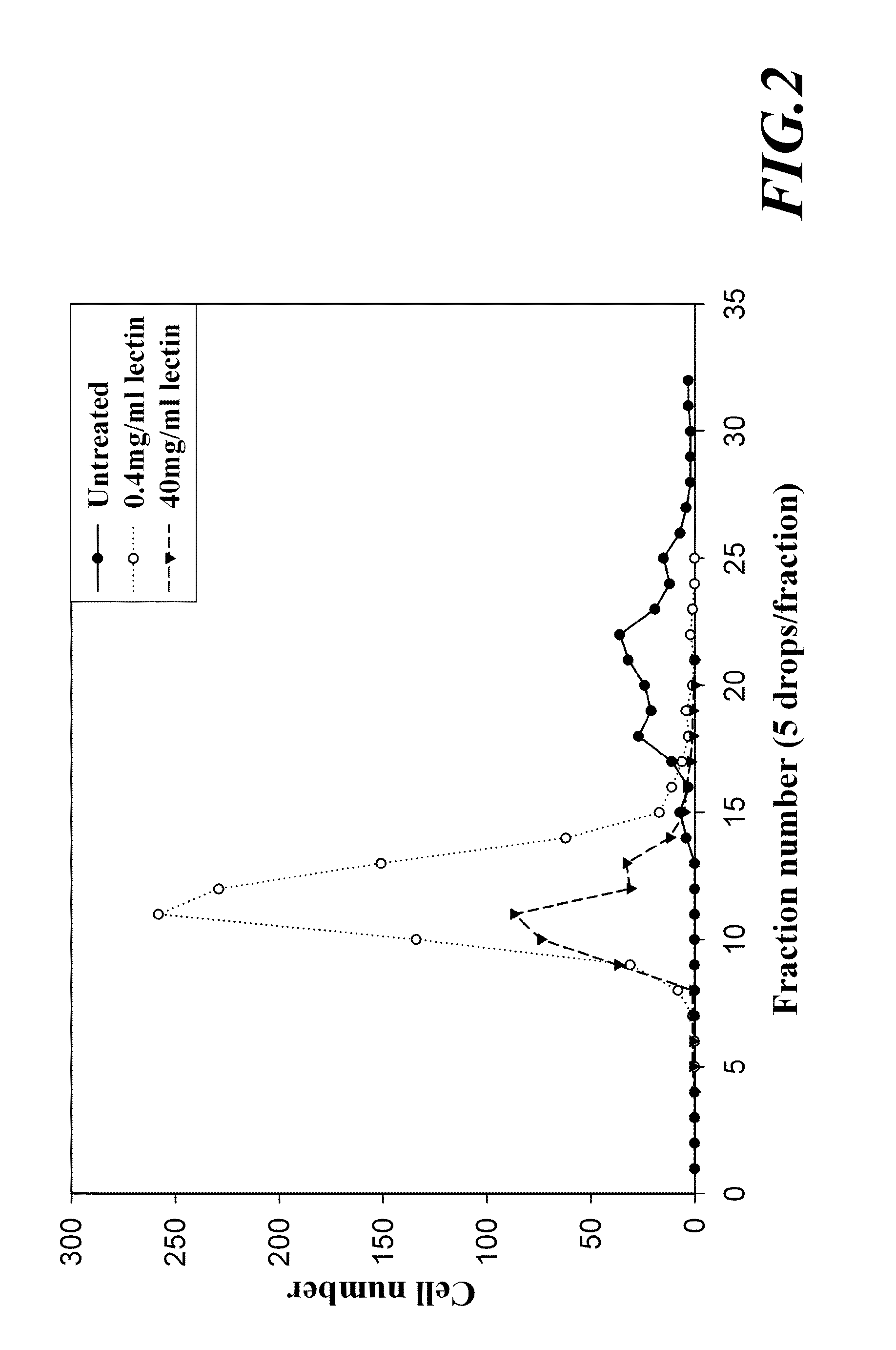
1. Lectin Altered the Rete...

example 2

Separation of Sub-Population of Mononuclear Cells

Example 2(A)

[0052]In example 2(A), the column also could be used for separating cell has an inner diameter of 6 mm, a length of 180 mm and a volume of 5 ml. This column was packed with resin particles and was washed first and thereafter, filled with a phosphate buffered saline (PBS).

[0053]A concentrated mononuclear cells suspension was diluted with PBS to a cell suspension at a concentration of 1×10⁶ mononuclear cells / ml, containing still a certain amount of erythrocytes. This cell suspension was loaded then on the above-described column. The column was eluted subsequently with PBS at a flow rate of 3 ml / min and cell fractions were collected in test tubes, respectively, in a manner that each test tube collected 5 drops of cell suspension eluent. Thereafter, the eluted cell suspension in each test tube was examined under an optical microscope and numbers of erythrocytes and leukocytes were counted by a cell counter, respectively. The r...

example 2 (

Example 2(B)

[0055]In example 2(B), the column also be used for separating cell was changed and has an inner diameter of 8 mm, a length of 200 mm and a volume of 10 ml. The procedure of example 2(A) was repeated, and the column was filled at a flow rate of 1.2 ml / min. The result was shown in FIGS. 7 and 8. FIG. 7 shows the number of mononuclear cells, while FIG. 8 shows the relative percentage of each sub-population of mononuclear cells.

[0056]The results obtained above suggest that the column could not achieve any separation effect against a single population of erythrocyte as shown in FIG. 4. For mononuclear cells in a same sample, several bands were eluted successively, as shown in FIGS. 5 and 7. After analyzing further by fluorescence immuno-staining, the column provided a partition effect with respect to various sub-populations of mononuclear cells such as, T lymphocyte, B lymphocyte and monocyte, and revealed a significantly difference variation, as shown in FIGS. 6 and 8.

[0057]...


PUM

PropertyMeasurementUnit
sizeaaaaaaaaaa
pHaaaaaaaaaa
diametersaaaaaaaaaa

Abstract

A method and an apparatus are provided for analyzing the interaction between small molecules and cells without using density gradient and antibody but using a column packed with identical resin particles. The interactions between cell surface and resin particles resulting in the different retention time of cells treating by different small molecules in the column contributed to examining whether the small molecule could interact with cell or not. This invention can also apply small molecule screening, drug screening through examining the retention time by using the column.

Description

RELATED APPLICATIONS[0001]This application is a continuation-in-part of U.S. patent application Ser. No. 11 / 119,908, filed on May 3, 2005.BACKGROUND OF THE INVENTION[0002]1. Field of the Invention[0003]The invention relates to an apparatus and a method for analysis of the interaction between small molecules and cells without using density gradient and antibody.[0004]2. Description of the Prior Art[0005]In current pharmaceutical studies, researchers wish to find the drug target site on cells so as to understand the action mechanism of drug on cells in order to develop more efficient or safer drugs. In the experimental design, homogeneous cells are used to demonstrate the action mechanism of drugs so as to understand the specific response from a particular cell population to a particular drug, and to identify the effect of a drug on different tissue. Thus, the separation and purification of cells play a highly important role in pharmacological experiments. Providing a unique populatio...

Claims


Application Information

Patent Timeline
Patent Type & Authority Applications(United States)
IPC IPC(8): G01N33/567C12M1/34
CPCG01N1/405G01N2500/10G01N2015/1486G01N33/569
Inventor CHANG, JIA-MING
Owner CHIH SHIN BIOMEDICAL TECH
Why Eureka
Try Eureka