Diagnostic system for the detection of skin cancer

a diagnostic system and skin cancer technology, applied in the field of skin cancer diagnostic system, can solve the problems of difficult early detection of malignant melanoma, fatal diagnosis errors, and more likely to spread, and achieve the effect of rapid evaluation and analysis of multiple skin lesions

Active Publication Date: 2009-09-03
ORLUCENT INC
View PDF35 Cites 28 Cited by
  • Summary
  • Abstract
  • Description
  • Claims
  • Application Information

AI Technical Summary

Benefits of technology

[0012]In some embodiments, a system for non-invasive diagnosis of neoplastic skin lesions is provided. The system comprises a compact illumination unit that provides illumination at a wavelength appropriate for image acquisition of a fluorescent probe; and a detection unit that receives the light emitted by the surface under investigation to provide a recording of the data. Optionally, the system further comprises a topical formulation comprising a diagnostically effective dose of a fluorescently labeled probe that specifically binds to a neoplastic marker. The fluorescently labeled probe may be provided in an excipient that enhances transdermal penetration. Optionally, the system further comprises an imaging unit that can capture and / or develop the image generated from the surface under investigation.
[0015]The invention provides a rapid evaluation tool to quickly analyze multiple skin lesions in a short time period, as well as in a non-invasive manner, to assess which lesions express markers associated with neoplasia. Additional options provide mapping of lesions, which may be archived for follow up screens to determine progression of growth.

Problems solved by technology

However if malignant melanoma is diagnosed at an early stage, it can usually be cured, but melanoma diagnosed at a late stage is more likely to spread and cause death.
Early detection of malignant melanoma is difficult because early melanomas, those having a Breslow thickness less than 1 mm, share many diagnostic features with benign lesions, such as dysplastic nevi or atypical melanocytic nevi.
The effectiveness of this procedure is dependent on the experience of the examiner, and errors in diagnosis can be fatal.
Occasionally, cancers have remained undetected which metastasize beyond the original site of occurrence and lead to a decrease in survival.
Thus high frequencies of unnecessary surgical procedures occur.
On several occasions the biopsy is carried out in the skin of the facial region and may result in some incidents of cosmetic scarring.
These detection methods are limited and cannot be used by the general physician's practice for screening individuals nor can they be used to determine the extent of the cancer lesion.

Method used


Image

  • Diagnostic system for the detection of skin cancer
  • Diagnostic system for the detection of skin cancer
  • Diagnostic system for the detection of skin cancer

Examples


Embodiment Construction

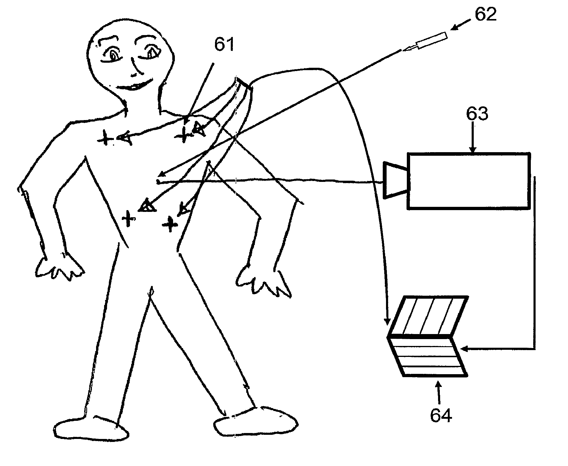
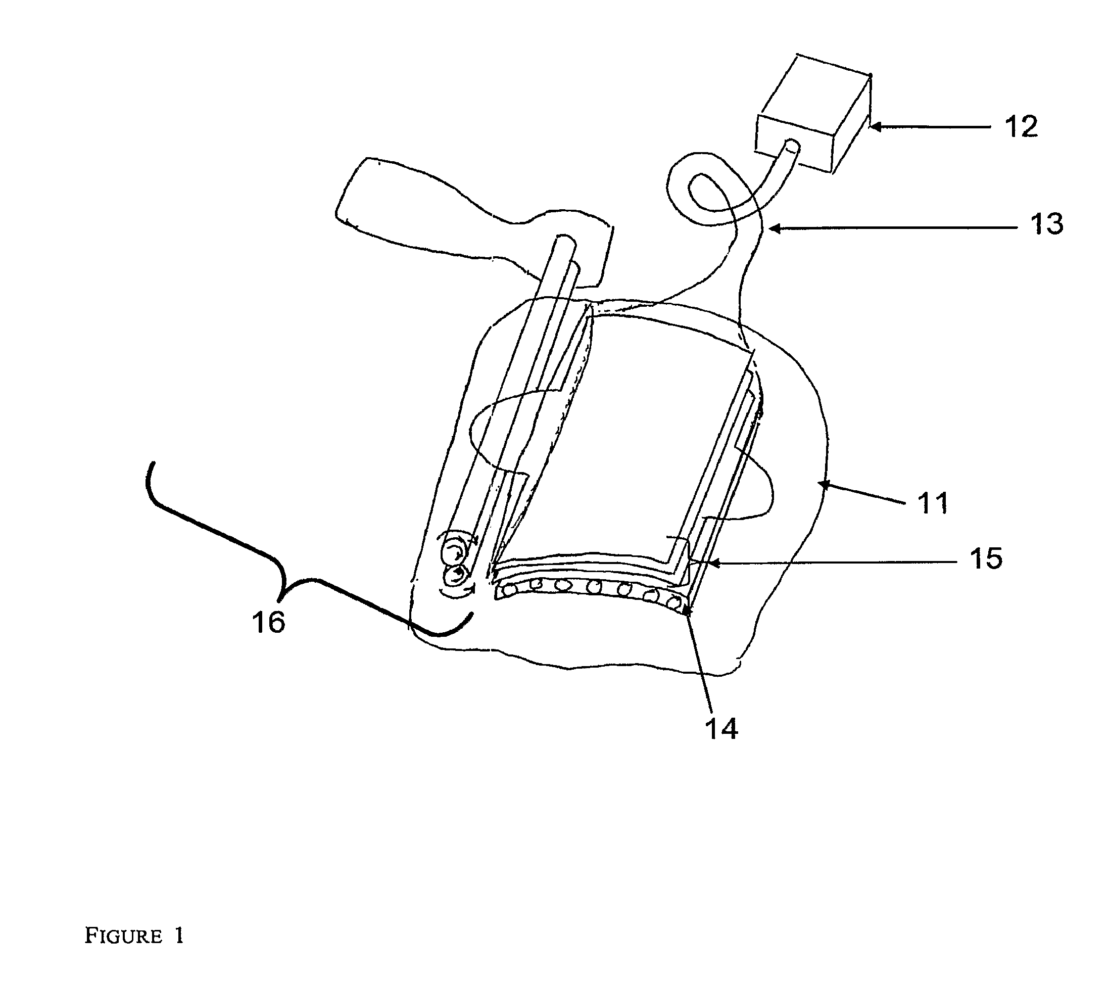
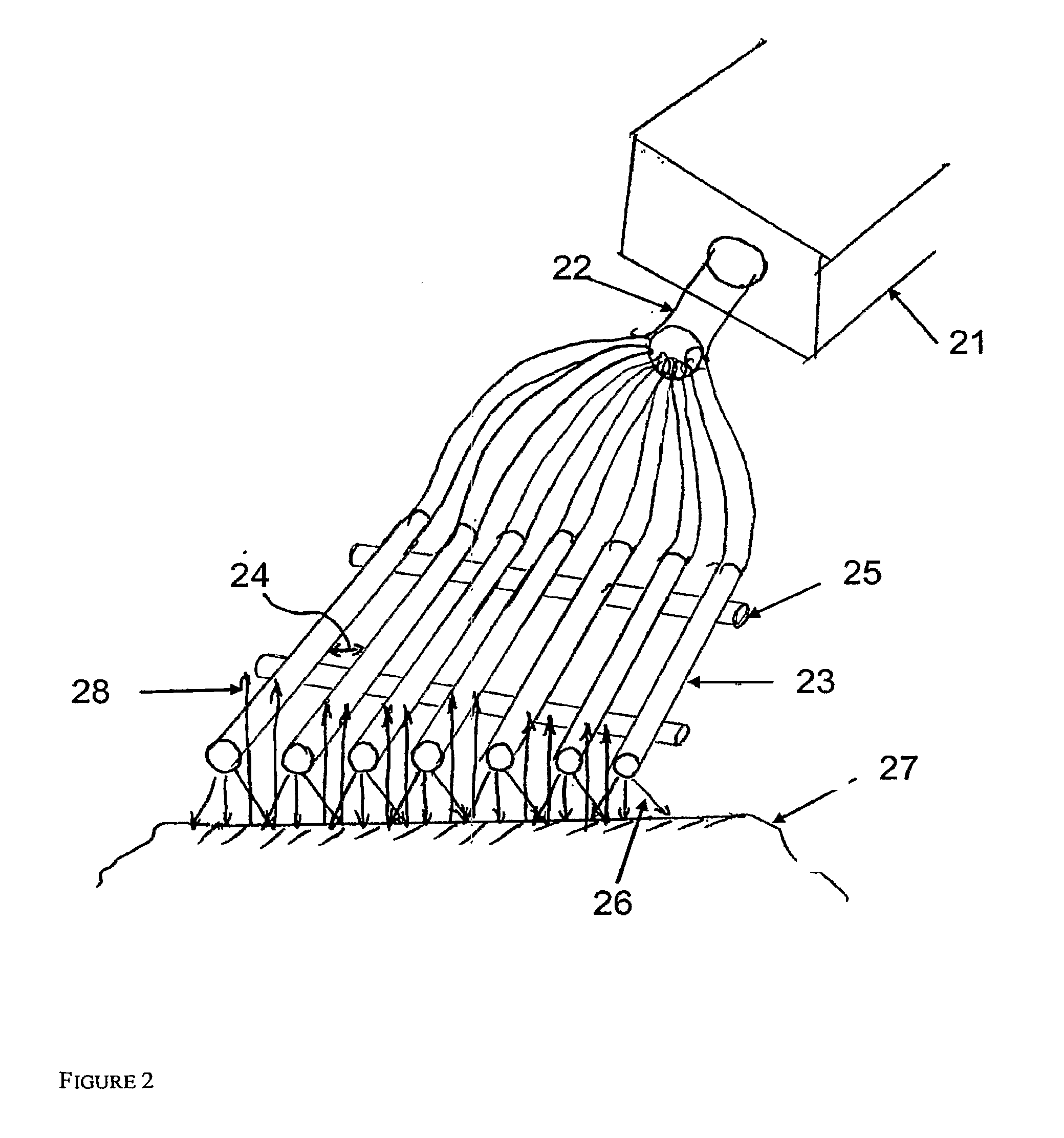
[0022]The present invention provide methods for identifying, characterizing and distinguishing between non-malignant and malignant skin lesions. In the methods of the invention, a fluorescently labeled probe that specifically binds to a neoplasia associated marker is topically administered to a lesion suspected of being neoplastic. In some embodiments, the probe is administered in a formulation that enhances transdermal penetration, e.g. in DMSO. Neoplasia associated markers of interest include, without limitation, integrin αvβ3 and / or integrin α5β1. The probe preferentially binds to markers present in neoplastic lesions, which binding is detected with a compact illumination unit that provides illumination at a wavelength appropriate for image acquisition.

[0023]Before the present compositions and methods are described in further detail, it is to be understood that this invention is not limited to particular methods described, as such may, of course, vary. It is also to be understood...


PUM


Abstract

Methods and compositions are provided for the analysis of skin surfaces to determine the presence of neoplastic tissue. In the methods of the invention, a composition comprising a florescent probe that binds to a specific neoplasia associated marker is applied topically to the area of interest. After topical administration, the probe preferentially binds to markers associated in neoplastic lesions in situ, which binding is detected with a compact illumination unit that provides illumination at a wavelength appropriate for image acquisition. The illumination unit comprises a light source and fiber optic bundle to direct the light towards the area of examination. A detection unit is used to capture and record an image of the area of investigation. The detection unit may be a digital camera, film camera, etc. A mapping module may also be provided to catalogue the site of examination.

Description

FIELD OF INVENTION[0001]This invention relates to test methods, devices and test kits for determining the presence of neoplastic cells or tissue in the skin or in proximity of the skin by using non invasive procedures.BACKGROUND OF THE INVENTION[0002]More than 1 million cases of basal cell or squamous cell cancer will be diagnosed annually. The most serious form of skin cancer, malignant melanoma, is expected to be diagnosed in 59,580 persons in 2005 in the US. Since 1981, the incidence of melanoma has increased approximately 3% per year. Melanoma is the most common cancer among people 25 to 29 years old. The three major types of skin cancer are basal cell carcinoma, squamous cell carcinoma, and melanoma.[0003]Although melanoma accounts for only 4% of all dermatological cancers, it causes more than 75% of all deaths related to skin cancer (Miller and Mihm, 2006, N Engl J Med, 355, 51-65). Metastatic melanoma can spread to other organs than the skin, most commonly the lungs and liver...

Claims


Application Information

Patent Timeline
Patent Type & Authority Applications(United States)
IPC IPC(8): A61K51/00A61B5/00
CPCA61B5/0071A61B5/416A61B5/444A61B5/445A61B2562/0233A61K51/00A61B2562/164A61K49/0006A61K49/0017A61K49/0004A61B2562/046A61B5/0077A61B2560/0431A61B2562/166A61B2576/02A61B2090/306
Inventor SHACHAF, CATHERINE M.SHACHAF, AMIT
Owner ORLUCENT INC
Why Eureka
Try Eureka