Optical unit for probe and optical unit producing method

Inactive Publication Date: 2009-10-01
FUJIFILM CORP
View PDF6 Cites 13 Cited by
  • Summary
  • Abstract
  • Description
  • Claims
  • Application Information

AI Technical Summary

Benefits of technology

[0026]Accordingly, an image of tissue can be stably obtained owing to an improved assembl

Problems solved by technology

It has been very difficult to position the lens optics with one another at very high precision.
An image in the tissue cannot be created with high quality if the contact between the first lens/lens group and the tissue is insufficient.
However, various problems arise with the struc

Method used


Image

  • Optical unit for probe and optical unit producing method
  • Optical unit for probe and optical unit producing method
  • Optical unit for probe and optical unit producing method

Examples


Embodiment Construction

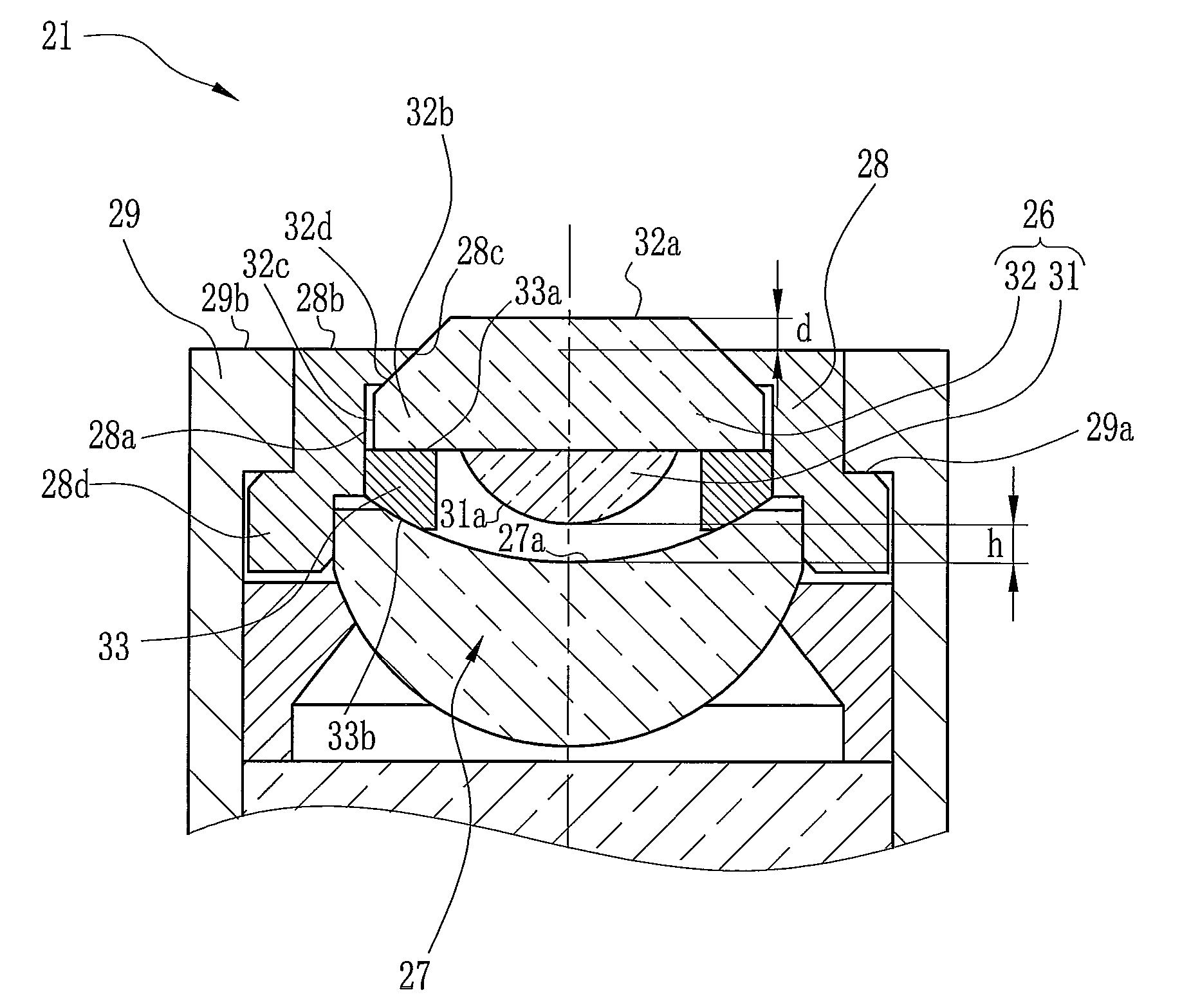
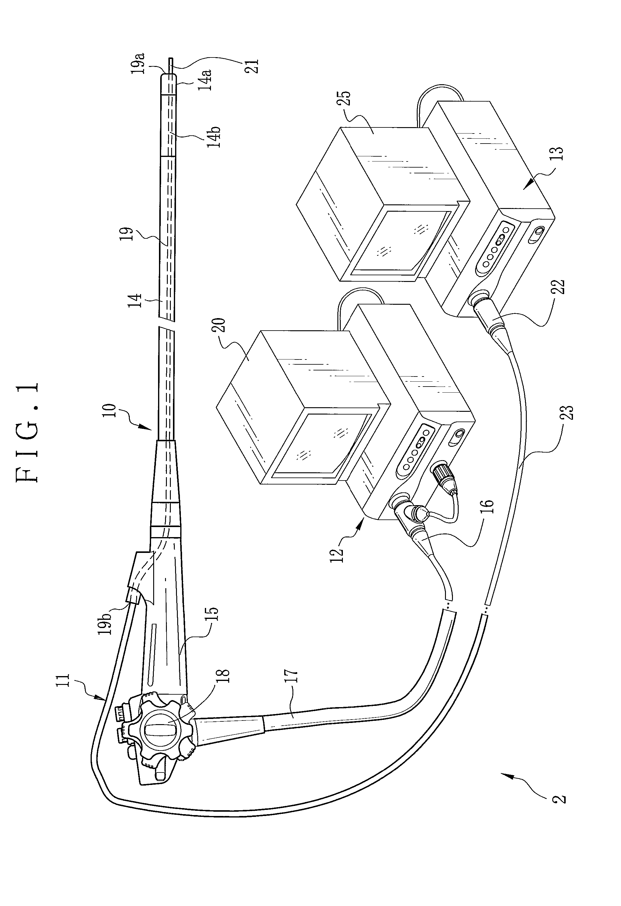
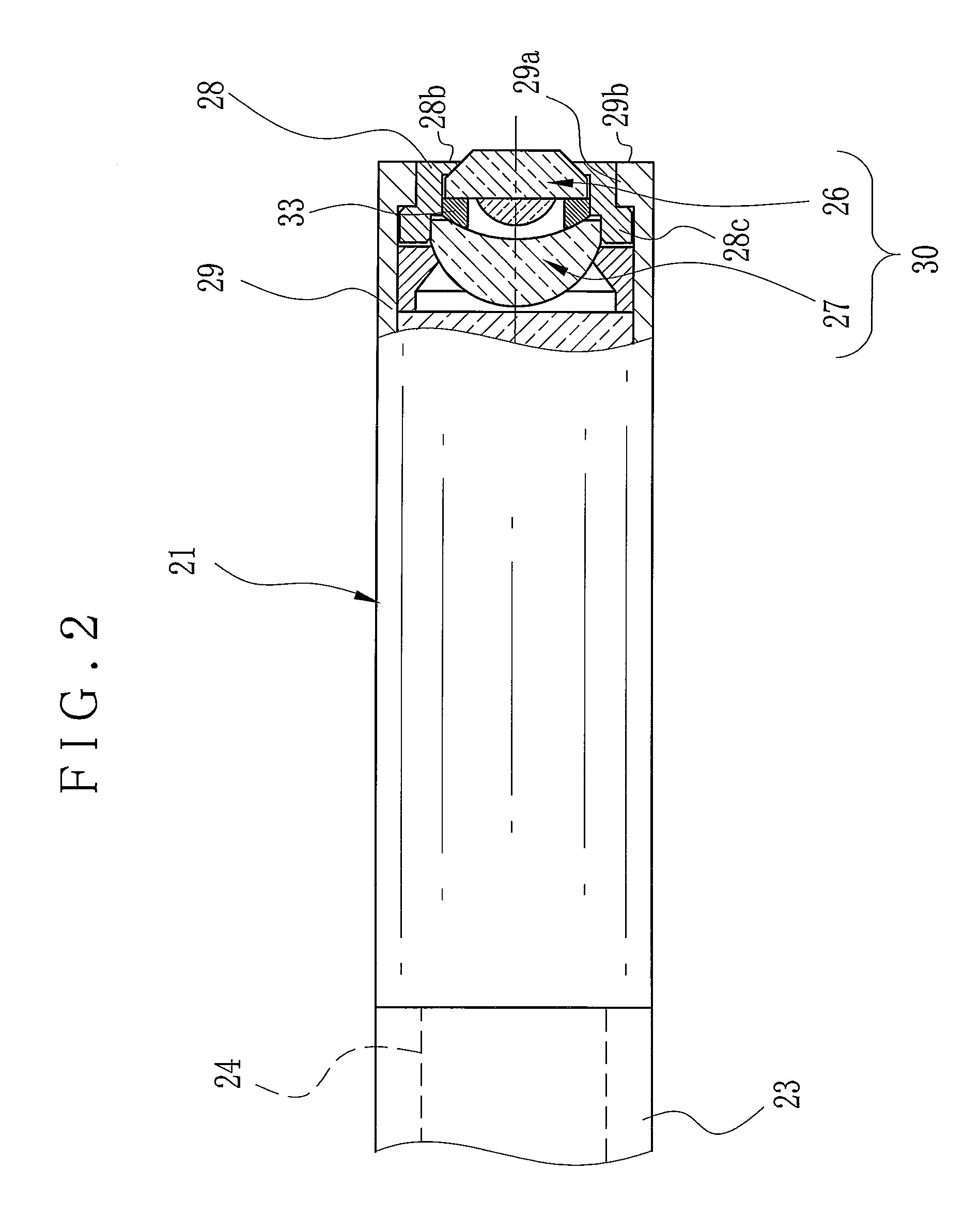
[0038]In FIG. 1, an endoscope system 2 includes an electronic endoscope 10 and a processor 12 in connection with the endoscope 10 for endoscopic imaging. Also, a probe apparatus is associated with the endoscope system 2, and includes a confocal laser probe 11 and a control processor 13. A forceps channel 19 is formed through the endoscope 10. The confocal laser probe 11 is inserted through the forceps channel 19 for use. The control processor 13 is connected with the confocal laser probe 11 for confocal laser imaging and observation. The endoscope 10 includes an insertion tube 14, a handle 15, a connector 16 and a universal cable 17. The insertion tube 14 is flexible and enters a human cavity or gastrointestinal tract of a patient's body. The handle 15 is disposed at a proximal end of the insertion tube 14. The connector 16 is connectable with the processor 12. The universal cable 17 extends between the handle 15 and the connector 16 for connection.

[0039]A head assembly 14a is dispo...


PUM


Abstract

A probe apparatus in combination with an endoscope includes a confocal laser probe for in-vivo imaging. The probe is insertable through a forceps channel of an endoscope, and has an optical unit. There is a lens barrel. Plural lens optics are mounted in the lens barrel. The plural lens optics include first lens optics disposed on an object side, and opposed to an object within a body. An optically inactive surface is formed with the first lens optics, and has at least one portion protruding from a barrel end surface of the lens barrel on the object side. Preferably, a height difference of protruding the optically inactive surface from the barrel end surface is 10-500 microns. The portion of the optically inactive surface protruding from the barrel end surface is coated with adhesive agent, for adhesion of the first lens optics thereto.

Description

BACKGROUND OF THE INVENTION[0001]1. Field of the Invention[0002]The present invention relates to an optical unit for a probe, and optical unit producing method. More particularly, the present invention relates to an optical unit for a probe, and optical unit producing method, in which the probe of a probe apparatus is used for in-vivo imaging of a tissue in a human cavity of a patient's body, and an image of the tissue can be stably obtained owing to an improved assembly of the optical unit.[0003]2. Description Related to the Prior Art[0004]A confocal laser probe of a probe apparatus is known, including an optical unit disposed at a distal end of single-mode optical fibers. The optical unit applies laser light to a tissue of a gastrointestinal tract in a patient Is body. Among components of the laser light reflected by the tissue, only reflected light at the focal plane of the optical unit on an object side, an object image of the tissue is obtained. U.S. Pat. Pub. No. 2004 / 156124 (...

Claims


Application Information

Patent Timeline
IPC IPC(8): G02B7/02
CPCA61B1/0011A61B1/00165A61B1/00188G02B23/2476A61B1/018G02B7/025G02B23/243A61B1/0125
Inventor ISHII, SHUICHIAYAME, DAISUKE
Owner FUJIFILM CORP
Why Eureka
Try Eureka