Method for Characterizing X-Ray Detector Materials Using a Raman Microscope

a raman microscope and detector material technology, applied in the field of microspectrometry, can solve the problems of affecting the free movement of electron hole pairs, crystals to be generated in selenium, and it is difficult to use existing ram an microspectrometers to analyze digital image panels in their entirety

Active Publication Date: 2010-01-28
HOLOGIC INC
View PDF4 Cites 0 Cited by
  • Summary
  • Abstract
  • Description
  • Claims
  • Application Information

AI Technical Summary

Benefits of technology

[0009]According to one aspect of the invention, a method for performing spectroscopy of a sample using a microspectrometer is described. The present invention enables spectroscopy to be performed across a desired analysis range that exceeds at least one of a size and travel capability of a stage of the microspectrometer to thereby allow analysis of oversized samples such as mammography panels using the existing microspectrometer hardware and software. The he method comprising the steps of controlling movement of a supplemental stage coupled to the microspectrometer to position an extended optical path of the microspectrometer over a point in the desired analysis range, wherein the sample is supported by the supplemental stage and wherein the movement of the sample enables spectral analysis of the sample at the point. The microspectrometer is then controlled to emit at least one laser pulse onto the point of the sample on the optical path and to perform spectral analysis of scatter wavelengths received in response to the laser pulse via the optical path. Such a process enables spectral analysis of oversized samples without destruction of the sample.

Problems solved by technology

However it is sometimes desirable to perform Raman analysis on samples having a size that exceeds that of an existing optical microscope stage.
However anomalies in the manufacturing process may give rise to defects within the amorphous selenium that impair the free movement of electron hole pairs.
For example, temperature changes or other processing procedures may cause crystals to be generated in the selenium.
However it is difficult to use existing Ram an microspectrometers to analyze digital image panels in their entirety because the size of the flat panel cannot be accommodated by the existing stage and travel capabilities of the microspectrometer.
In addition, even if the travel of the existing stage could be adjusted, the physical space constraints between the microscope 20, optical transfer tube 33, and spectrometer 30 limit the ability to properly examine the entire panel.
As a result, inspection of problem coordinates of a mammography panel requires destruction of the panel.
This process was time consuming, destructive and concomitantly expensive.

Method used


Image

  • Method for Characterizing X-Ray Detector Materials Using a Raman Microscope
  • Method for Characterizing X-Ray Detector Materials Using a Raman Microscope
  • Method for Characterizing X-Ray Detector Materials Using a Raman Microscope

Examples


Embodiment Construction

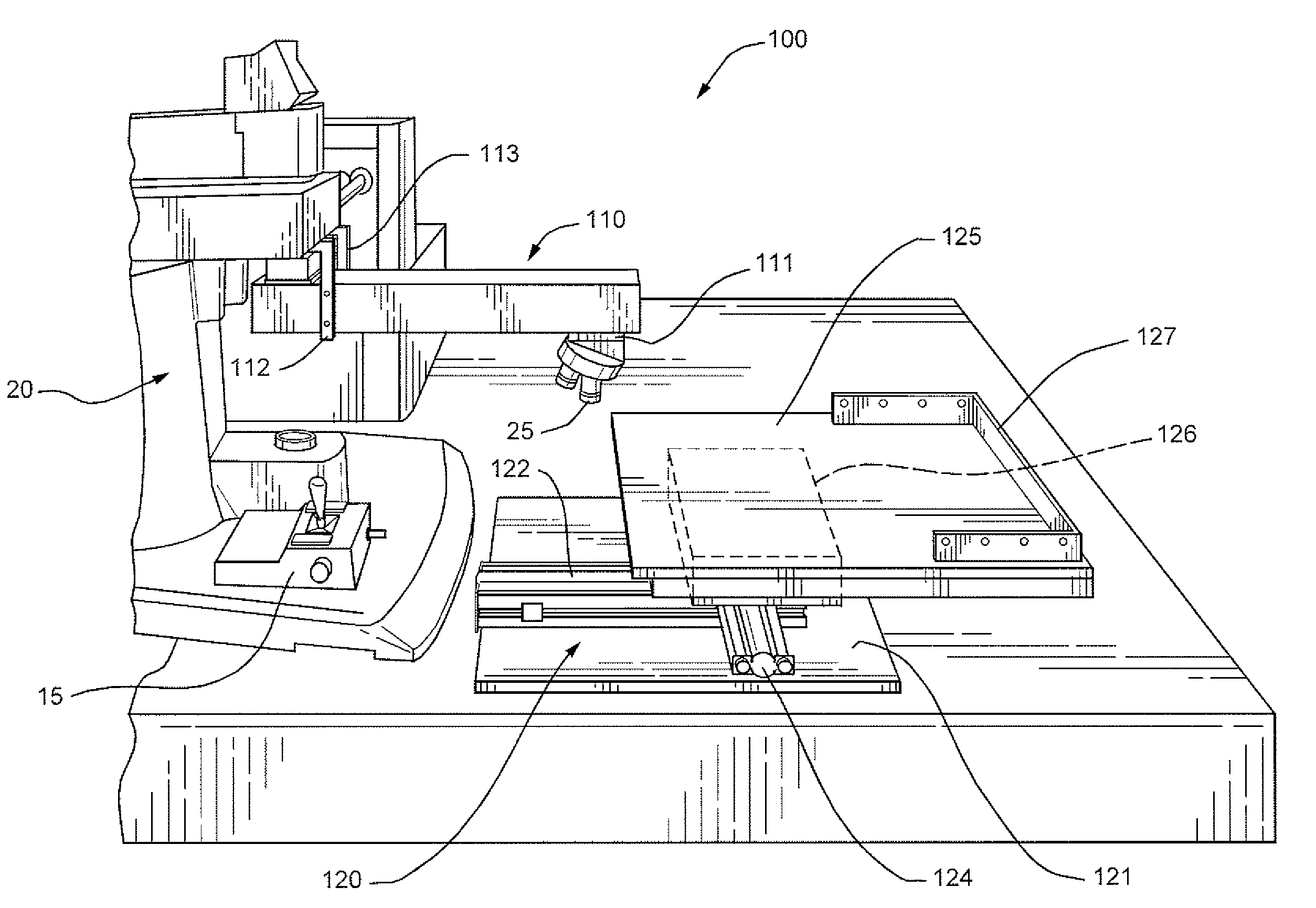
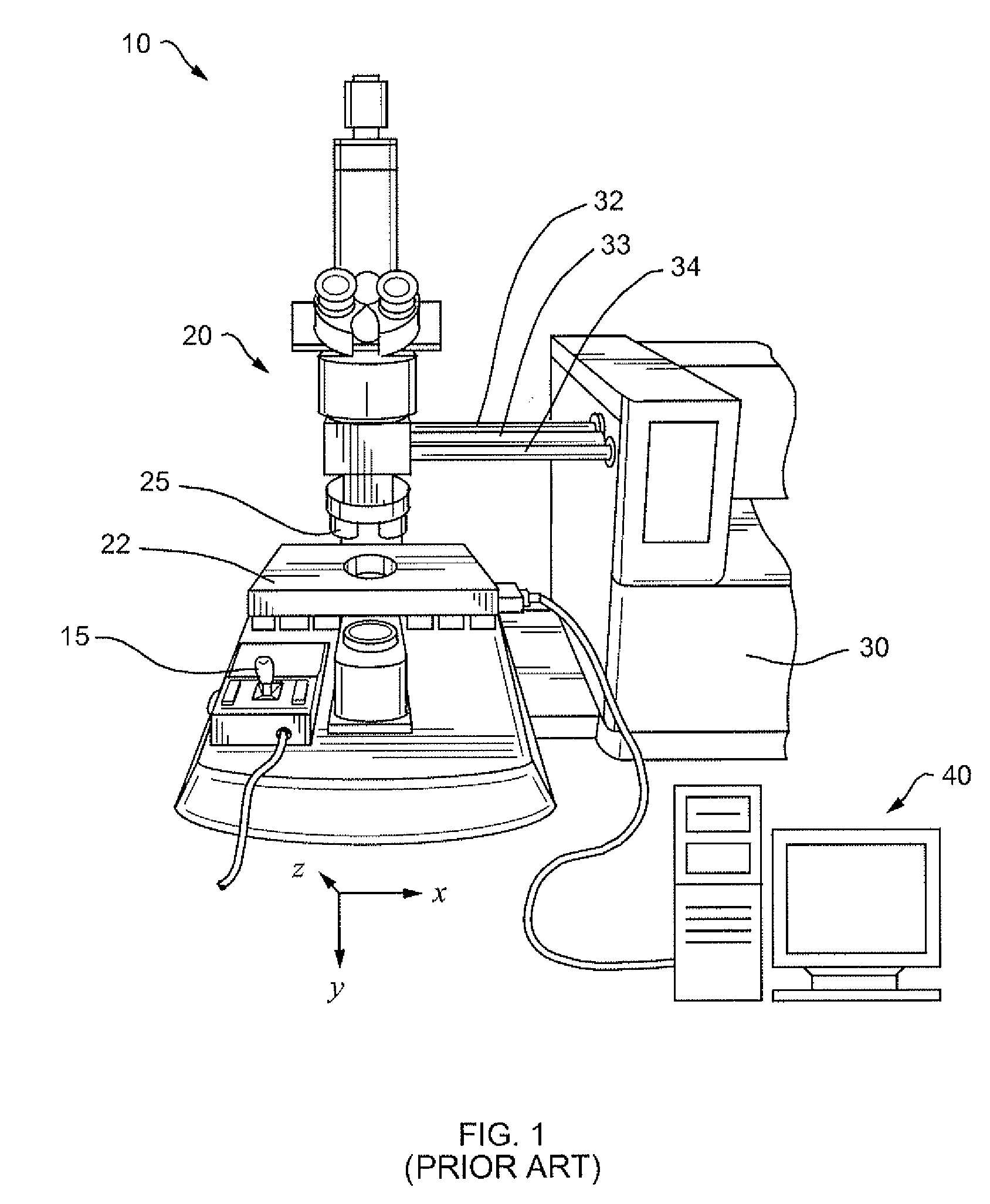
[0024]According to one aspect of the invention, an improved Raman microspectrometer system extends the optical reach and analysis range of an existing Raman microspectrometer to allow analysis and / or repair of an oversized sample. For the purposes of this application, an oversized sample shall mean any sample that exceeds the travel capabilities of an existing stage of the existing Raman microspectrometer in any one of an x, y or z dimensions. The improved Raman microspectrometer system includes an extender for extending the optical reach of the existing microspectrometer and a supplemental stage which extends the analysis range of the existing microspectrometer by providing travel capabilities for non-destructive analysis of an entire oversized sample. Such an arrangement decreases manufacturing costs associated with testing oversized samples such as mammography panels, enabling analysis and / or repair to be performed without destruction. In addition, as will be described further be...


PUM


Abstract

An improved Raman microspectrometer system extends the optical reach and analysis range of an existing Raman microspectrometer to allow analysis and / or repair of an oversized sample. The improved Raman microspectrometer system includes an extender for extending the optical reach of the existing microspectrometer and a supplemental stage which extends the analysis range of the existing microspectrometer by providing travel capabilities for non-destructive analysis of an entire oversized sample. Such an arrangement decreases manufacturing costs associated with testing oversized samples such as mammography panels, enabling analysis and / or repair to be performed without destruction.

Description

FIELD OF THE INVENTION[0001]This invention relates generally to the field of micro-spectrometry and more particularly to a non-destructive method and apparatus for identifying, analyzing and repairing digital imaging panels using a microspectrometer.BACKGROUND OF THE INVENTION[0002]FIG. 1 illustrates a typical Raman microspectrometer 10. The Raman microspectrometer 10 includes an optical microscope 20, coupled via supports 32 and 34 to a combined excitation laser source / spectrometer 30. The Raman microspectrometer is used to analyze the molecular structure of a sample that is disposed on the microscope stage 22. During analysis the sample is secured to the stage 22 and laser beam pulses are directed via the optical transfer tube 33 through the lens of the microscope 20 onto points in the sample. Resulting Raman and Rayleigh scatter from the sample is forwarded back through the microscope lens and optical transfer tube 33 to the spectrometer. The spectrometer filters out the Rayleigh...

Claims


Application Information

Patent Timeline
Patent Type & Authority Applications(United States)
IPC IPC(8): G01J3/44
CPCG02B21/26G02B21/24
Inventor MAZZIO, VICTOR
Owner HOLOGIC INC
Why Eureka
Try Eureka