
Magnetic resonance method and apparatus for generating a perfusion image

a magnetic resonance and perfusion image technology, applied in the field of magnetic resonance perfusion image generation methods and apparatuses, can solve the problems of inability to immediately obtain final combined perfusion images, and achieve the effect of reducing the effect of motion artifacts and improving the accuracy and quality of the resulting perfusion imag

Active Publication Date: 2010-02-11
SIEMENS MEDICAL SOLUTIONS USA INC
View PDF7 Cites 8 Cited by
  • Summary
  • Abstract
  • Description
  • Claims
  • Application Information

AI Technical Summary

Benefits of technology

[0010]It is an object of the present invention to provide a method and apparatus for acquiring magnetic resonance perfusion images wherein the effect of motion artifacts is reduced, thereby improving the accuracy and quality of the resulting perfusion images.
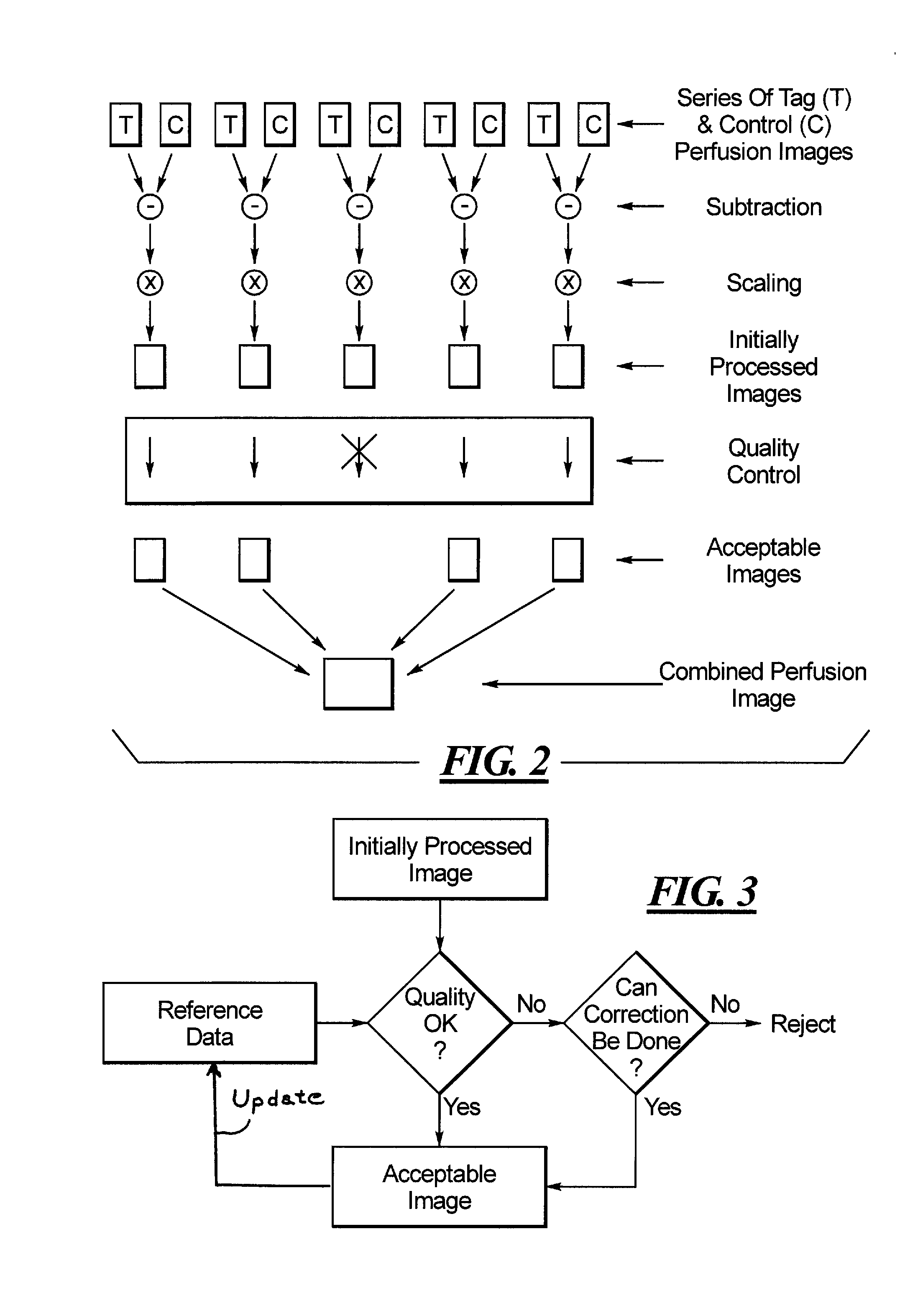
[0011]The above object is achieved in accordance with the present invention in a method and apparatus for magnetic resonance perfusion imaging wherein the initially processed perfusion images, that respectively result from a processed combination, such as subtraction, of a tag image and a control image, are subjected to a quality control analysis before the initially processed images are combined to form the resultant perfusion image. In the quality control analysis, any initially processed image that fails to satisfy predetermined quality control criteria is rejected, and is not used in the subsequent averaging, or combining procedure, that is used to generate the combined perfusion image. Initially processed images that exhibit excessive motion artifacts therefore do not make a contribution to the combined perfusion image, so that the overall quality of the combined perfusion image is significantly improved.
[0012]The quality control can take place fully automatically in computerized fashion, or can be conducted with user interaction, so that a user can make his or her own judgment as to the level of quality of the images that will enter into the combining procedure to produce the combined perfusion image. Even if the procedure is implemented automatically, a user or operator still can be informed by an alarm or some other type of indicator if an excessive number of initially processed perfusion images of unacceptable quality are habitually occurring, so that the user or operator can take corrective steps in the settings of the magnetic resonance data acquisition system that is being employed for generating the magnetic resonance data.

Problems solved by technology

In this embodiment, however, the final, combined perfusion images are not immediately available while the patient is still in the magnetic resonance data acquisition chamber.

Method used


Image

  • Magnetic resonance method and apparatus for generating a perfusion image
  • Magnetic resonance method and apparatus for generating a perfusion image
  • Magnetic resonance method and apparatus for generating a perfusion image

Examples


Embodiment Construction

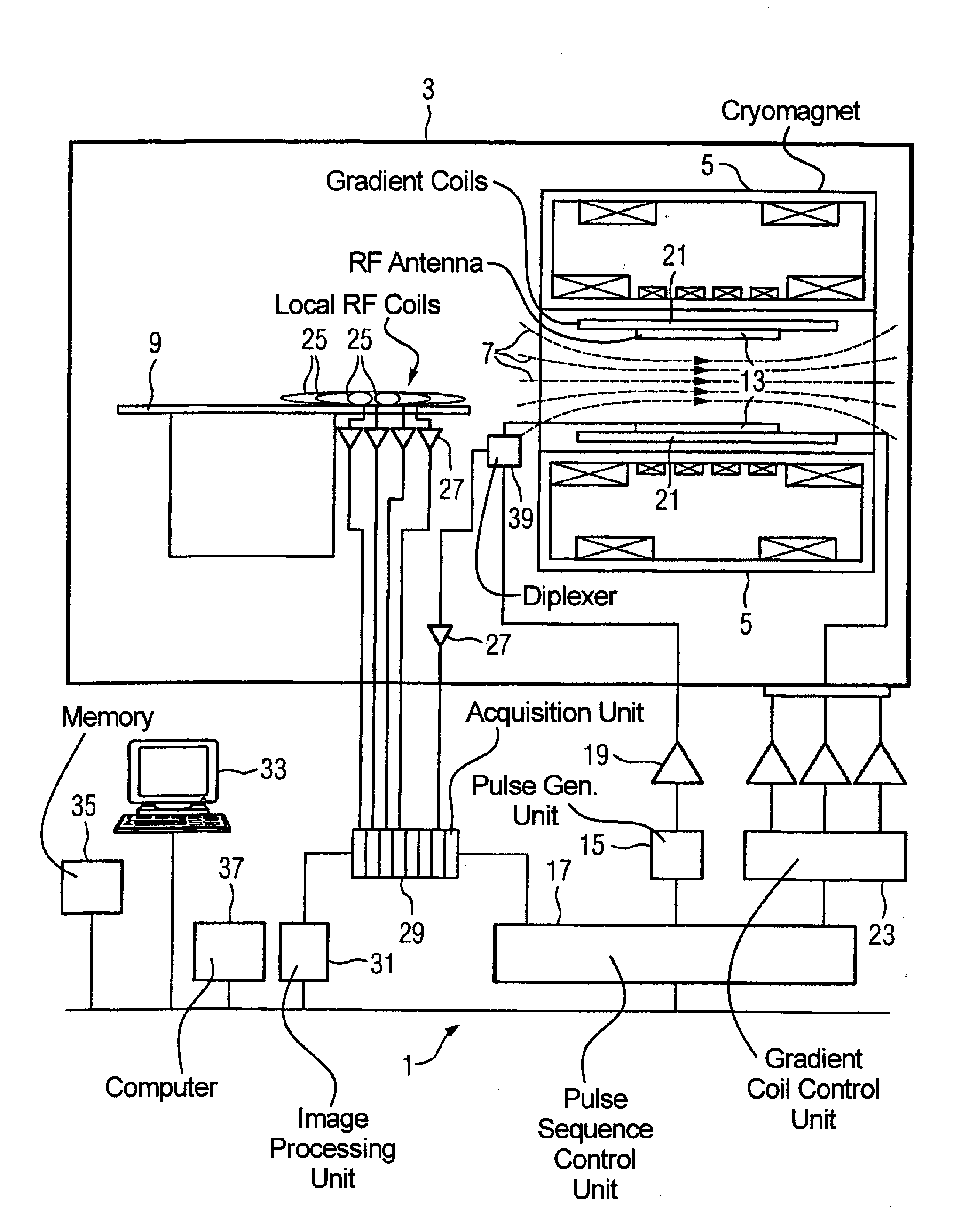
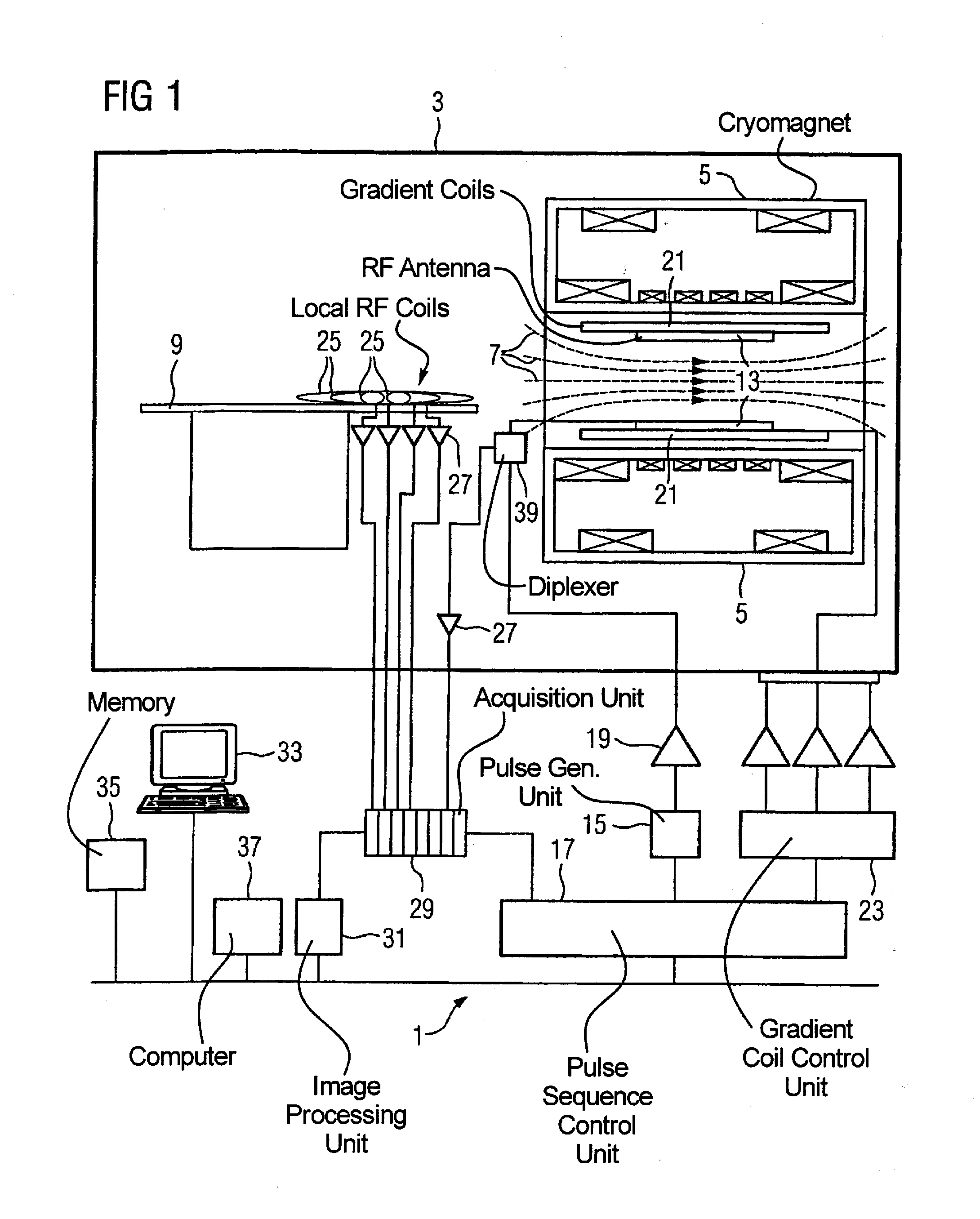
[0017]FIG. 1 schematically shows the design of a magnetic resonance apparatus 1 with its basic components. In order to examine a body by means of magnetic resonance imaging, various magnetic fields tuned to one another as precisely as possible in terms of their temporal and spatial characteristics are applied.

[0018]A strong magnet (typically a cryomagnet 5 with a tunnel-shaped opening) arranged in a radio-frequency shielded measurement chamber 3 generates a static, strong basic magnetic field 7 that typically amounts to 0.2 Tesla to 3 Tesla and more. A body or a body part (not shown here) to be examined is borne on a patient bed 9 and positioned in the homogeneous region of the basic magnetic field 7.

[0019]The excitation of the nuclear spins of the body ensues via magnetic radio-frequency excitation pulses that are radiated via a radio-frequency antenna (shown here as a body coil 13). The radio-frequency excitation pulses are generated by a pulse generation unit 15 that is controlle...


PUM


Abstract

In a magnetic resonance method and apparatus for generating perfusion images, a perfusion series of magnetic resonance perfusion images is acquired that includes tag images and at least one control image, that are grouped in pairs. From each pair an initially processed perfusion image is obtained, such as by subtraction. Each initially processed image is subjected to a quality control review by analysis with respect to at least one image quality criterion. Any initially processed image that does not satisfy the quality criterion is rejected. Only initially processed images that satisfy the quality criterion are combined to form a resultant magnetic resonance perfusion image. Artifacts in the resultant perfusion image are thereby reduced or avoided.

Description

BACKGROUND OF THE INVENTION[0001]1. Field of the Invention[0002]The present invention relates to a method and an apparatus for generating magnetic resonance images, and in particular to a method and an apparatus for generating magnetic resonance perfusion images.[0003]2. Description of the Prior Art[0004]Magnetic resonance technology has been increasingly used in recent years to generate angiographic images since, relative to other medical imaging modalities such as, for example, radioscopy with x-rays or computed tomography, it has among other things, the advantage that patient and medical personal are subjected to no radiation exposure.[0005]Magnetic resonance (MR) technology is a known technology with which images of the inside of an examination subject can be generated. For this purpose, the examination subject is positioned in a strong, static, homogeneous basic magnetic field (field strengths of 0.2 Tesla to 7 Tesla and higher) in an MR apparatus so that the subject's nuclear ...

Claims


Application Information

Patent Timeline
IPC IPC(8): A61B5/055
CPCA61B5/055G01R33/4833G01R33/56333G01R33/5635G01R33/56366G06T2207/30168G06T5/50G06T7/0002G06T7/0085G06T2207/10088G01R33/56509G06T7/13
Inventor PFEUFFER, JOSEF
Owner SIEMENS MEDICAL SOLUTIONS USA INC
Learn More