Method of Screening Antibacterial Drug Compounds

a technology of antibacterial drug and compound, applied in the field of medicine and agriculture, can solve problems such as the expression of bacterial proteins, and achieve the effect of greater toxicity

Inactive Publication Date: 2010-06-03
TEMASEK LIFE SCIENCES LABORATORY
View PDF2 Cites 3 Cited by
  • Summary
  • Abstract
  • Description
  • Claims
  • Application Information

AI Technical Summary

Benefits of technology

[0010]One embodiment of the invention provides an in vitro method for identifying antibacterial compounds that have significantly greater toxicity to a specific bacterial protein than to eukaryotic cells, which comprises providing a eukaryotic cell culture comprising eukaryotic cells, transfecting these cells with a gene encoding the bacterial protein, expressing the bacterial protein in the cells, screening these transfected eukaryotic cells with at least one candidate antibacterial compound for toxicity against or inhibition of the bacterial protein and simultaneously assessing the transfected eukaryotic cells for indica...

Problems solved by technology

As used herein, toxicity to or toxic effects against the expressed bacterial protein m...

Method used


Image

  • Method of Screening Antibacterial Drug Compounds
  • Method of Screening Antibacterial Drug Compounds
  • Method of Screening Antibacterial Drug Compounds

Examples


example 1

Fusion-Protein Construction and Transformation of the Yeast Schizosaccharomyces pombe

[0059]Transformations with the plasmids were carried out by using the Lithium-acetate method on S. pombe strain MBY192 (h− leu1-32 ura4-D18) (lab collection) or S. pombe mutants MBY241 (h− arp3-1c leu1-32 ura4D-18 ade6-210), MBY311 (h− cdc12-112 leu1-32 ura4D-18), MBY598 (h90 arp2-1a mam2::leu2 leu1-32 ura4D-18 ade6-216), MB-Y1446 (h− alp4-1891 leu1-32), MYB1565 (wee1-50 ura4D-18), MBY2527 (h−Δfor3::kan leu1-32 ura4D18 ade6), MBY415 (h− orb6-25 ade6-M210 leu1-32), and MBY3197 (h− tea1Δ946::kanR leu1-32 ura4-D18 ade6-M216). GFP-MreB, its mutants, and FtsZ-GFP were all expressed from the medium-strength nmt42 promoter. The following fusions were constructed: gfp-mreB, mrfp-mreB, ftsZ-gfp, and sulA-HA.

[0060]The ORF corresponding to GFP was amplified from pREP42-GFP with primers MOH2411 and MOH2412 (for fusion to mreB) and MOH1017 and MOH1276 (for fusion to ftsZ). See Table I, below for primer sequence...

example 2

Expression of GFP Fusion Proteins and Fluorescence Microscopy

[0062]GFP-MreB and FtsZ-GFP were expressed in S. pombe by growing the transformed cultures in the absence of thiamine for 16-20 hours. In the case of fission-yeast temperature-sensitive mutants carrying the GFP-MreB plasmid, cultures were grown in the absence of thiamine for 25-30 hours at 24° C. to allow sufficient expression of the protein. However, prior to the formation of MreB-polymers, the cultures were shifted to restrictive temperatures for a period of 4 hours. The cells were subsequently fixed and stained for actin or tubulin.

[0063]The cells were-either fixed with formaldehyde 0.7%) or observed live under a fluorescence microscope. Inhibition of MreB polymerization was achieved by the addition of A22 at a final concentration of 50 μg / ml after 14-16 hours of growth in the absence of thiamine. Cultures were allowed to grow further for a period of 6-8 hours before washing of A22 for time-lapse microscopy. The cells w...

example 3

Bacterial Actin Expression

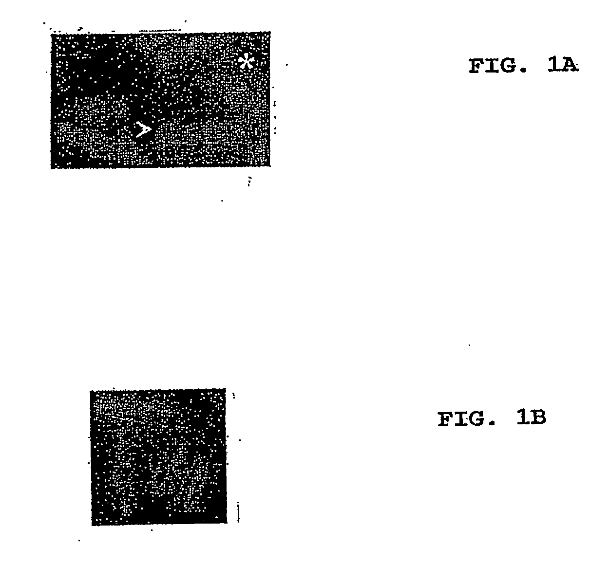
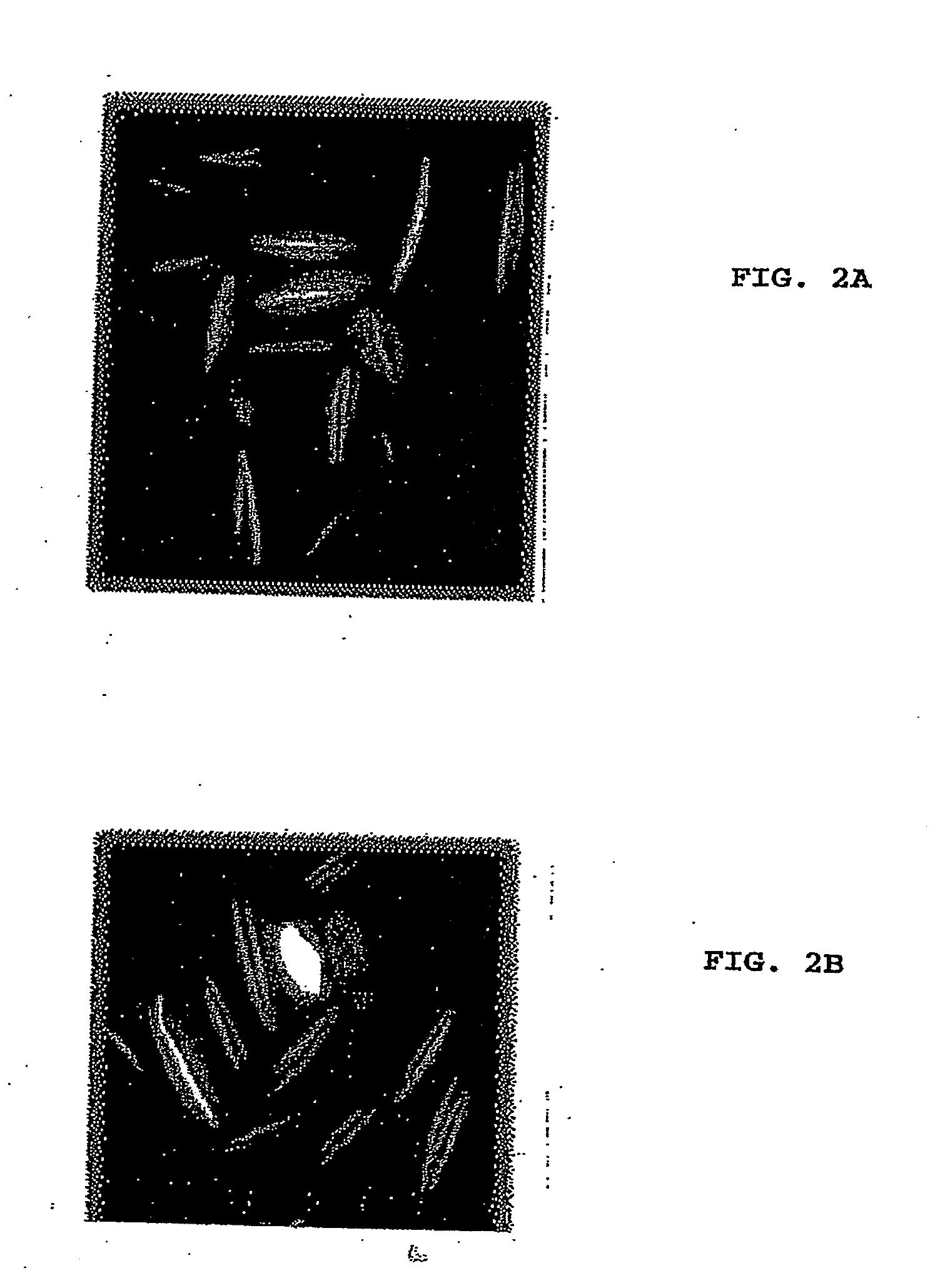
[0065]The plasmid containing the GFP-MreB gene fusion (pREP42-gfp-mreB) was transformed into S. pombe cells using lithium acetate. The transformants were grown in the absence of thiamine for 18-22 hours to induce the expression of GFP-MreB. The polymeric structures formed by MreB were observed under a fluorescent microscope.

[0066]FIGS. 2A and 28 show MreB (a bacterial actin) expressed in S. pombe as a fusion with GFP. Filaments produced in the yeast are clearly visible.


PUM


Abstract

Compounds are identified simultaneously as having antibiotic activity targeting a specific microbial protein and having no or limited toxicity against eukaryotic cells by expressing the microbial protein in eukaryotic cells by expressing the microbial protein in eukaryotic cells which then are used to screen candidate antibiotic compounds. Preferably, yeast cells such as Schizosaccharomyces pombe are transfected with and express a target bacterial protein such as FtsZ or MreB, optionally as a fusion with a reporter protein, and these transfected cells are used to screen libraries of compounds simultaneously for activity against the bacterial protein and lack of toxicity against the yeast cell.

Description

[0001]This application claims the benefit of U.S. provisional application Ser. No. 60 / 815,605, filed Jun. 22, 2006, the entire contents of which is hereby incorporated by reference.BACKGROUND OF THE INVENTION[0002]1. Technical Field[0003]The invention relates to the fields of medicine and agriculture, and specifically to screening methods to identify antibiotic compounds that have activity to combat bacteria in vitro, in vivo in patients such as mammals, including humans, and in planta.[0004]2. Description of the Background Art[0005]Bacteria remain the greatest cause of infectious disease. Bacterial diseases result in both considerable human morbidity and mortality, and in tremendous losses in agriculture due to diseases of plants and animals. Decades after the discovery and extensive use of antibiotics, bacteria, particularly those that have acquired resistance to conventional antibiotics, have re-emerged as major threats. The discovery of new classes of anti-bacterial compounds is...

Claims


Application Information

Patent Timeline
IPC IPC(8): C40B30/00C12Q1/18C12N1/20
CPCG01N33/9446C12Q1/18
Inventor SRINIVASAN, RAMANUJAMBALASUBRAMANIAN, MOHANHORI, MAKI
Owner TEMASEK LIFE SCIENCES LABORATORY
Why Eureka
Try Eureka