Wearable photoacoustic vascular imaging system

a vascular imaging and wearable technology, applied in the field of vascular imaging and detection, can solve the problems of inability to clearly identify prone to guesswork in needle insertion, and inability to accurately measure the location of veins,

Inactive Publication Date: 2010-06-24
IBOKSUNNYVALE EFFIONG ETUKUDO +1
View PDF5 Cites 7 Cited by
  • Summary
  • Abstract
  • Description
  • Claims
  • Application Information

AI Technical Summary

Benefits of technology

[0007]A method of vascular imaging using photo-acoustic principle is described. Additionally, a wearable micro-display component is also described. The method includes pulsed laser as an energy source at a wavelength range at which both the blood and the adjoining tissue absorb the light and convert the energy into kinetic energy which heats the tissue. The temperature of the tissue rises and falls in sympathy with the frequency of the impinging light. The alternative heating and cooling of the illuminated region causes alternating sympathetic expansion and contraction of the region with corresponding rising and falling of the tissue surface and of the air in contact with the surface, thereby generating sound waves. A sensor enables the differentiation between the venous and surrounding tissues from which imaging of the venous tissue is extracted. In addition to the acoustic sensors, the sympathetic rising and falling of the tissue surface are converted into color characteristic signals representative of the relative displacement of the tissue surface. Additionally, the differential heating of the tissues enables the visual detection of the venous tissues by appropriate filter lenses. These embodiments also present miniaturization of the generation, imaging and display components, enabling the detection of the blood vessels in a wearable form factor.

Problems solved by technology

The lack of clarity in vein location and identification quite often leads to guess-work in needle insertion by the medical practitioner.
An exercise which for the patient is quite often a matter of painful experimentation, and for the practitioner an embarrassing practice of trial and error.
For the purpose of this application this methodology would be inappropriate in many of its scopes from size to contamination of the blood.
However, all these methods of vascular imaging involve very expensive and large radiology equipment which while widely in use is neither portable nor practical in the level of venous or arterial imaging and detection for the purpose described herein.

Method used


Image

  • Wearable photoacoustic vascular imaging system
  • Wearable photoacoustic vascular imaging system
  • Wearable photoacoustic vascular imaging system

Examples


Embodiment Construction

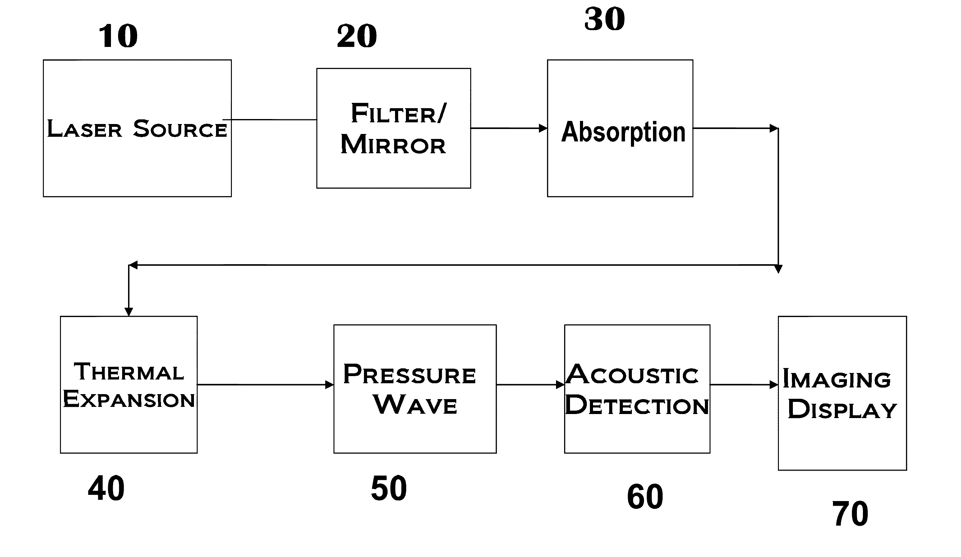
[0019]FIG. 1 shows the schematics of the general principle of the invention. Excitation light or laser pulses in the visible or NIR optical wavelengths are generated from any semiconductor laser source 10. The wavelengths are such as are transparent to body tissue, typically 600 to 1300 nm. The signals are transmitted through the optical fiber line 15 to the tissue where absorption takes place, 20. The large variation in the optical absorption and scattering of the various tissues, particularly hemoglobin is exploited to provide a broad optical contrast and heating differential 30. The differential in heating coupled with a differential in coefficient of expansion of the adjoining tissues result in a variation in the thermal expansion of the tissues, 40. The pulsed irradiation generates periodic expansion and contraction of the irradiated tissue in sympathy with the frequency of the irradiation and contrasting components of the irradiated tissue. If the temperature rises in any port...


PUM


Abstract

A non-invasive wearable imaging apparatus for vascular detection employing photo-acoustic principle is disclosed. Additionally, a wearable micro-display component is also described. The method includes pulsed laser as an energy source at a wavelength range at which both the blood and the adjoining tissue absorb the light and convert the energy into kinetic energy which heats the tissue. The temperature of the tissue rises and falls in sympathy with the frequency of the impinging light. The alternative heating and cooling of the illuminated region causes alternating sympathetic expansion and contraction of the region with corresponding rising and falling of the tissue surface and of the air in contact with the surface, thereby generating sound waves. A sensor enables the differentiation between the venous and surrounding tissues from which imaging of the venous tissue is extracted. In addition to the acoustic sensors, the sympathetic rising and falling of the tissue surface are converted into color characteristic signals representative of the relative displacement of the tissue surface. Additionally, the differential heating of the tissues enables the visual detection of the venous tissues by appropriate filter lenses. These embodiments also present miniaturization of the generation, imaging and display components, enabling the detection of the blood vessels in a wearable form factor.

Description

FIELD OF INVENTION[0001]The present invention relates to the area of vascular imaging and detection for all forms of medical treatment requiring access of patients' veins either for delivery of medicine or the extraction of blood.BACKGROUND OF INVENTION[0002]Vascular detection is a critical component of any medical delivery regimen. Whether in the extraction of blood for analysis, the intravenous delivery of medication or the injection of contrast dyes for imaging, vein detection is critical to that component of medical care. The lack of clarity in vein location and identification quite often leads to guess-work in needle insertion by the medical practitioner. An exercise which for the patient is quite often a matter of painful experimentation, and for the practitioner an embarrassing practice of trial and error. Proficiency is quite often a skill acquired from years of practice tinged with luck. Additionally, some fast action medications require intra-venous (I-V) delivery. A fast ...

Claims


Application Information

Patent Timeline
Patent Type & Authority Applications(United States)
IPC IPC(8): A61B6/00
CPCA61B5/0059A61B5/0095A61B8/00A61B5/742A61B5/489A61B5/7445
Inventor IBOK, EFFIONG ETUKUDO
Owner IBOKSUNNYVALE EFFIONG ETUKUDO
Why Eureka
Try Eureka