Systems and methods for anesthetizing ear tissue

a technology of ionophoretic drugs and anesthesia, applied in the field of ionophoretic drug delivery methods and systems, can solve the problems of limited success of prior ionophoretic devices and systems, inability to be used in all patients, and difficulty in children's tm, etc., and achieve the effect of sufficient flexibility

Active Publication Date: 2010-08-05
TUSKER MEDICAL
View PDF83 Cites 104 Cited by
  • Summary
  • Abstract
  • Description
  • Claims
  • Application Information

AI Technical Summary

Benefits of technology

[0009]In one aspect of the invention, a system for use in iontophoretic substance delivery to the tympanic membrane of an ear of a human or animal subject may include an elongate, flexible tube with a proximal portion and a distal portion, a first flexible sealing element shaped like an umbrella to form a seal within the ear canal, a second flexible sealing element shaped like an umbrella to form a seal within the ear canal, a distal stiffening tube located within the distal portion of the elongate tube distal to the sealing member, a luer fitting coupled with the proximal portion of the tube and including a side vent in fluid communication with the main lumen of the tube, and an electrode device. The flexible tube may include a main lumen extending therethrough. The distal portion may include an inner lip at the distal end of the distal portion and a sealing member proximal to the inner lip. The elongate tube may have sufficient flexibility to bend to conform to the shape of an ear canal. The first flexible sealing element may be integral to and disposed on an exterior of the elongate tube and being offset a distance from a distal most portion of the elongate tube. The second flexible sealing element may be integral to and disposed on the exterior of the elongate tube and proximal to the first sealing element. The distal stiffening tube may prevent the distal portion of the elongate tube from bending. The electrode device may include an elongate shaft. The electrode tip may have a diameter greater than that of the elongate shaft. The electrode device may be movable within the tube lumen of the earplug from a retracted position, in which fluid may pass around the electrode through the tube, to an advanced position, in which the electrode tip may fit within the distal portion of the elongate tube between the inner lip and the sealing member to form a fluid-tight seal.
[0010]In one aspect of the present invention, a method of anesthetizing a tympanic membrane of an ear of a patient using iontophoresis may involve delivering an anesthetizing drug solution to an ear canal of the patient, inserting an iontophoresis device into the ear canal filled with anesthetizing drug solution, venting excess anesthetizing drug solution through the lumen while inserting and while the electrode is in the first position, moving the electrode from the first position to the second position, and activating the electrode in the second position. The iontophoresis device may include an electrode moveable from a first position to a second position inside a lumen. The first position of the iontophoresis device may vent the ear canal. The second position of the iontophoresis device may seal the ear canal.

Problems solved by technology

Prior iontophoresis devices and systems have had limited success and often cannot be used in all patients.
Prior devices generally do not seal the drug solution in an ear canal, thus requiring a patient to recline and tilt his / her head during an iontophoresis procedure.
Using currently available iontophoresis methods, the patient must remain relatively motionless in this reclined, head-tilted position for 5-15 minutes while the iontophoresis procedure provides adequate anesthesia to the TM, which can be especially difficult for children.
Furthermore, using the currently available systems it is only possible to anesthetize one ear at a time, thus making iontophoretic anesthesia of both TMs in a patient a relatively lengthy, uncomfortable process.
Currently available and previously described earplugs generally do not conform adequately to the curved anatomy of the ear canal and thus do not form a good seal in the ear canals of at least some (and in some cases all) patients.
Thus, current earplugs typically allow fluid to leak out of the ear, which makes iontophoretic anesthesia delivery difficult if not impossible with the patient in an upright position.
Furthermore, previously described earplug devices for use in iontophoresis have not addressed issues such as bubble formation in the iontophoretic drug solution, which bubbles may interfere with the contact between an iontophoretic electrode and the solution.

Method used


Image

  • Systems and methods for anesthetizing ear tissue
  • Systems and methods for anesthetizing ear tissue
  • Systems and methods for anesthetizing ear tissue

Examples


Embodiment Construction

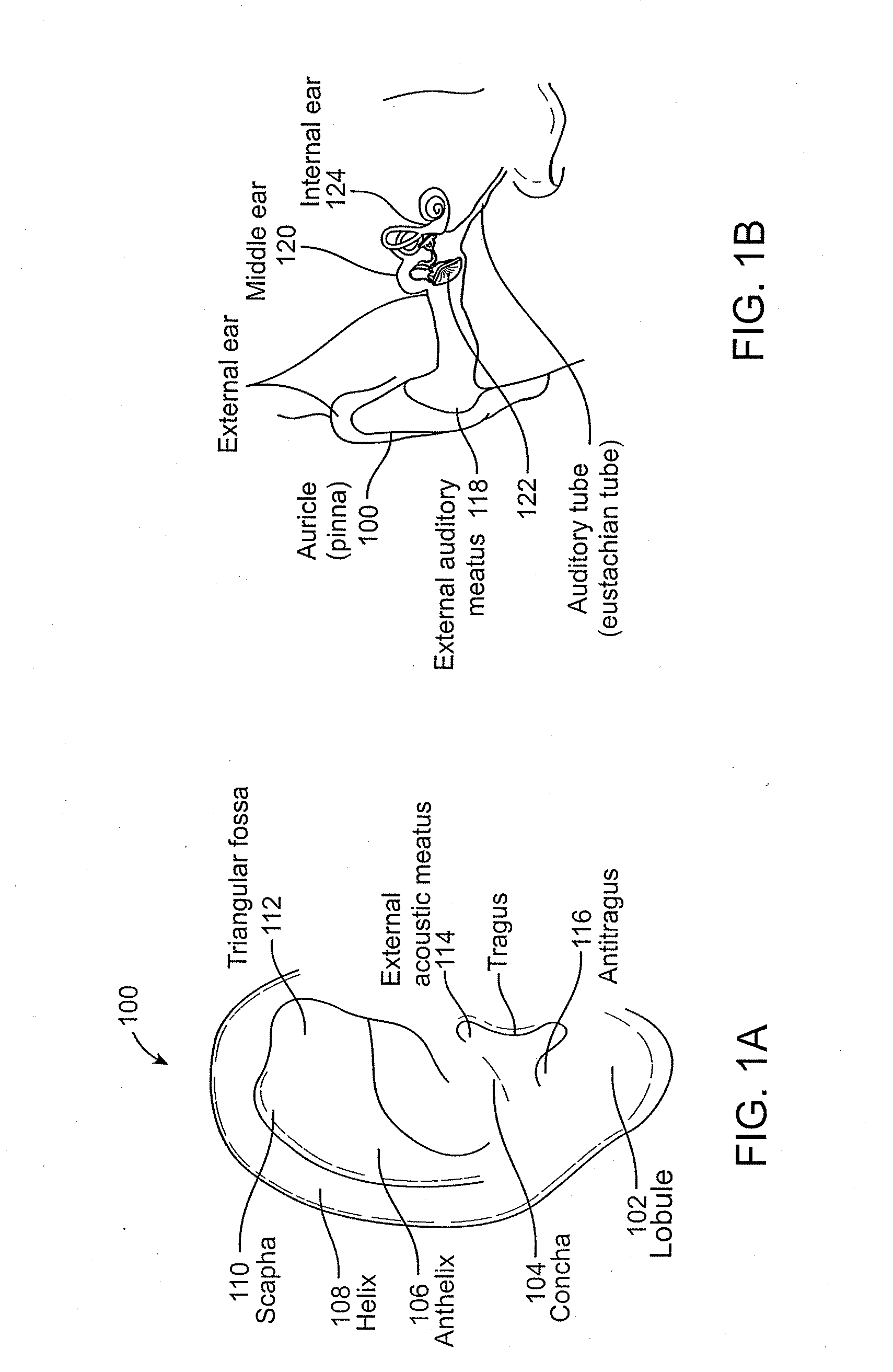
[0062]FIG. 1A shows a view of an outer ear. The outer ear includes a major element known as the auricle or pinna 100. The outer ear serves as a funnel for directing sounds into the internal portions of the ear. The major physical features of the ear include the lobule 102, concha 104, anthelix 106, helix 108, scapha 110, triangular fossa 112, external acoustic meatus 114, tragus 116, and antitragus 118.

[0063]FIG. 1B shows a cross-section of the inner and outer portions of the ear. The pinna 100 is shown connected to the external auditory meatus 118, or ear canal. The ear canal 118 is shown as a relatively straight passage, but is often a more curved, tortuous passageway. The ear canal 118 is connected to the middle ear 120, which includes the ear drum 122. The middle ear 120 in turn is connected to the internal ear 124. The ear drum 122 normally has a pocket of air behind an outer portion called the tympanic membrane. When the middle ear 120 becomes infected, fluid swells inside the...


PUM


Abstract

A system and method for use in iontophoretic anesthesia of a tympanic membrane are disclosed. The system generally includes an earplug and an electrode device. The earplug includes at least one sealing member for sealing the earplug in an ear canal. The sealing member includes microholes which vent fluid above a certain pressure threshold. A headset may connect the earplug to a second earplug. The method involves using the system on a human or animal subject.

Description

CROSS-REFERENCES TO RELATED APPLICATIONS[0001]This application is a continuation-in-part of U.S. application Ser. No. 12 / 510,217, filed on Jul. 27, 2009, which claims the benefit of U.S. Provisional Application No. 61 / 085,360, filed on Jul. 31, 2008, the entireties of which are incorporated by reference herein for all purposes.FIELD OF THE INVENTION[0002]The present invention is related to iontophoretic drug delivery methods and systems. In particular, the present invention is related to novel and advantageous iontophoretic drug delivery methods and systems for anesthetizing ear tissue.BACKGROUND OF THE INVENTION[0003]Iontophoresis is a method for delivering a drug across a biological membrane, such as the skin or, in the case of certain ear surgery procedures, the tympanic membrane (TM). By applying low-level electrical current to a similarly charged drug solution, iontophoresis repels ions of the drug, thus transporting them across the skin or other membrane. In ear procedures, at...

Claims


Application Information

Patent Timeline
Patent Type & Authority Applications(United States)
IPC IPC(8): A61N1/30
CPCA61F11/00A61F2011/085A61K9/0009A61K9/0046A61M1/0088A61M3/0254A61M3/0279A61M19/00A61M2210/0662A61M2210/0668A61N1/303A61N1/36021A61M2205/3341A61F11/12A61M31/002A61M2202/048A61N1/0526A61N1/325A61F11/08A61M37/00A61M1/90
Inventor MORRISS, JOHN H.LIU, GREGGIROTRA, ROHITANDREAS, BERNARD H.BARON, SCOTT J.HAYTER, PAUL G.JENKINS, THOMASNEWHAUSER, JR., RICHARD R.WALKER, JEFFREY A.
Owner TUSKER MEDICAL
Why Eureka
Try Eureka