Radiographic image capture system, radiation generation device, image capture control device and radiographic image capture device

a radiographic image and control device technology, applied in the direction of x/gamma/cosmic radiation measurement, radiography control devices, instruments, etc., can solve the problems of reducing image quality, unable to achieve timing synchronization, and failure of imaging attempts, so as to achieve the effect of stably capturing radiographic images and suppressing radiation exposure to an investigation subj

Inactive Publication Date: 2010-08-19
FUJIFILM CORP
View PDF1 Cites 26 Cited by
  • Summary
  • Abstract
  • Description
  • Claims
  • Application Information

AI Technical Summary

Benefits of technology

[0015]The present invention is made in consideration of the above circumstances and provides a radiographic image capture system, radiation generation device, image capture control device, and radiographic image capture device that are able to stably capture radiographic images while suppressing radiation exposure to an investigation subject.
[0016]A first aspect of the present invention is a radiographic image capture system including: a radiation source control component that, when capturing a radiographic image, controls a radiation source such that radiation for image capture is irradiated after radiation for notification of radiation exposure commencement has been irradiated; a detection component that is provided within an irradiation range of radiation irradiated from the radiation source and that detects radiation irradiated from the radiation source; and a radiographic image capture device. The radiographic image capture device includes: a generation component having a sensor section with sensitivity to radiation and generating image data denoting a radiographic image representing radiation irradiated onto the sensor section; and an image capture control component that, when radiation for notification has been detected, controls the generation component such that a radiographic image is captured with the radiation for image capture irradiated after detection.
[0017]A second aspect of the present invention is a radiation generation device including: a radiation source capable of irradiating radiation; and a radiation source control component that, when capturing a radiographic image, controls the radiation source such that radiation for image capture is irradiated after radiation for notification of radiation exposure commencement has been irradiated.
[0018]A third aspect of the present invention is an image capture control device including: a radiation source control component that, when capturing a radiographic image, controls a radiation source such that radiation for image capture is irradiated after radiation for notification of radiation exposure commencement has been irradiated.
[0019]A fourth aspect of the present invention is a radiographic image capture device including: a generation component that has a sensor section with sensitivity to radiation, the generation component generating image data denoting a radiographic image representing the radiation irradiated onto the sensor section; a detection component, the detection component being provided within an irradiation range of radiation irradiated from a radiation source that irradiates radiation for image capture after radiation for notification of radiation exposure commencement has been irradiated, and the detection component detecting radiation irradiated from the radiation source; and an image capture control component that controls the generation component such that a radiographic image is captured with the radiation for image capture irradiated after detection when the radiation for notification has been detected by the detection component.

Problems solved by technology

However, in such FPD's, charge is generated and accumulated in each pixel even in a state in which X-rays are not irradiated, due to dark current and the like.
If this reset operation is stopped midway then a line aberration is generated in the radiographic image at the position at which the extraction operation of the reset mode was stopped, with a reduction in image quality.
However, in the technique of JP-A No. 2005-13272, since the imaging timing is limited and a radiographic image cannot be captured at any desired timing, this sometimes leads to cases when timing synchronization is not achieved and an imaging attempt fails.
In addition, when communication between the electronic cassette and the console is performed by wireless communication, sometimes the communication state becomes unstable and delays in data transmission arise.
Therefore, even though timing data of the commencement and completion of radiation irradiation is sent by wireless communication to an electronic cassette, as in the technique described in JP-A No. 2006-25832, sometimes delays arise in wireless communication, synchronization of timing is not achieved, radiation irradiation cannot be performed when the FPD is in the charge accumulation state, and image capture fails.
In addition, in the technique of JP-A No. 2008-132216, the investigation subject is exposed to unnecessary irradiation during transition to the charge accumulation state of the FPD after the sensor detects the irradiation of radiation for image capture.

Method used


Image

  • Radiographic image capture system, radiation generation device, image capture control device and radiographic image capture device
  • Radiographic image capture system, radiation generation device, image capture control device and radiographic image capture device
  • Radiographic image capture system, radiation generation device, image capture control device and radiographic image capture device

Examples


first exemplary embodiment

[0028]Explanation will first be given of the configuration of a Radiology Information System 10 according to an exemplary embodiment.

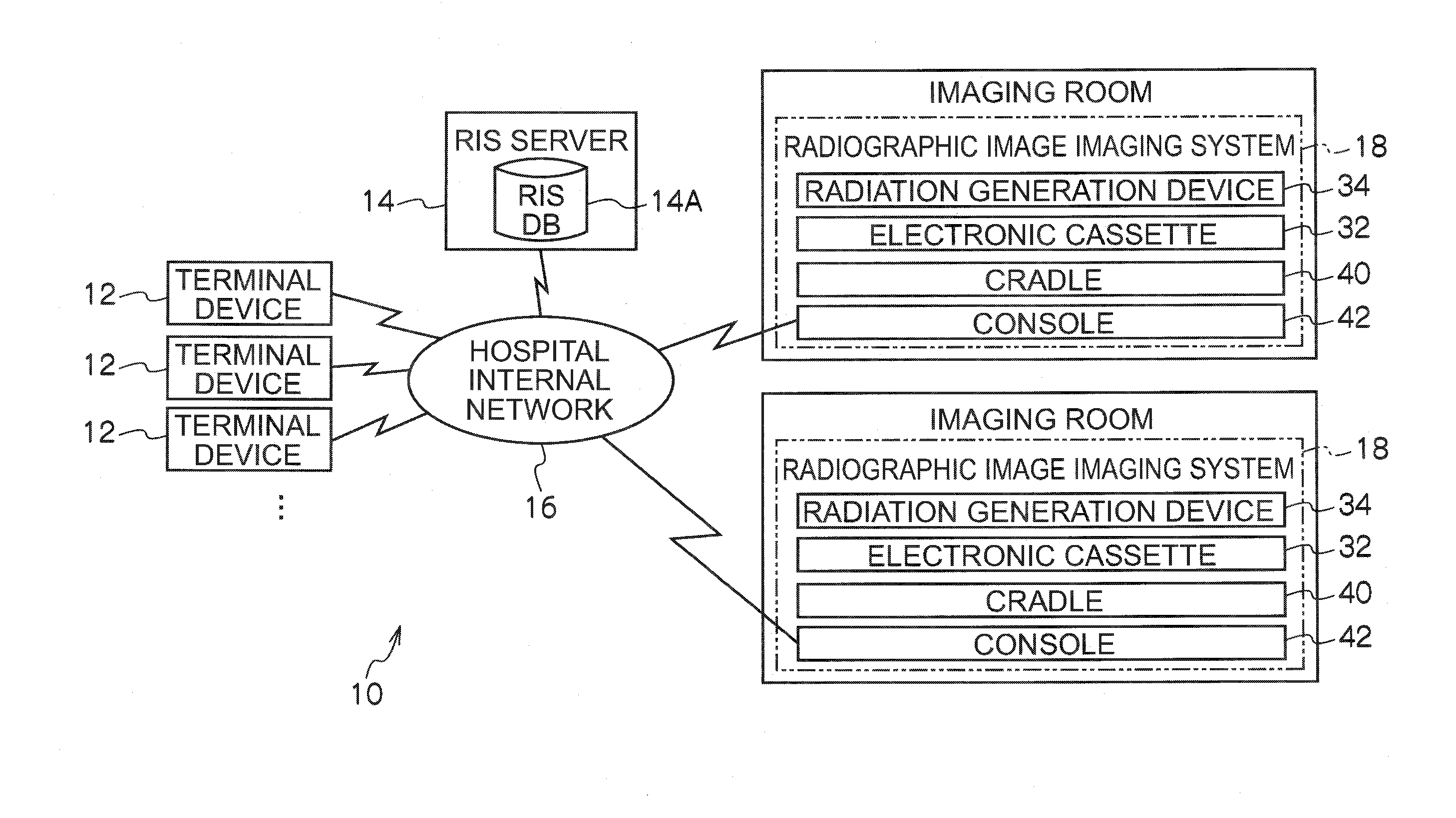
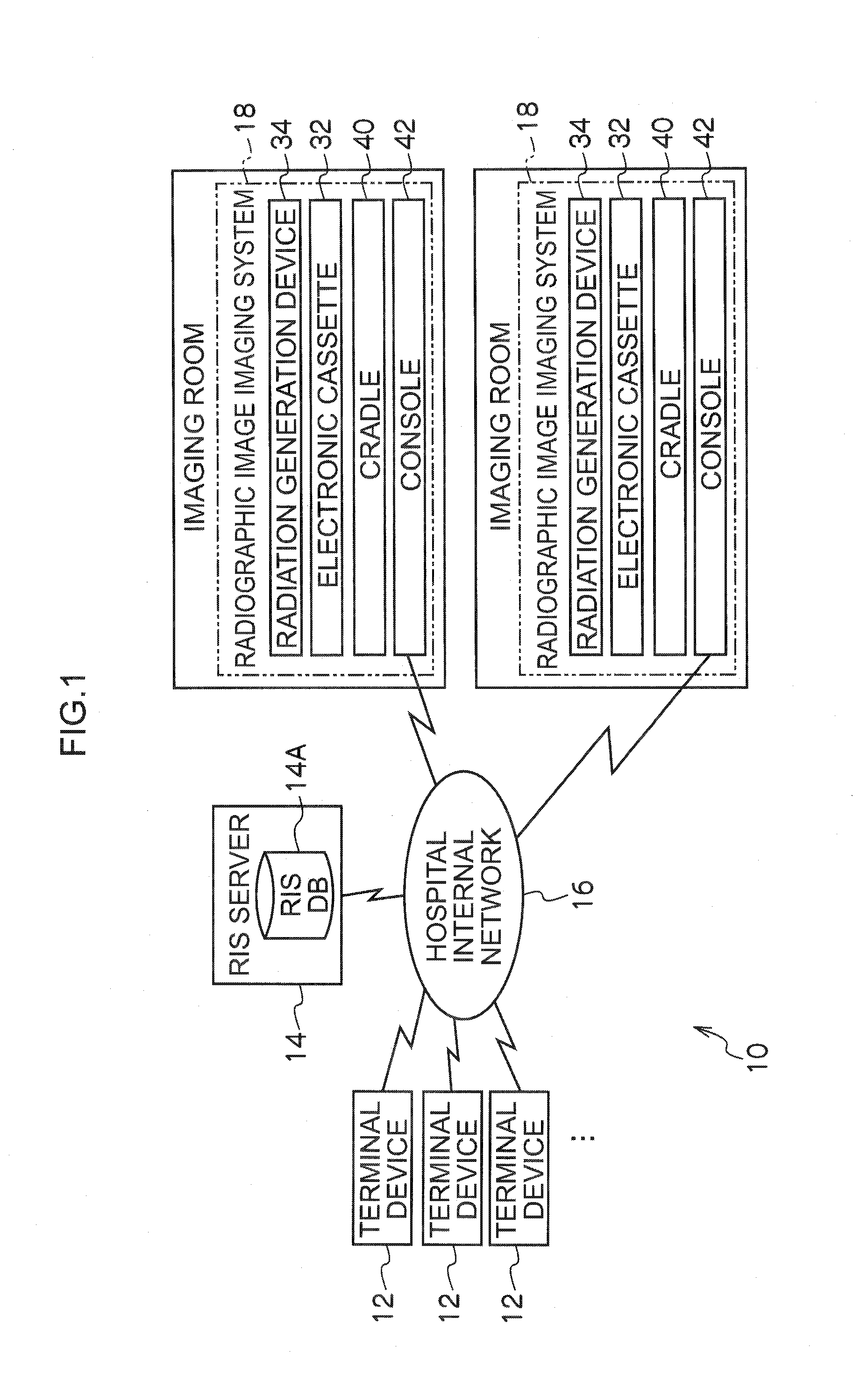
[0029]A block diagram is shown in FIG. 1 illustrating main portions of the configuration of the Radiology Information System 10 according to the present exemplary embodiment (referred to below as RIS 10).

[0030]The RIS 10 is a systems for performing data management in a radiology department, such as management of consultation appointments, consultation record, and the like. The RIS 10 configures part of an HIS (Hospital Information System).

[0031]The RIS 10 is configured with plural imaging request terminal devices 12 (referred to below as terminal devices 12), an RIS server 14, radiographic image imaging systems 18 placed in each individual radiographic imaging room (or operating theater) in the hospital, and a hospital internal network 16, formed from a wired or wireless LAN (Local Area Network) and connecting together each of the radiographic image im...

second exemplary embodiment

[0099]Explanation now follows of a second exemplary embodiment of the present invention.

[0100]The configuration of the radiology information system 10, the imaging system18, and the electronic cassette 32 according to the second exemplary embodiment are similar to those of the above first exemplary embodiment (see FIG. 1 to FIG. 5), and so explanation thereof is omitted.

[0101]In FIG. 7, a timing chart is illustrated, showing the flow of operation when imaging a radiographic image with the imaging system 18 according to the second exemplary embodiment. Note that portions similar to those of the first exemplary embodiment (see FIG. 6) are allocated the same reference numerals and explanation thereof abbreviated, and portions that are different thereto are explained, allocated with the suffix “A”.

[0102]In the electronic cassette 32 according to the second exemplary embodiment, when radiation X1 for notification is detected, reset operation is performed to extract and remove charge accu...


PUM


Abstract

A radiographic image capture system is provided. The system includes: a radiation source control component that, when capturing a radiographic image, controls a radiation source such that radiation for image capture is irradiated after radiation for notification of radiation exposure commencement has been irradiated; a detection component that is provided within an irradiation range of radiation irradiated from the radiation source and that detects radiation irradiated from the radiation source; and a radiographic image capture device that includes: a generation component that has a sensor section with sensitivity to radiation and that generates image information denoting a radiographic image represented by radiation irradiated onto the sensor section, and an image capture control component that, when radiation for notification has been detected by the detection component, controls the generation component such that a radiographic image is captured with the radiation for image capture irradiated after detection.

Description

CROSS-REFERENCE TO RELATED APPLICATION[0001]This application claims priority under 35 USC 119 from Japanese Patent Applications No. 2009-035379 filed on Feb. 18, 2009 and No. 2010-024363 filed on Feb. 5, 2010, the disclosures of which are incorporated by reference herein.BACKGROUND OF THE INVENTION[0002]1. Field of the Invention[0003]The present invention relates to a radiographic image capture system, radiation generation device, image capture control device, and radiographic image capture device.[0004]2. Description of the Related Art[0005]Recently radiation detectors of FPD's (Flat Panel Detectors) and the like that can directly convert radiation into digital data have been put into practice, the FPD's having a radiation sensitive layer disposed on a TFT (Thin Film Transistor) active matrix substrate. Portable radiographic image capture devices (referred to below as “electronic cassettes”) have also recently been put into practice using such radiation detectors housed in a case a...

Claims


Application Information

Patent Timeline
Patent Type & Authority Applications(United States)
IPC IPC(8): G01T1/161G01T1/24
CPCG01T1/17
Inventor TSUBOTA, KEIJIYOSHIDA, YUTAKAKITANO, KOUICHINISHINO, NAOYUKIOHTA, YASUNORI
Owner FUJIFILM CORP
Why Eureka
Try Eureka