Method and Apparatus for Monitoring Fluid Content within Body Tissues

a technology for monitoring and body tissues, applied in the field of patient monitoring, can solve the problems of insufficient monitoring of patients, cardiac decompensation or heart failure, and measurement alone may not be sufficient to reliably detect and/or avoid an impending cardiac decompensation, so as to improve the accuracy and reliability of fluid determination, the effect of determining the hydration of the target tissu

Inactive Publication Date: 2010-09-16
MEDTRONIC MONITORING
View PDF0 Cites 351 Cited by
  • Summary
  • Abstract
  • Description
  • Claims
  • Application Information

AI Technical Summary

Benefits of technology

[0008]Embodiments of the present invention provide improved methods and devices for monitoring the fluid of body tissues, such as the lungs, with an adherent device. The adherent device comprises an ultrasonic transducer and at least one patient measurement sensor. The ultrasonic transducer can be configured to target a tissue of interest, for example lung tissue, and the at least one sensor can be coupled with the ultrasonic transducer to determine the fluid of the tissue, so as to improve the accuracy and reliability of the fluid determination. An amount of fluid of the tissue may be determined in many ways, and the amount may comprise a relative amount of fluid, for example based on a relative intensity of the ultrasound signal. In many embodiments, the hydration of the target tissue can be determined without imaging, which can benefit both in home monitoring in measurements in hospitals. The adherent device may comprise a processor coupled to the ultrasonic transducer and the at least one sensor to determine the fluid of the tissue. For example the processor can be configured to determine when the patient is positioned on a fluid sensitive orientation, and transmit the ultrasound signal in response to the fluid sensitive orientation. The ultrasonic transducer and at least one measurement sensor can be coupled to a support and configured to measure the patient data when the support is adhered to the patient. For example, the ultrasonic transducer and an electrocardiogram electrode can be positioned on the support so as to couple to the patient with a gel pad in contact with the electrode and the ultrasonic transducer.

Problems solved by technology

While such long term care may be at least partially effective, many patients may not be sufficiently monitored and may eventually succumb to cardiac decompensation or heart failure.
Although such a device may be effective in measuring electrocardiography, such measurements alone may not be sufficient to reliably detect and / or avoid an impending cardiac decompensation.
In at least some instances, cardiac decompensation can be difficult to detect, for example in the early stages.
At least some of the known devices may not collect the right kinds of data to treat patients optimally.
For example, although successful at detecting and storing electrocardiogram signals, devices such as the Holter monitor can be somewhat bulky and may not collect all of the kinds of data that would be ideal to diagnose and / or treat a patient, for example to detect decompensation.
Although the build-up of liquid in the patient may be a symptom of compromised physiologic status of the patient, at least some of the current methods and apparatus used to measure patient hydration may not be well suited and / or effective for long term monitoring of the patient.
Although impedance may be used to measure patient hydration, at least some of the current methods of measuring patient hydration with impedance may be somewhat indirect and may be less than ideal to detect an impending patient decompensation.
Although in hospital measurements based on tissue imaging may be used to measure hydration, for example tissue imaging with x-rays and ultrasound, such instrumentation can be complex and may not be well suited for the patient to use in his or her home in at least some instances.
For example, the ultrasound and x-ray devices may be relatively large, expensive and complex, and may also require trained personnel for use in a dedicated care center in at least some instances.
Because clinical signs related to increased tissue hydration such as distressed breathing may not be present until a substantial volume of liquid has accumulated in the lungs of the patient, at least some patients who have been released from the hospital may not seek care until the accumulated liquid has reached a significant and / or dangerous size in at least some instances.
Also, patients who are in a hospital setting may not be properly diagnosed until an imaging study is completed, such that proper diagnoses of the patient in the hospital setting may not occur as quickly as would be ideal in at least some instances.

Method used


Image

  • Method and Apparatus for Monitoring Fluid Content within Body Tissues
  • Method and Apparatus for Monitoring Fluid Content within Body Tissues
  • Method and Apparatus for Monitoring Fluid Content within Body Tissues

Examples


Embodiment Construction

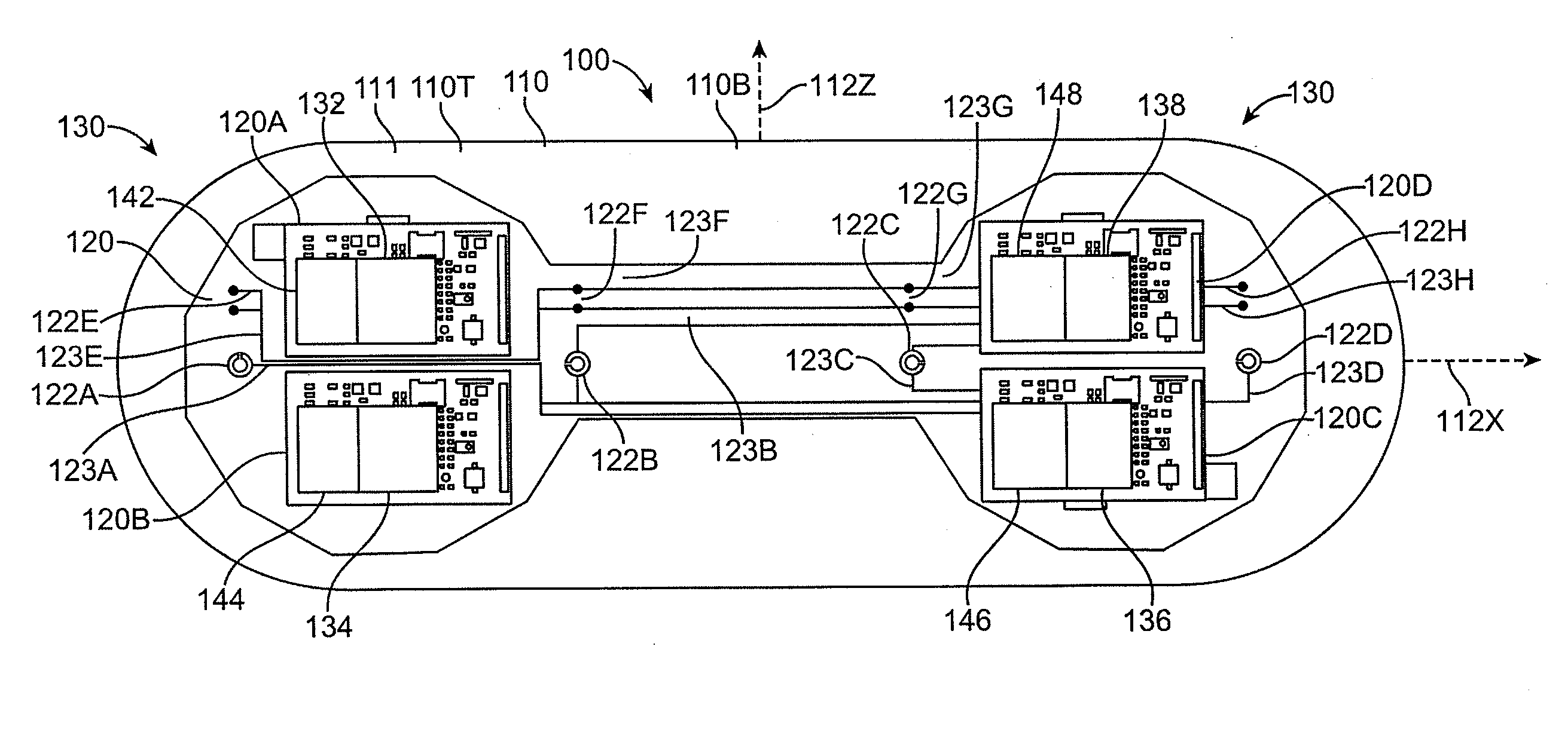
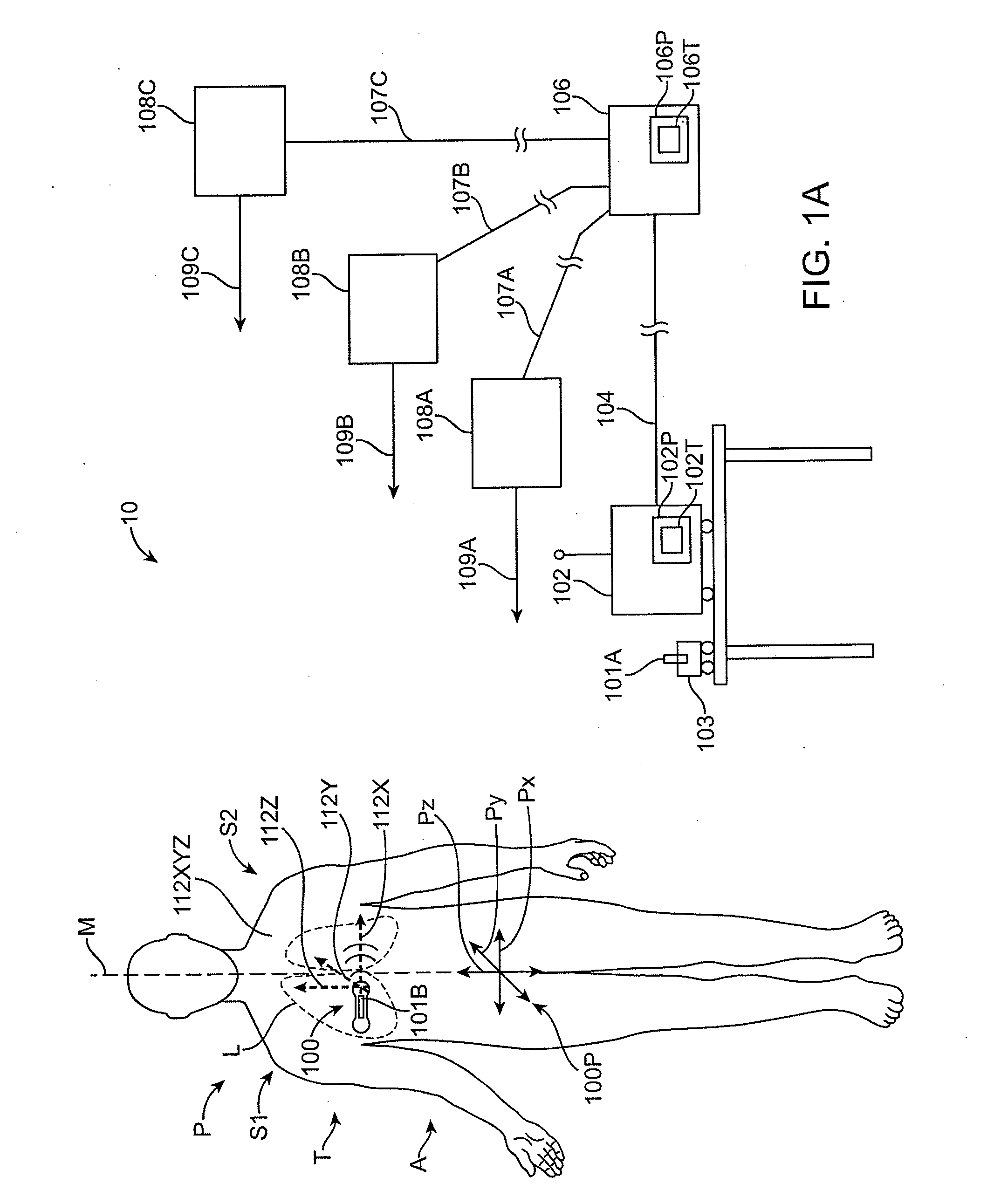
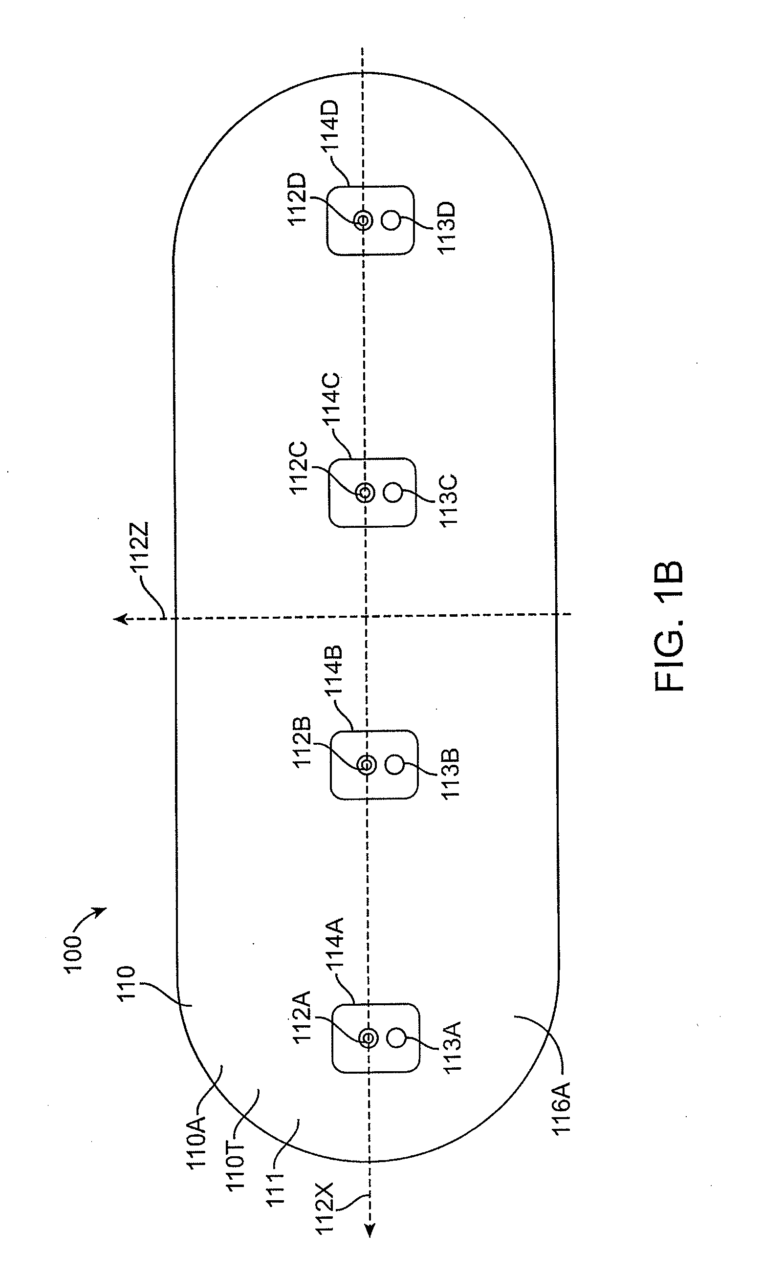
[0077]Embodiments of the present invention relate to patient monitoring. Although embodiments make specific reference to monitoring fluid content and electrocardiogram signals with an adherent patch, the system methods and device described herein may be applicable to any application in which physiological monitoring is used, for example wireless physiological monitoring for extended periods.

[0078]Embodiments of the present invention provide methods and devices for monitoring fluid content within body tissues, such as the lungs, via impingement of ultrasonic signals into the body through an adherent device and by analysis of a return signal. Changes in fluid content can manifest as altered return signals when compared to data collected over the life of the adherent device or a series of adherent devices. Differences in ultrasound propagation may be used to determine fluid content in body tissues, and to monitor fluid content, and thus monitor the progress of various clinical conditio...


PUM


Abstract

Methods and devices for monitoring fluid content within body tissues. An adherent device having a support configured to transmit a signal into a body of a patient, and receive a reflected portion of the signal, and adhere to the skin of the patient. In many embodiments, the adherent device includes an ultrasonic transducer and other sensors. In many embodiments, the ultrasonic transducer is used in coordination with the other sensors to predict a cardiac decompensation.

Description

CROSS-REFERENCES TO RELATED APPLICATIONS[0001]This application claims the benefit of U.S. Provisional Patent Application No. 61 / 159,733, filed on Mar. 12, 2009, the entirety of which is incorporated herein by reference for all reasons.BACKGROUND OF THE INVENTION[0002]The present invention relates to patient monitoring. Although embodiments make specific reference to monitoring patients with an adherent patch device to detect fluid in the lungs, the system methods and device described herein may be applicable to many applications in which physiological monitoring is used, for example wireless physiological monitoring with devices for extended periods.[0003]Patients are often treated for diseases and / or conditions associated with a compromised status of the patient, for example a compromised physiologic status such as heart disease. In some instances a patient may have suffered a heart attack and require care and / or monitoring after release from the hospital. While such long term care...

Claims


Application Information

Patent Timeline
Patent Type & Authority Applications(United States)
IPC IPC(8): A61B8/14A61B5/04
CPCA61B5/02055A61B5/0402A61B5/4878A61B5/6833A61B7/003A61B2562/0219A61B8/08A61B8/4236A61B8/4472A61B8/488A61B7/04A61B5/318A61B5/4875
Inventor ENGEL, JONATHAN
Owner MEDTRONIC MONITORING
Why Eureka
Try Eureka