
Self-adjusting endourethral device & methods of use

a self-adjusting, endotracheal technology, applied in the field of medical devices, can solve the problems damage to the epithelium and detrusor muscles, and discharge of bladder contents can be a source of serious and distressing problems, so as to improve urine drainage, facilitate urination, and facilitate urination

Inactive Publication Date: 2010-09-23
ABBEYMOOR MEDICAL INC
View PDF7 Cites 40 Cited by
  • Summary
  • Abstract
  • Description
  • Claims
  • Application Information

AI Technical Summary

Benefits of technology

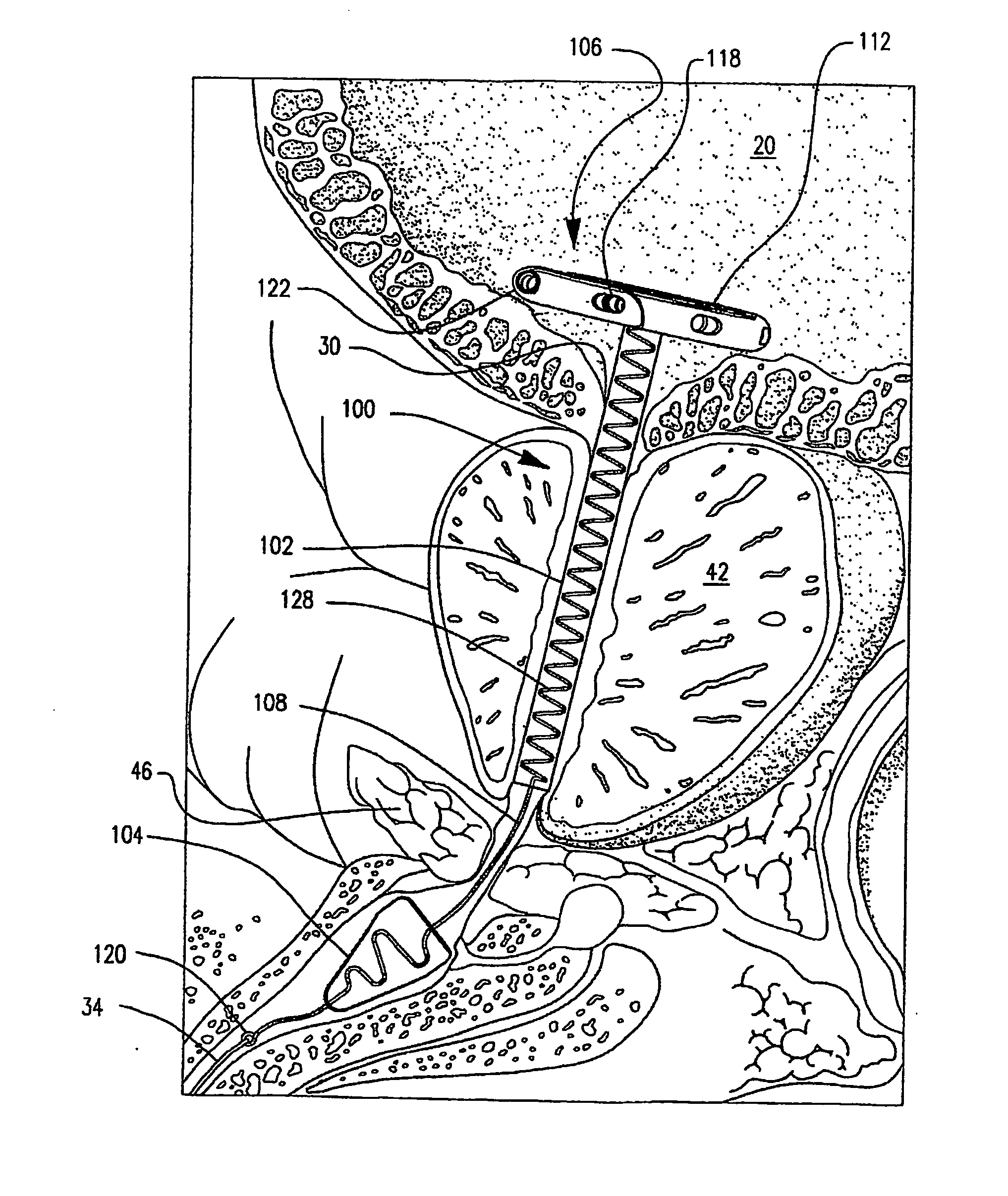
[0032]The novel devices described hereafter support the male urethra in an open condition, and permit unencumbered urination. The subject devices also offer relief from the discomforts of obstruction and diagnostic utility. Several of the embodiments are selectively useful for relief of symptoms associated with overactive bladder, prostatitis, and treatment of infection and cancer within the urogenital tract.
[0033]Further still, the subject devices offer many clinical advances. First, all embodiments provide improved urine drainage in patients with obstruction by supporting the prostatic urethra in an “open” status regardless of the reason for the obstruction. Second, all embodiments provide ease of insertion and self adjustment of the device. Third, all embodiments provide for convertible features allowing for continuous drainage or normal physiological drainage. Fourth, all embodiments provide two point anchoring. Fifth, all embodiments provide for an ideal platform for medicament, e.g., solution, delivery. Sixth, use of the subject devices provide for treatment and diagnostic opportunities that do not presently exist.
[0035]Each device embodiment may be easily positioned to accommodate the prostate length and sphincteric anatomy of the patient. Devices are stabilized in the urethra by two anchoring elements. The first anchor is preferably positioned in the bladder; a second anchor is positioned within the bulbous urethra. The anchor portions are spaced apart by a body which selectively supports a portion of the urethra without inhibiting the normal function of the external sphincter. The devices therefore re-enable physiologically normal urination. The device configurations are easy to insert, stable, and easy to remove. In connection to removal, a preformed or pretensioned free end of the device may be readily manipulated so as to “release” the configuration associated therewith in furtherance of device removal ease and / or patient comfort.

Problems solved by technology

Discharge of bladder contents can be a source of serious and distressing problems for persons whose anatomy is temporarily, or over time, incapable of completely controlling the outflow of urine from the bladder, a clinical condition known as urinary retention.
Urinary problems can have serious consequences, particularly when the problem is one of retention, incomplete emptying, or dysuria.
Failure to do so can result in damage of the epithelium and detrusor muscles associated with the bladder, and an increased potential for bacterial invasion which is commonly thought to contribute to urinary tract infection (UTI) potentially leading to life-threatening kidney failure.
As individuals age, particularly males, the frequency of difficulties experienced within the intricate urogenital system increases.
Problems range in severity from minor inconvenience, which lowers the quality of life, to life threatening disease.
Second, it provides the pressure necessary to discharge the urine through the urethra.
The causes for retention are either excess outlet obstruction within the urethra, or inability of the micturition process to progress in a normal coordinated manner.
Individuals who cannot easily and completely empty their bladders almost always experience a reduction in their quality of life.
The symptoms are virtually always suspected to be caused by the intrusion of an enlarged prostate gland upon the urethra, however, symptoms are often caused by irregularities in bladder function, or sphincter deficiencies.
As previously noted, bladder outlet obstructions are primarily caused by the enlargement of the prostate gland which results in radial compression of the urethra surrounded thereby (i.e., the prostatic urethra), thus obstructing (i.e., constricting) urine flow, resulting in incomplete emptying of the bladder (i.e., there being what is clinically referred to as a “post void residual” (PVR) remaining in the bladder).
Furthermore, diseases within the prostate may also affect function of the reproductive system by obstructing the ejaculatory ducts that pass through the interior of the prostate.
The orientation of the prostate, which is located anterior of the rectum, and seminal vesicles, can result in physiological interdependencies which become complex to diagnosis and treat.
This is particularly true with casual diuretics such as coffee may cause urgency and frequency.
Another common imbalance, constipation, places pressure on the posterior of the prostate adding to the effect of obstruction, with many drugs known to have a side effect of constipation.
The capsule may however, enlarge and produce similar voiding symptoms to those of benign enlargement.
For this reason prostate cancer is difficult for the urologist or oncologist to known when to treat.
Problems and disadvantages of heretofore known devices include the deleterious effects (i.e., pitting, depositions, etc.) associated with the urethral environment upon critical device components (e.g., valve actuators, flow conduits, etc.) which at a minimum render such devices less effective, and which at a maximum, cause device component failure, or render the device wholly ineffective, which necessitates emergent removal and, as the case may' be, urinary tract damage repair.
Problems of device leakage, or less than complete emptying of the bladder are also widely known.
Furthermore, issues surrounding device deployment and fit, positioning, repositioning, and retention (i.e., sufficient anchoring) have also been well documented.
Based upon clinical findings, many have been shown to be uncomfortable, thus severely retarding their utility as a therapy.

Method used


Image

  • Self-adjusting endourethral device & methods of use
  • Self-adjusting endourethral device & methods of use
  • Self-adjusting endourethral device & methods of use

Examples


Embodiment Construction

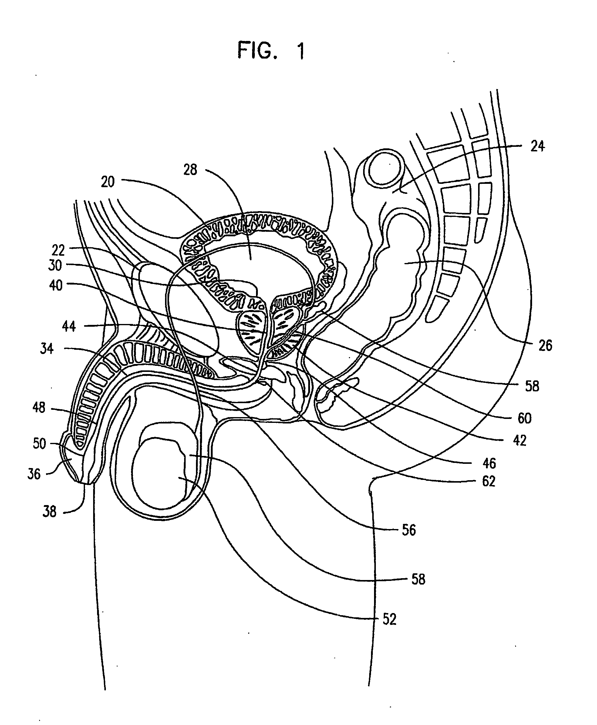
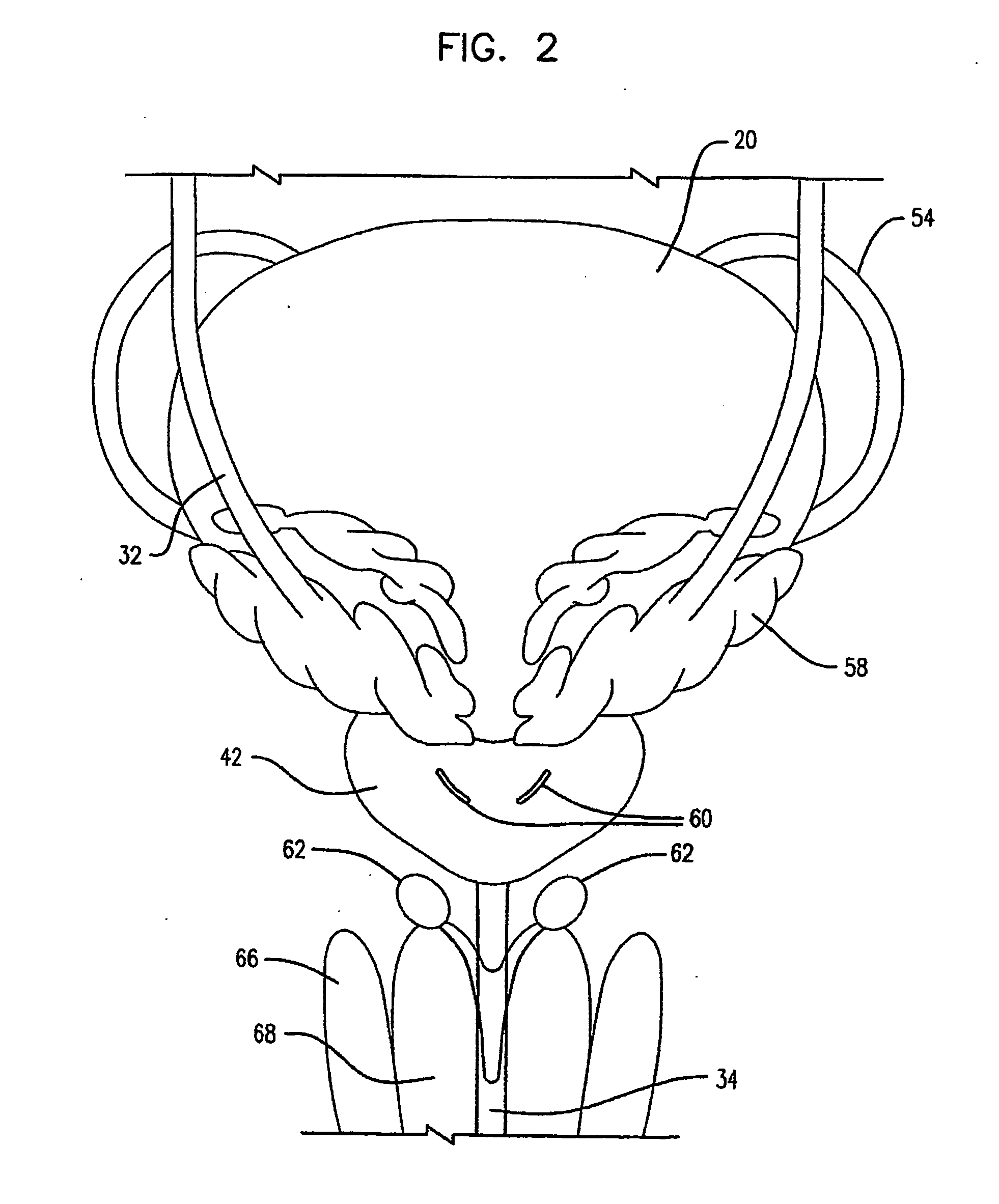
[0052]Prior to a detailed discussion of the subject device, its several embodiments, and attendant systems, an abbreviated description of the anatomical environment of same is helpful. In furtherance thereof, FIGS. 1 & 2 generally illustrate the physiologic structures of the male urinary system, as well as the male reproductive system. A sectional side view of the male urinary system is presented in FIG. 1 with a front view of select structures of FIG. 1 illustrated in FIG. 2.

[0053]In connection to the urinary system, the bladder 20, generally centrally located and residing posterior of the pubic bone 22 and anterior of the sigmoid colon 24 and rectum 26 (FIG. 1), temporarily stores urine 28, and periodically expels it when the bladder neck 30 (i.e., the lower base of the bladder) opens, as the bladder 20 contracts. Urine, produced by the kidneys (not shown), passes into the bladder via dedicated ureters 32 (FIG. 2), and periodically exists therefrom via the urethra 34, a continuous...


PUM


Abstract

A self adjusting and / or positioning indwelling urethral device is provided. The device generally includes a prostatic urethral stent body and a urethral anchoring element. The prostatic urethral stent body includes a preconfigured end portion for anchored receipt within a bladder with the urethral anchoring element extending from the prostatic urethral stent body via a linkage. Engagement of the proximal anchor, delimited by the preconfigured end portion of the stent body, with the bladder is no or minimal impact upon the trigone region.

Description

[0001]This is an international application filed under 35 USC §363, claiming priority under 35 USC §119(e) of U.S. Prov. Appl. Nos. 60 / 691,635 and 60 / 691,636, each filed Jun. 20, 2005 and each incorporated herein by reference in its entirety.TECHNICAL FIELD[0002]The present invention generally relates to medical devices for use within a cavity of the human body, more particularly, indwelling drainage devices, and still more particularly, to urinary stents that dwell in the male prostatic urethra.BACKGROUND OF THE INVENTION[0003]Discharge of bladder contents can be a source of serious and distressing problems for persons whose anatomy is temporarily, or over time, incapable of completely controlling the outflow of urine from the bladder, a clinical condition known as urinary retention. Traditionally, indwelling urethral catheters (i.e., Foley catheters, or the like), in which a free passage is created between the bladder and the outside of the human body so as to ensure the permanent...

Claims


Application Information

Patent Timeline
IPC IPC(8): A61F2/04
CPCA61M2025/0681A61M27/008
Inventor WILLARD, LLOYD K.WHALEN, MARK J.REID, JOHN M.SMITH, CHAD S.EUTENEUER, CHARLES L.
Owner ABBEYMOOR MEDICAL INC
Learn More