Photoacoustic detection of analytes in solid tissue and detection system

a solid tissue and analyte technology, applied in the field of analyte detection in solid tissue and tissue analysis systems, can solve the problems of large increase in the number of sections, difficult to detect when a lymph node is sectioned, and inability to perform biopsy

Inactive Publication Date: 2010-11-11
UNIVERSITY OF MISSOURI
View PDF31 Cites 80 Cited by
  • Summary
  • Abstract
  • Description
  • Claims
  • Application Information

AI Technical Summary

Problems solved by technology

A weakness in the sentinel node mapping technique is the biopsy used to determine whether an extracted sentinel lymph node has metastasized cancer cells.
Such cells make up a very small volume of a lymph node, and are difficult to detect when a lymph node is sectioned during a biopsy.
Even a large increase in the number of sections, impractical in reality, would still rely largely upon chance to detect micro-metastasized cells.
Immunohistochemical staining for the melanoma ma

Method used


Image

  • Photoacoustic detection of analytes in solid tissue and detection system
  • Photoacoustic detection of analytes in solid tissue and detection system
  • Photoacoustic detection of analytes in solid tissue and detection system

Examples


Embodiment Construction

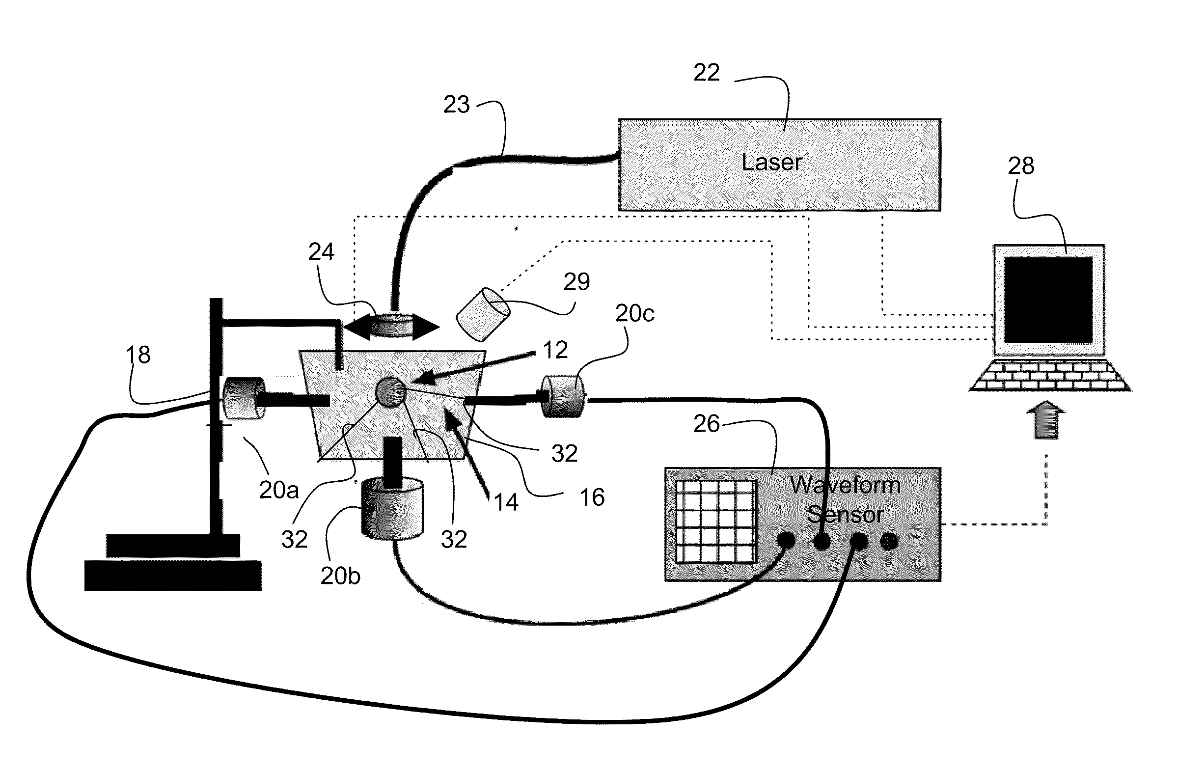
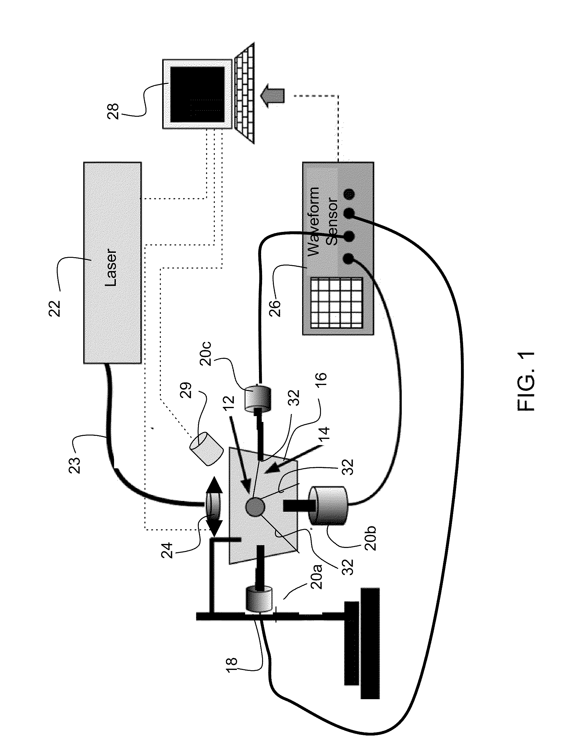
[0012]The invention provides methods and systems for photoacoustic detection of analytes in extracted solid tissues, such as sentinel lymph nodes. Methods and systems of the invention are capable of detecting and geographically locating microscopic analytes in lymph nodes. Any analyte that is a light absorber can be detected in solid tissues, but methods and system of the invention are especially useful to detect the presence and position of micro-metastases in lymph nodes. Methods and systems of the invention can, for example, detect the presence or absence of and geographically locate in three dimensions the position of micro-metastases in extracted sentinel lymph nodes as replacement or aid to a traditional sentinel node biopsy.

[0013]A preferred system for detecting an analyte in solid tissue, such as an intact lymph node in vitro, includes a laser arranged to generate a pulsed laser beam into the solid tissue, which can be a fully intact lymph node. An acoustic sensor, and prefe...


PUM


Abstract

A preferred system for detecting an analyte in solid tissue, such as an intact lymph node, in vitro includes a laser arranged to generate a pulsed laser beam into solid tissue, which can be a fully intact lymph node. An acoustic sensor, and preferably at least three acoustic sensors are arranged in different positions to span a three dimensional space, such as in an X, Y and Z coordinate system, to detect photoacoustic signals generated within the lymph node. At least one computer receives signals from the acoustic sensor(s). The computer determines the presence or absence of, and preferably the position of analyte, from the signals and the timing of the signals. A preferred method for detecting an analyte in a lymph node in vitro includes exposing an extracted lymph node to a pulsed laser beam. A photoacoustic signal is sensed. The photoacoustic signal is analyzed to confirm the presence or absence of an analyte in the lymph node. Preferably, multiple photoacoustic signals are sensed from sensors that span a three dimensional space and the position of analyte is also determined.

Description

PRIORITY CLAIM AND REFERENCE TO RELATED APPLICATION[0001]The application claims priority under 35 U.S.C. §119 from prior provisional application Ser. No. 61 / 170,880, which was filed Apr. 20, 2009.FIELD OF THE INVENTION[0002]Fields of the invention is analyte detection in solid tissue and tissue analysis systems. A preferred application of the invention is the in vitro detection of melanoma micro-metastasis in intact excised sentinel lymph nodes, and a preferred system of the invention is a photoacoustic system that can detect both the presence and geographical location of melanoma micro-metastasis in extracted lymph nodes.BACKGROUND[0003]A sentinel lymph node is the node from a first group of nodes that is reached by metastasizing cancer cells that travel from a cancerous tumor through the lymphatic system. Sentinel lymph node mapping involves detecting a sentinel lymph node, typically via dye injection, and removing the sentinel lymph node for a biopsy to determine the presence or ...

Claims


Application Information

Patent Timeline
IPC IPC(8): C12Q1/02C12M1/34
CPCG01N21/1702G01N33/4833A61B5/0095A61B5/418A61B8/4477G01N2291/02809A61B8/0841A61B8/15G01N29/2418
Inventor VIATOR, JOHN A.MCCORMACK, DEVINDALE, PAUL S.
Owner UNIVERSITY OF MISSOURI
Why Eureka
Try Eureka