Assessing tissue rejection

a tissue and tissue technology, applied in the direction of material testing goods, biochemistry equipment and processes, instruments, etc., can solve the problems of insufficient training of pathologists, inability to produce a picture of active events, and inherent in the diagnosis of pathology of tissue rejection, etc., to achieve the effect of suppressing tissue rejection

Inactive Publication Date: 2010-11-18
HALLORAN PHILIP F
View PDF2 Cites 0 Cited by
  • Summary
  • Abstract
  • Description
  • Claims
  • Application Information

AI Technical Summary

Benefits of technology

[0005]This document provides methods and materials involved in assessing tissue rejection (e.g., organ rejection) in mammals. For example, this document provides methods and materials involved in the early detection of tissue rejection (e.g., kidney rejection) and the assessment of the extent of rejection of a tissue, e.g., a transplanted organ, in a mammal. Early diagnosis of patients rejecting transplanted tissue (e.g., a kidney) can help clinicians determine appropriate treatments for those patients. For example, a clinician who diagnoses a patient as rejecting transplanted tissue can treat that patient with medication that suppresses tissue rejection (e.g., an immunosuppressant).

Problems solved by technology

The diagnosis of allograft rejection remains an important issue in kidney transplantation.
This is particularly problematic at the important interface that separates TCMR from borderline changes, which is the point that also defines where treatment decisions are made (Furness et al., Nephrol Dial Transplant, 12(5):995-100 (1997)).
Other limitations are inherent in diagnostic pathology of rejection, including sampling error, intra-observer variation, and a shortage of trained pathologists.
In addition, describing morphology does not produce a picture of active events such as active inflammation and active injury, and provides a qualitative assessment of tissue after damage has occurred or even progressed.
For example, although fibrosis can be observed with pathology, pre-fibrotic events are not detectable with a standard Banff pathology assessment.

Method used


Image

  • Assessing tissue rejection
  • Assessing tissue rejection
  • Assessing tissue rejection

Examples


example 1

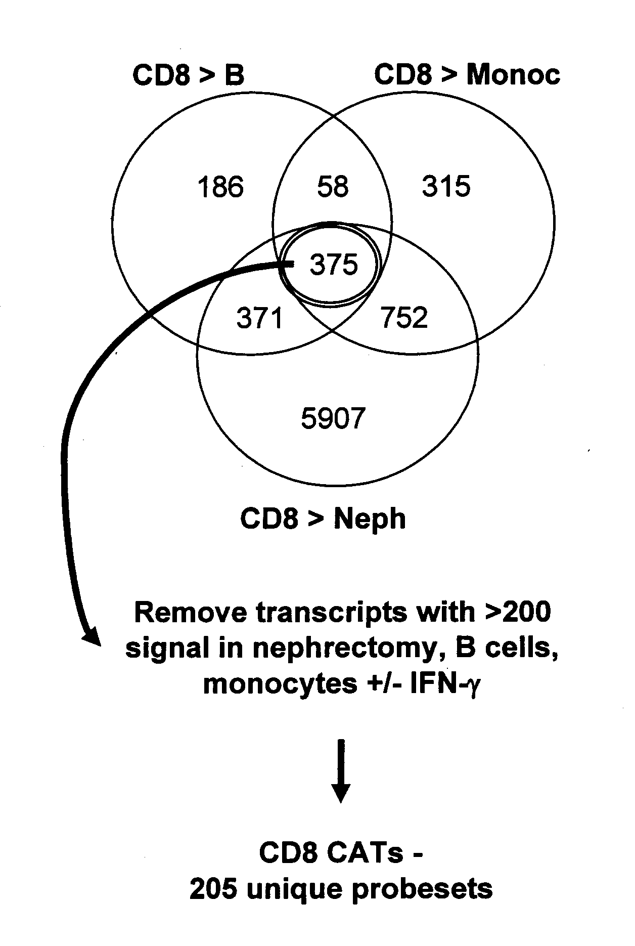
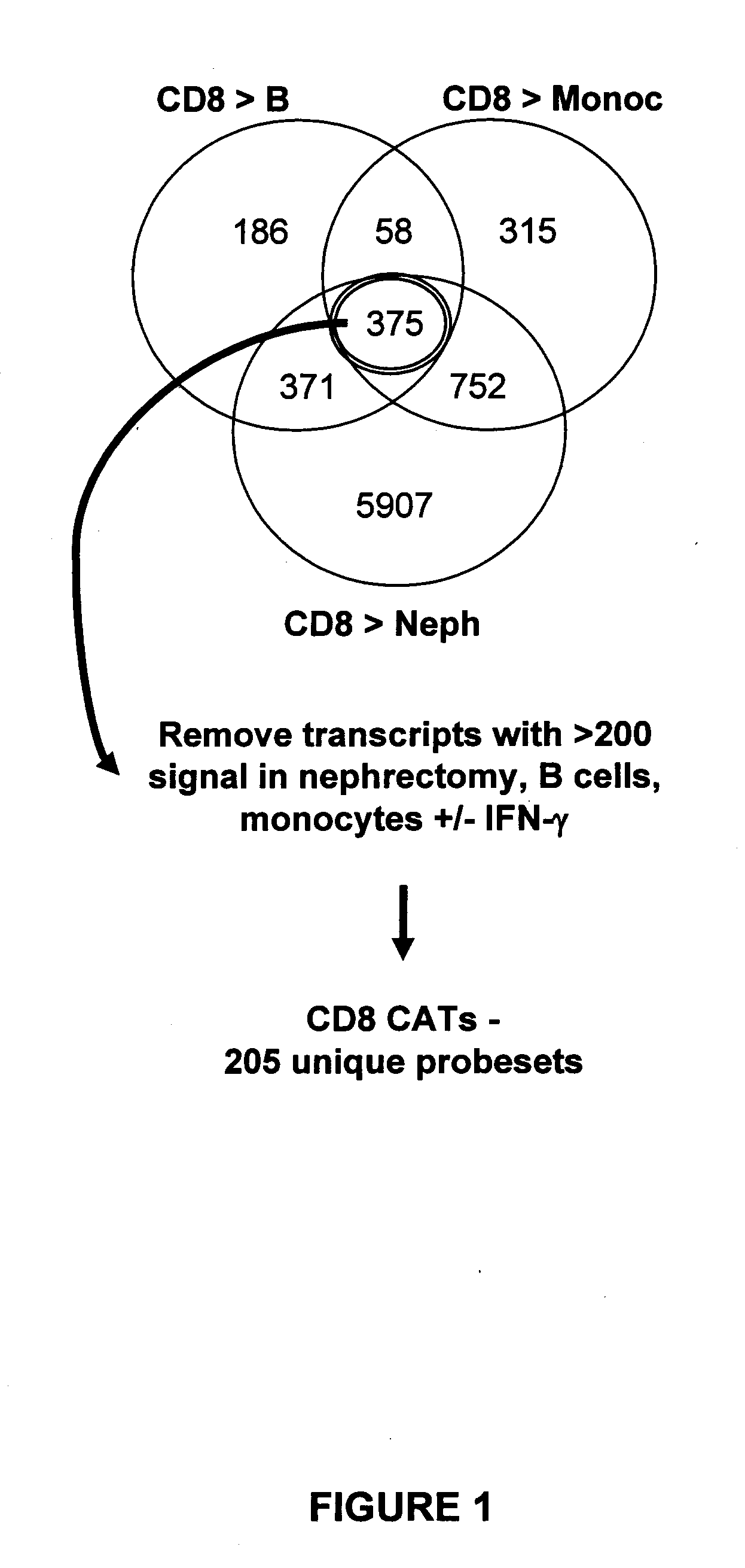
Characterizing Human Cytotoxic T Lymphocyte-Associated Transcripts (CATs)

[0039]Cell cultures: Cell cultures were maintained in complete RPMI (RPMI 1640 supplemented with 10% FBS (Invitrogen Life Technologies, Carlsbad, Calif.), 2 mM L-glutamine, β-mercaptoethanol, non-essential amino acids, sodium pyruvate, and antibiotic-antimycotic solution). The cultures were incubated at 37° C. in the presence of 5% CO2.

[0040]Isolation and generation of cell populations: Whole blood samples were obtained from healthy volunteers. Peripheral blood mononuclear cells (PBMCs) were isolated from the whole blood samples by density gradient centrifugation using Ficoll® (GE Healthcare, Piscataway, N.J.). The PBMCs were used to prepare purified cell populations. Effector CD4 and CD8 cells were generated through several rounds of MLR stimulations. PBMCs were first cultured at a ratio of 1:1 with RPMI8866 cells treated with mitomycin (Sigma, St. Louis, Mo.). The mitomycin-treated RPMI8866 cells served as st...

example 2

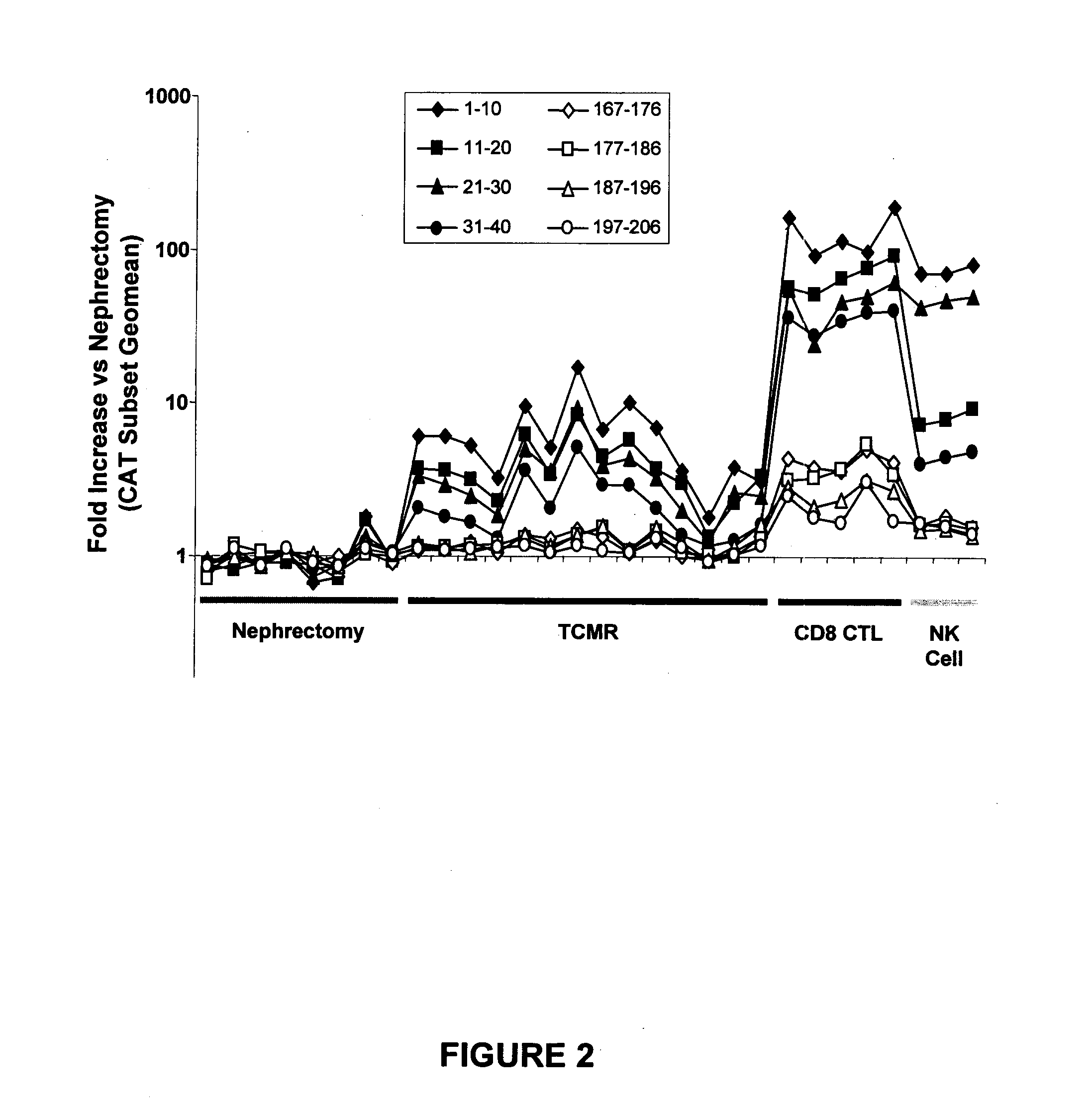
Assessing CAT Expression in Human Kidney Transplants with TCMR

[0048]Human kidney transplant biopsies: Human kidney transplant biopsies (n=16) diagnosed as TCMR (T cell-mediated rejection) were selected from a patient cohort. The diagnosis of TCMR was based on histopathology using Banff criteria (Racusen et al., Kidney Int., 55:713-723 (1999)) and the clinical diagnosis of a rejection “episode” based on retrospective assessment of clinical course, independent of transcriptome analysis. Kidney tissues (n=8) from macroscopically and histologically unaffected areas of the cortex of native nephrectomies performed for renal carcinoma served as controls. In addition to the cores obtained for conventional diagnostic assessment, an 18-gauge biopsy core was collected for gene expression analysis (see Example 1), immediately placed in RNALater solution, kept at 4° C. for 4-24 hours, and then stored at −20° C. RNA was isolated as described in Example 1 above, and an average of 4 μg of RNA was o...


PUM

PropertyMeasurementUnit
morphologyaaaaaaaaaa
timeaaaaaaaaaa
lengthaaaaaaaaaa

Abstract

This document relates to methods and materials involved in assessing tissue rejection (e.g., organ rejection) in mammals. For example, methods and materials involved in detecting tissue rejection (e.g., kidney rejection) are provided, as are methods and materials for determining the extent of rejection in mammals (e.g., humans).

Description

BACKGROUND[0001]1. Technical Field[0002]This document relates to methods and materials involved in assessing tissue rejection (e.g., organ rejection) in mammals. For example, this document relates to methods and materials involved in detecting tissue rejection (e.g., kidney rejection) in mammals and determining the burden or extent of rejection in mammals (e.g., humans).[0003]2. Background Information[0004]The diagnosis of allograft rejection remains an important issue in kidney transplantation. Rejection can manifest as an acute episode or as subtle loss of function, proteinuria, scarring, and graft loss (Meier-Kriesche et al., Am J Transplant, 4(3):378-383 (2004)). Two mechanisms of rejection are recognized in the Banff histologic classification: T cell mediated rejection (TCMR) and antibody-mediated rejection (ABMR; Solez et al., Am J Transplant, 7(3):518-526 (2007); Racusen et al., Am J Transplant, 4(10):1562-1566 (2004)). TCMR can be diagnosed by scoring interstitial inflammati...

Claims


Application Information

Patent Timeline
Patent Type & Authority Applications(United States)
IPC IPC(8): C12Q1/68C12Q1/02G01N33/68
CPCC12Q1/6883G01N2800/245C12Q2600/158
Inventor HALLORAN, PHILIP F.
Owner HALLORAN PHILIP F
Why Eureka
Try Eureka