
Repair and treatment of bone defect using agent produced by chondrocytes capable of hypertrophication and scaffold

Inactive Publication Date: 2010-11-25
HOYA CORP
View PDF5 Cites 5 Cited by
  • Summary
  • Abstract
  • Description
  • Claims
  • Application Information

AI Technical Summary

Benefits of technology

[0157]According to the present invention, it is possible to provide a composite material containing an induced osteoblast differentiation inducing agent produced by chondrocytes capable of hypertrophication and a scaffold, which can promote or induce osteogenesis in a biological organism, and a method of producing the composite material and a method of utilizing the composite material.
[0158]Such a composite material can promote or induce the osteogenesis in the biological organism. By using it, it is possible to induce the osteogenesis even in a region where bone does not exist in the vicinity thereof. Such a composite material could not be provided using the prior art, but is, first, provided using the present invention.

Problems solved by technology

However, in the case of human, sources of the autologous bone are limited to a patient itself and an amount thereof which can be collected from the patient is limited.
Further, an additional operation is required for collecting the autologous bone, and the collection thereof becomes high costs and causes pain to the patient.
In addition, the use of the autologous bone causes a new bone defect to a region (normal bone region) from which the bone is collected.
However, as compared with the autologous bone, each of the conventional artificial bone implant and bone supply material also has disadvantages in that it hardly forms bone due to a poor osteogenic ability thereof and is easily broken on impact due to low rigidity thereof.
Therefore, prognosis after these surgical treatments is not always so good, this often requires multiple operations.
On the other hand, most Japanese people dislike use of cadaveric tissues, and thus the cadaveric tissues are not used so often.
Although bone banks are an alternative way of providing the autologous bone, so far, development thereof is insufficient.
However, these methods are artificial, but are not natural (non-patent document 1, that is, Maniatopoulos et al.
As a result, there is an anxiety for a property and function of differentiated osteoblasts.
Therefore, the homologs do not always have an ability of inducing differentiation into the osteoblasts.

Method used


Image

  • Repair and treatment of bone defect using agent produced by chondrocytes capable of hypertrophication and scaffold
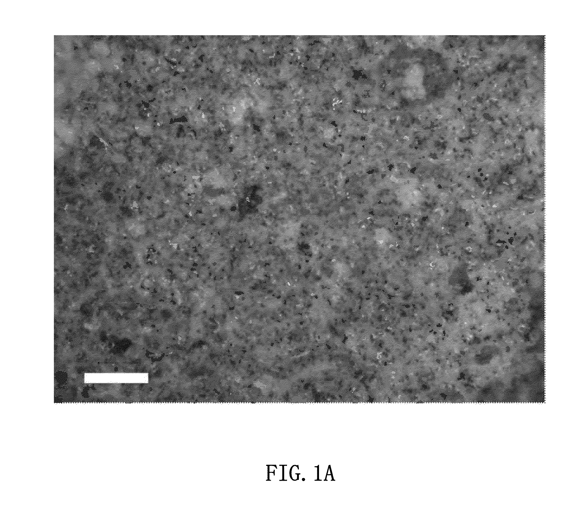
  • Repair and treatment of bone defect using agent produced by chondrocytes capable of hypertrophication and scaffold
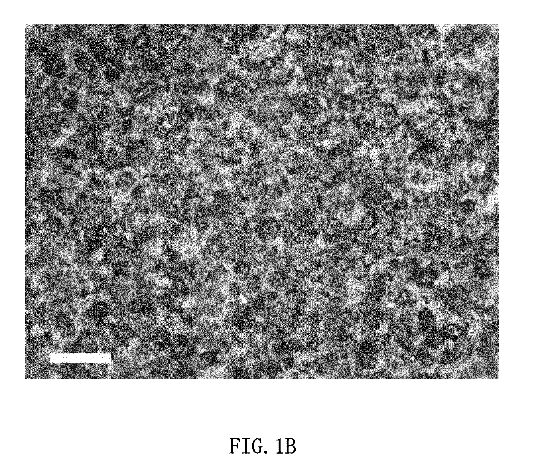
  • Repair and treatment of bone defect using agent produced by chondrocytes capable of hypertrophication and scaffold

Examples


example 1

Preparation and Detection of Cellular Function Regulating Agent Produced by Culturing Chondrocytes Capable of Hypertrophication Derived from Costa / Costal Cartilage in MEM Differentiation Agent Producing Medium

[0506](Preparation of Chondrocytes Capable of Hypertrophication from Costa / Costal Cartilages)

[0507]Four week-old male rats (Wistar) and 8 week-old male rats (Wistar) were, respectively, divided into groups, and examined in this Example. These rats were sacrificed using chloroform. The rats' chests were shaved using a razor and their whole bodies were immersed into a Hibitane solution (10-fold dilution) to be disinfected. The rats' chests were incised and costa / costal cartilages were collected aseptically.

[0508]Translucent growth cartilage regions were collected from boundary regions of the costa / costal cartilages. The growth cartilage regions were sectioned and stirred in a 0.25% trypsin-EDTA / dulbecco's phosphate buffered saline (D-PBS) at 37° C. for 1 hour. Next, the sections ...

example 2

Preparation and Detection of Cellular Function Regulating Agent Produced by Culturing Chondrocytes Capable of Hypertrophication Derived from Sternal Cartilage in MEM Differentiation Agent Producing Medium

[0618](Preparation of Chondrocytes Capable of Hypertrophication from Sternal Cartilages)

[0619]Eight week-old male rats (Wistar) are sacrificed using chloroform. The rats' chests are shaved using a razor and their whole bodies are immersed into a Hibitane solution (10-fold dilution) to be disinfected. The rats' chests are incised and inferior portions of sternal cartilages and processus xiphoideus are collected aseptically. Translucent growth cartilage regions are collected from the inferior portions of sternal cartilages and the processus xiphoideus.

[0620]The growth cartilage regions are sectioned and stirred in a 0.25% trypsin-EDTA / dulbecco's phosphate buffered saline (D-PBS) at 37° C. for 1 hour. Next, the sections are rinsed by centrifugation (at 170×g for 3 minutes), and then st...

example 3

Preparation and Detection of Cellular Function Regulating Agent Produced by Culturing Chondrocytes Capable of Hypertrophication Derived from Costa / Costal Cartilage in HAM Differentiation Agent Producing Medium

[0637](Detection of Agent Produced by Chondrocytes Capable of Hypertrophication Collected from Costa / Costal Cartilage)

[0638]Chondrocytes capable of hypertrophication were collected from costa / costal cartilages in the same manner as Example 1. A HAM differentiation agent producing medium (containing a HAM medium, 10% FBS (fetal bovine serum), 10 nM dexamethasone, 10 mM β-glycerophosphate, 50 μg / mL ascorbic acid, 100 U / mL penicillin, 0.1 mg / mL streptomycin and 0.25 μg / mL amphotericin B) was added to the chondrocytes capable of hypertrophication so that they were diluted so as to become a density of 4×10⁴ cells / cm2. The chondrocytes capable of hypertrophication were cultured, and then a supernatant of the medium (culture supernatant) was collected on a time course (4 days, 7 days,...


PUM

PropertyMeasurementUnit
Sizeaaaaaaaaaa
Molecular weightaaaaaaaaaa
Biocompatibilityaaaaaaaaaa

Abstract

The present invention provides a composite material for promoting or inducing osteogenesis in a biological organism. The composite material includes: A) an induced osteoblast differentiation inducing agent obtained by culturing chondrocytes capable of hypertrophication in a differentiation agent producing medium containing dexamethasone, β-glycerophosphate, ascorbic acid and a serum component; and B) a biocompatible scaffold. The present invention also provides a method of producing this composite material and a method of promoting or inducing osteogenesis in a biological organism.

Description

TECHNICAL FIELD[0001]The present invention is directed to a composite material containing an osteoblast differentiation inducing agent induced (produced) by chondrocytes capable of hypertrophication (chondrocytes with an ability of hypertrophication) and a scaffold, and a method of producing the composite material and a method of utilizing the composite material.BACKGROUND ART[0002]Osteogenesis is a preferred method of treating diseases associated with decrease of osteogenesis, bone injuries or bone defects. In the case where a bone tissue sustains injury such as a bone fracture or abscission due to a bone tumor, osteoblasts which are bone forming cells proliferate and differentiate to form bone, so that the bone fracture or a bone defective region is cured. In the case of mild injury, by immobilizing bone at an affected area, the osteoblasts can be activated so that the affected area is cured.[0003]In the case where osteoblasts cannot be effectively activated in circumstances such ...

Claims


Application Information

Patent Timeline
IPC IPC(8): A61K9/00A61K35/12A61K38/18A61P19/00
CPCA61L27/12A61L27/22A61L27/3817A61L27/3847A61L27/3895A61L2430/02A61L27/58A61L2300/252A61L2300/412A61L2300/414A61L27/54A61P19/00
Inventor OKIHANA, HIROYUKI
Owner HOYA CORP
Learn More