Cornea storage container to optimize cornea health

Inactive Publication Date: 2011-01-13
NUMEDIS
View PDF4 Cites 9 Cited by
  • Summary
  • Abstract
  • Description
  • Claims
  • Application Information

AI Technical Summary

Benefits of technology

[0017]The present invention is a novel cornea container that can improve the health of corneas, as determined by quantitative specular microscope analysis of the human corneal epithelium with respect to endothelial cell shape and corneal thickness. Accordingly, it is an object of the present invention to provide improved conventional corneal containers that overcome the problems of conventional corneal containers in order to provide superior cornea health.
[0018]In one aspect of the present invention, projections emanate from the lid to prevent the cornea from becoming suctioned against the lid.
[0019]In another aspect of the present invention, a corneal basket comprised of a plurality of prongs and disc support surfaces allows the area between the disc support surfaces and the container base to have an open area greater than 38%, and more preferably at least 50%, to allow improved movement of solutes residing within the preservation medium.
[0020]In another aspect of the present invention, the corneal basket includes upper and lower disc support surfaces to allow a greater range of cornea sizes to reside in the container. The corneal basket is structured to allow the area between the upper disc support surfaces and the container base to have an open area greater than 38%, and more preferably at least 50%, to allow improved movement of solutes residing within the preservation medium.

Problems solved by technology

Corneas with extremely low endothelial cell densities can no longer maintain a dehydrated state.
The corneas may decompensate, swell, and become cloudy over time, with an associated loss of visual acuity.
Unfortunately, cornea containers that are used, or have been conceived, are suboptimal.
However, there was no control over the position of the cornea, causing problems that included trapping the endothelium in a position that cut it off from the surrounding medium, allowing the epithelium to make contact with the walls of the vial, letting gas bubbles contact the cornea, and preventing lack of controlled positioning for specular microscopy and slit lamp evaluation.
Although it was easy to deposit the cornea into the vial, the ability to easily retrieve the cornea was difficult.
Although this configuration resolves some of the positioning problems of the suture approach, such as preventing the endothelium from being cut off from its media supply, the epithelium is forced to be in direct contact with the dividers that reside in the vial.
Direct physical contact between the dividers and the epithelium can cutoff media access, affecting the health of the cells that comprise the epithelium, and can physically damage the epithelium as it is dragged across the dividers when the cornea is removed for surgical implantation.
That process can add contaminants to the container as the technician is likely to place their gloved hands directly upon the alligator clip to open it during the process rather than find a clever way to actuate the alligator clip with a sterile tool.
Touching a component that resides within the container, even with gloves, is not good practice because bioburden level is dependent on what the technician's gloves have contacted previously and is also impacted by the skill level of the technician.
Thus, the process of using this storage container increases contamination risk and is highly dependent on the skill and patience of the technician.
Manipulation of the tissue by the technician may also damage the non-regenerating endothelium.
Also, there is no geometry to prevent gas from contacting the cornea as the container is shipped, subjecting the cornea to potential damage in transit.
However, the harvesting process currently used to obtain donated corneas often leads to corneas of various diameters and rarely results in a completely circular excision.
The apparatus '242 does not easily accommodate corneas of various diameters, or those that are not circular, since the support ring and the retaining lugs only allow about a 12% variation in cornea diameter before extra trimming is required.
The more the cornea is handled for trimming, the more potential problems arise.
For example, twisting, stretching, additional contact with forceps, and extra cutting increase the chances of damage to the tissue, particularly at its edges and on the endothelial cell surface.
Furthermore, the outcome can vary from technician to technician since cutting the corneas to match the limited diameters accepted by the apparatus of '242 requires patience, time, and a high level of skill.
Moreover, the act of using forceps to press the cornea into the retaining lugs of the support ring can inflict further damage to the cornea.
Still another problem with the apparatus of '242 is that gas in the container has the potential to make contact with the cornea during shipping, and can even become trapped in direct contact with the endothelium depending on the orientation of the container.
The basket design included prongs which obstructed the ability for slit lamp observation of the epithelium.
Although the conventional cornea container has many advantages over any other proposed or previously tried cornea container, we have discovered that the design acts to limit cornea health.
One problem, detailed within, is that the design of the corneal basket impedes the effective use of preservation medium within the container and as a result is suboptimal for maintaining corneal health.
The other problem is that the lid design allows the sclera to become suctioned to it, thereby cutting off solute movement to the endothelium, and in some cases, even trapping gas against the endothelium.
An additional problem exists with the width of the prongs, as measured from the inner diameter to the outer diameter of their basket arrangement.

Method used


Image

  • Cornea storage container to optimize cornea health
  • Cornea storage container to optimize cornea health
  • Cornea storage container to optimize cornea health

Examples


example 1

The Effect of Altering Cornea Container Geometry on Corneal Health as Determined by Quantitative Specular Microscope Analysis

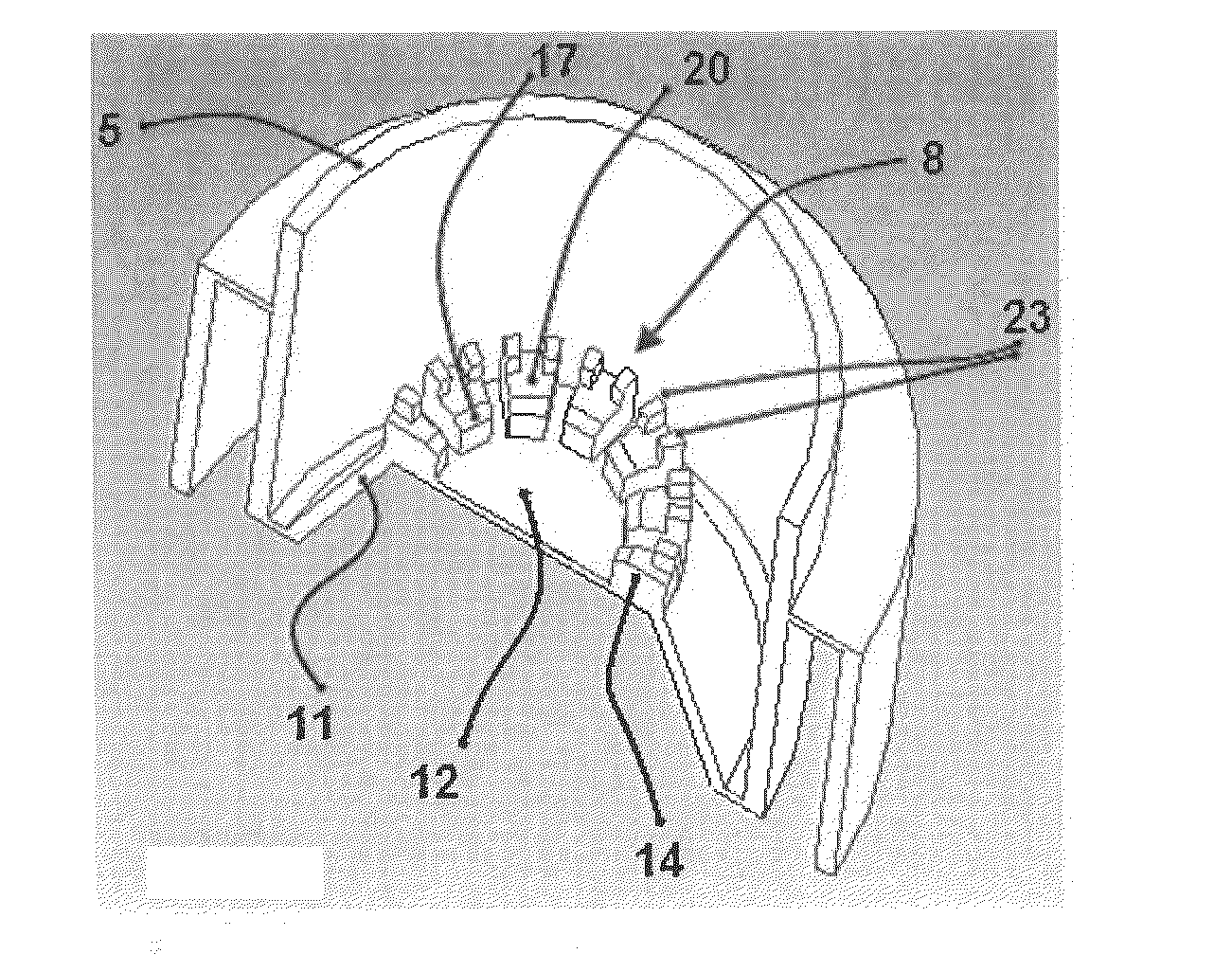
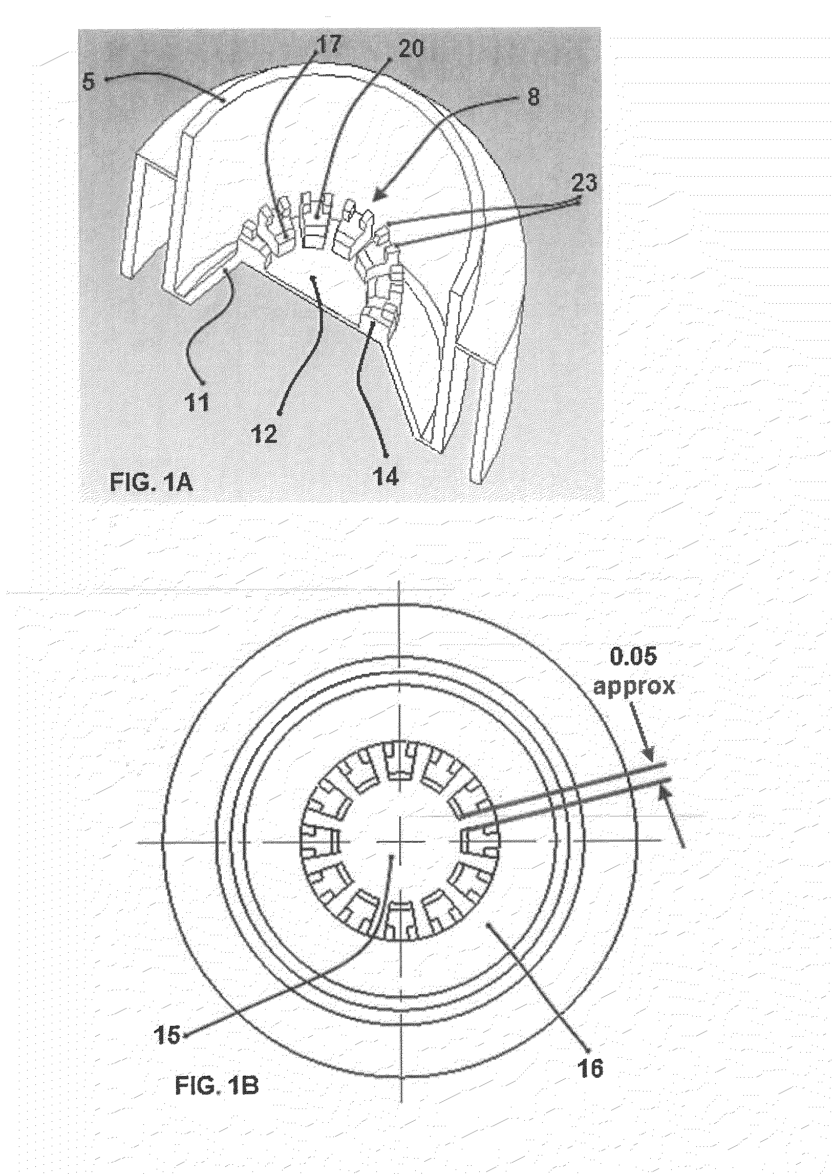
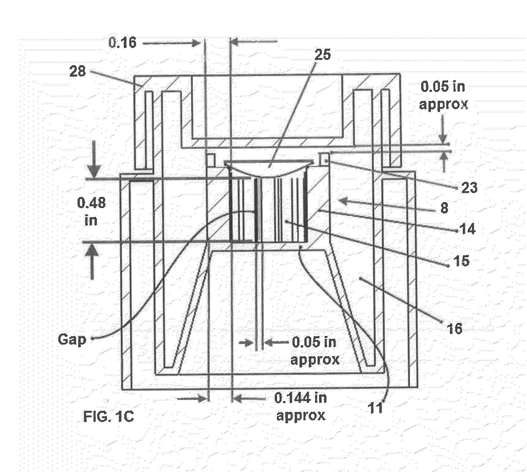
[0069]The aim of quantitative specular microscopic analysis is to assign values to endothelial cells that can provide a measure of their functional status or health of the human cornea. One of the parameters of quantitative specular microscopic analysis is determining the shape of the corneal endothelial cell. In a perfect cornea, endothelial cells demonstrate a perfect 6-sided hexagonal cell. This 6-sided configuration allows for the cell to function optimally. The normal human corneal endothelium is a monolayer of uniformly sized cells with a predominately hexagonal shape. Human corneal endothelial cells that demonstrate great variability in shape or hexagonality are considered to be under physiological stress and abnormal. Corneas that exhibit increased swelling during storage are also considered to be under physiological stress.

[0070]Maintenance of corneal...

example 2

[0079]The ability for dye to disperse within a cornea container of the present invention, constructed as described in Example 1, was compared to that of an Independent Corneal Viewing Chamber™. Cornea container devices resided upon a stationary surface with their lids removed and the container base of each device was filled with water. Then, trypan blue was dispensed into each device in proximity of the center of corneal basket in the area where the cornea would reside. Photographs were taken. The photograph of FIG. 12 shows a typical example of the pattern of trypan blue dye dispersion in each apparatus. As clearly shown in FIG. 12, trypan blue easily dispersed throughout cornea container 300 (i.e. the apparatus of the present invention). To the contrary, the majority of trypan blue remained trapped in the corneal basket of Independent Corneal Viewing Chamber™301, with a relatively small amount moving into the surrounding liquid in a poorly distributed pattern forced by the small g...


PUM


Abstract

An apparatus for shipping, storing, and viewing a cornea. The device offers an improvement to cornea health relative to conventional cornea containers.

Description

RELATED APPLICATION[0001]This application claims the benefit of priority of U.S. Provisional Application No. 61 / 187,937 filed Jun. 17, 2009, the disclosure of which is hereby incorporated by reference.TECHNICAL FIELD[0002]The technical field of this invention is related to devices and methods that improve cornea preservation.BACKGROUND OF THE INVENTION[0003]Each of the applications, patents, and papers cited in this application and in as well as each document or reference cited in each of the applications, patents, and papers (including during the prosecution of each issued patent; “application cited documents”), and each of the PCT and foreign applications or patents corresponding to and / or claiming priority from any of these applications and patents, and each of the documents cited or referenced in each of the application cited documents, are hereby expressly incorporated herein.[0004]The cornea is the transparent structure that forms the anterior one sixth of the outer coat of th...

Claims


Application Information

Patent Timeline
IPC IPC(8): A01N1/02
CPCA01N1/0263A61F2/142A61F2/0095A01N1/0273
Inventor SKELNIK, DEBRAWILSON, ROGERWELCH, DANIEL P.WILSON, JOHN R.
Owner NUMEDIS
Why Eureka
Try Eureka