

Method for detection of microorganism and kit for detection of microorganism
a microorganism and kit technology, applied in the field of microorganism detection and kit for microorganism detection, can solve the problems of difficult operation, high cost of flow cytometers, and difficulty in obtaining plate culture results, so as to achieve convenient and quick distinction
- Summary
- Abstract
- Description
- Claims
- Application Information
AI Technical Summary
Benefits of technology
Problems solved by technology
Method used
Image

Examples
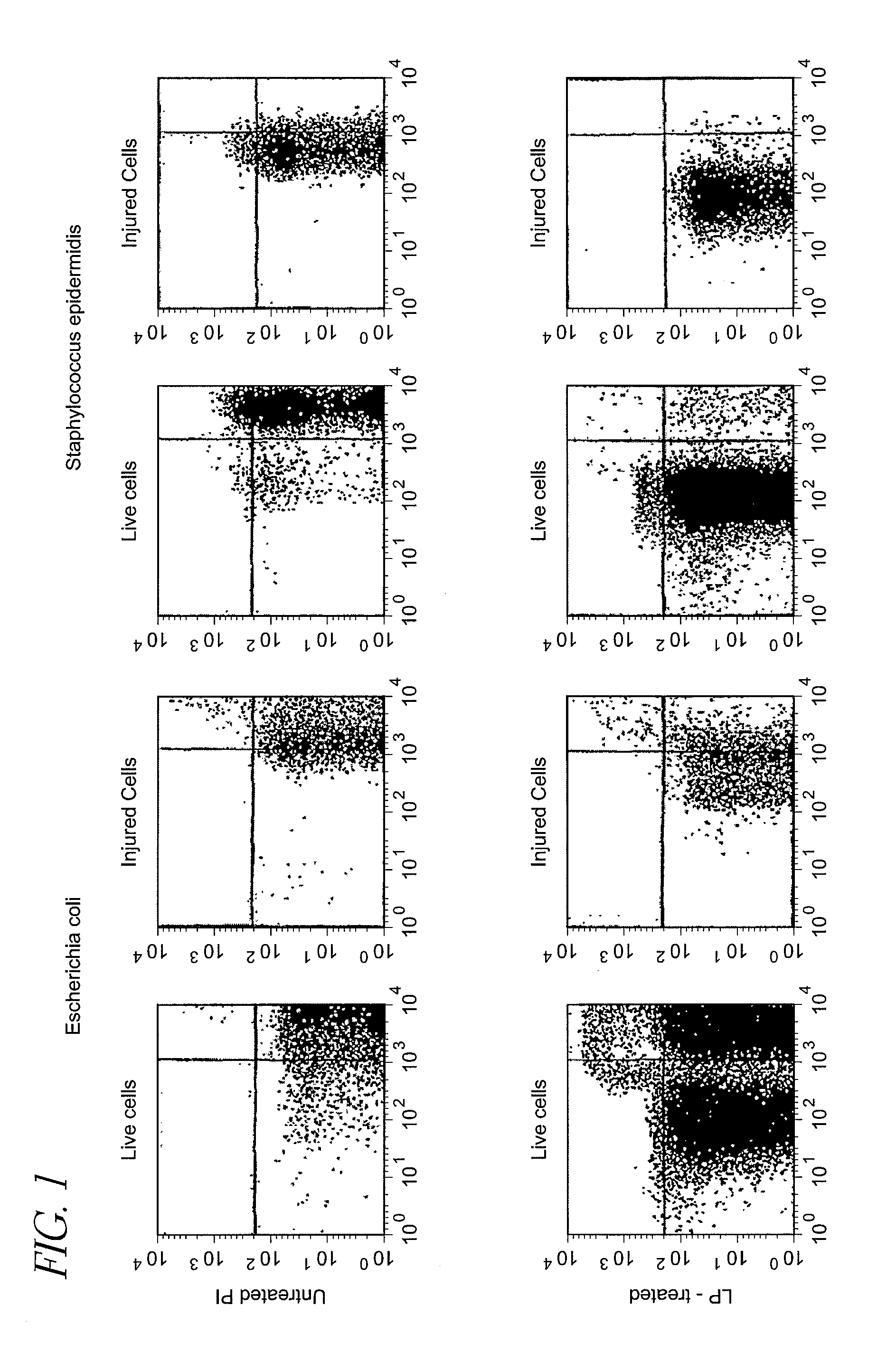
control example 1
[0133]Detection of live cells and injured cells of Escherichia coli and Staphylococcus epidermidis suspended in physiological saline (LP-treated or untreated) with flow cytometer
(1) Preparation of Samples
1-1. Preparation of Suspensions of Live Cells and Injured Cells
1-1-1. Escherichia Coli Suspensions
[0134]Escherichia coli DH5a was inoculated into L broth, and cultured at 10° C. for 12 hours as stationary culture, and then the culture was subjected to refrigerated centrifugation at 4° C. and 3,000×g for 10 minutes to harvest the cells. The harvested cells were suspended in physiological saline (Otsuka Pharmaceutical, the same shall apply to the following description), and the suspension was centrifuged to harvest the cells again. The same procedure was repeated once more to wash the cells.
[0135]The washed cells were suspended in physiological saline, the suspension was appropriately diluted, and 0.1 ml of the suspension was applied on L agar medium and cultured to measure live cells...
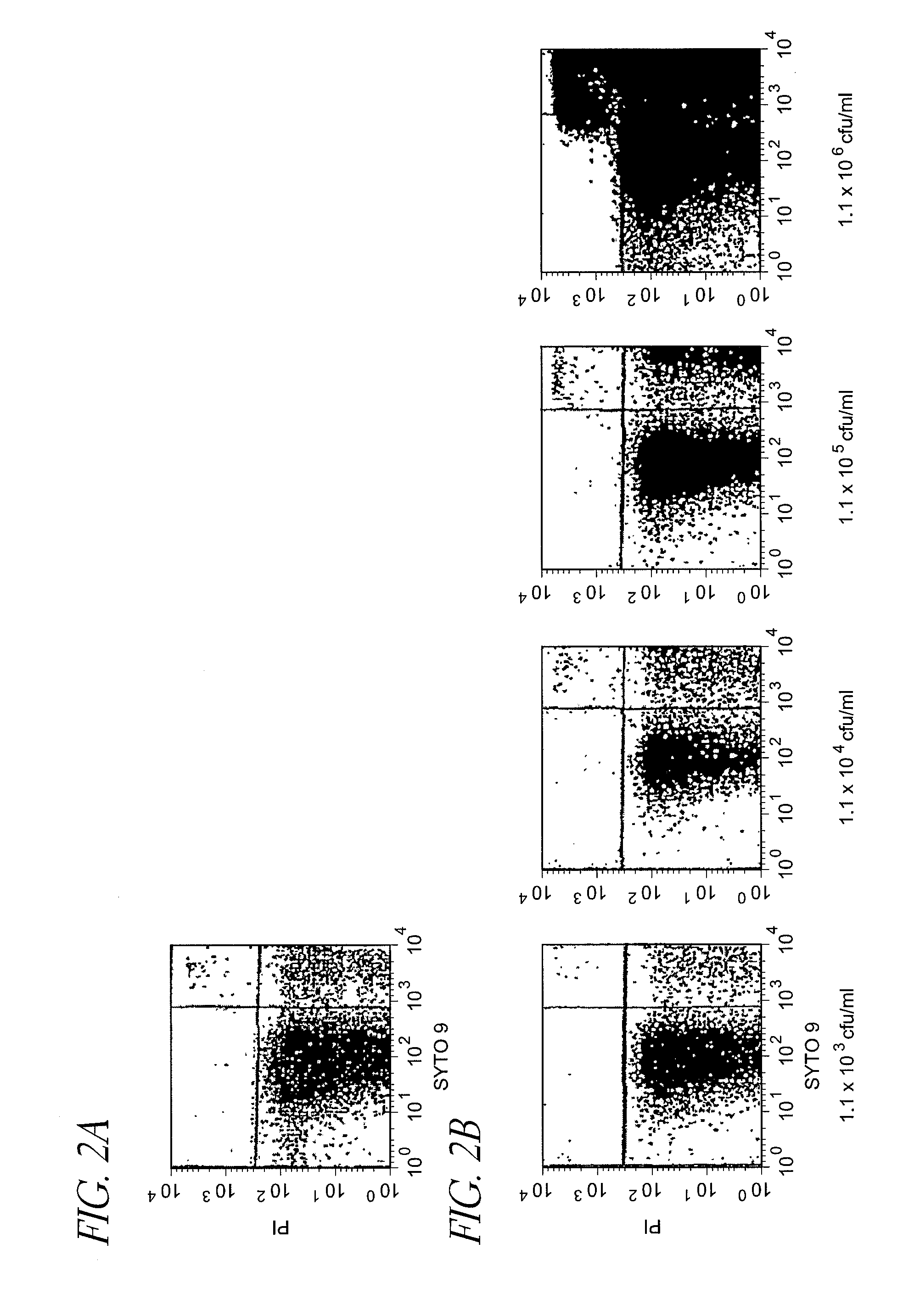
control example 2
[0149]Detection of Escherichia coli live cells (LP-treated) suspended in homogenized milk subjected to ultra high temperature pasteurization with flow cytometer
(1) Preparation of Samples
[0150]To 1 ml of 1.1×10⁷ cfu / ml Escherichia coli suspension (live cells) prepared in the same manner as that in Control Example 1, 9 ml of commercial homogenized cow's milk (subjected to ultra high temperature pasteurization (130° C., 2 seconds) henceforth also referred to as the “UHT homogenized milk”) was added to dilute the suspension 10 times, and the diluted suspension was serially diluted in the same manner to prepare UHT homogenized milk inoculated with 1.1×10² to 1.1×10⁶ cfu / ml Escherichia coli (live cells). As the aforementioned UHT homogenized milk, one confirmed not to form colonies when it was incubated at 37° C. for 48 hours and at 25° C. for 72 hours on an agar medium was used.
[0151]Then, the UHT homogenized milk of various dilution rates inoculated with Escherichia coli (live cells) an...
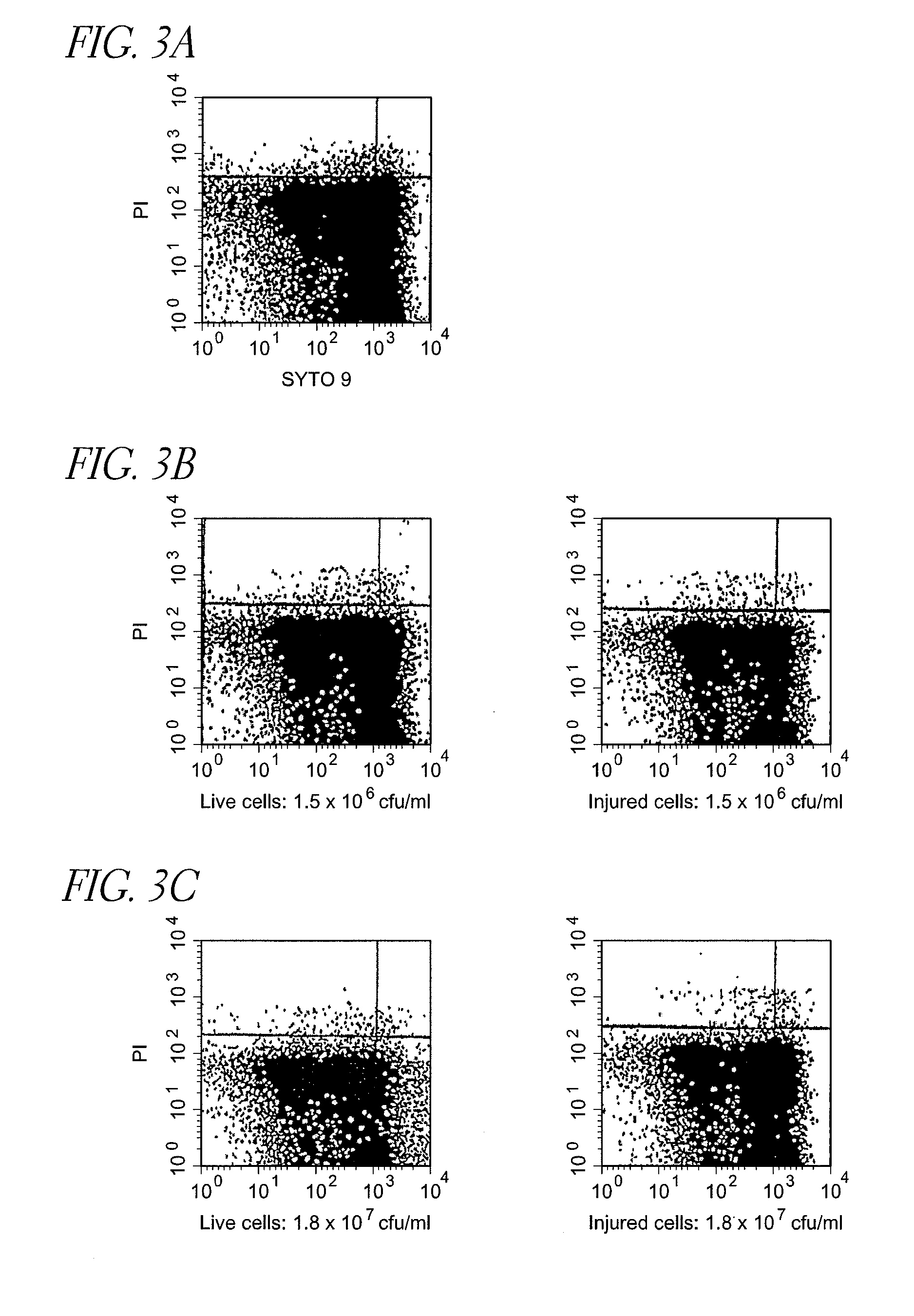
control example 3
[0157]Detection of live cells and injured cells of Escherichia coli and Staphylococcus epidermidis (LP-treated) suspended in non-homogenized milk subjected to low temperature long time pasteurization (LTLT) with flow cytometer
(1) Preparation of Samples
[0158]To 1 ml each of 1.5×10⁷ cfu / ml Escherichia coli suspensions (live cells and injured cells) prepared in the same manner as that in Control Example 1, 9 ml of commercial cow's milk not subjected to a homogenization treatment (non-homogenized, subjected to low temperature long time pasteurization (63° C., 30 minutes), henceforth also referred to as the “LTLT non-homogenized milk”) was added to dilute the suspensions 10 times, and diluted suspensions were serially diluted in the same manner to prepare LTLT non-homogenized milk inoculated with 1.5×10² to 1.5×10⁶ cfu / ml Escherichia coli (live cells and injured cells). As the aforementioned LTLT non-homogenized milk, one confirmed not to form colonies when it was incubated at 37° C. for...
PUM
| Property | Measurement | Unit |
|---|---|---|
| time | aaaaa | aaaaa |
| temperature | aaaaa | aaaaa |
| concentration | aaaaa | aaaaa |
Abstract
Description
Claims
Application Information