
Method for detection of microorganism and kit for detection of microorganism

a microorganism and kit technology, applied in the field of microorganism detection and kit for microorganism detection, can solve the problems of difficult operation, high cost of flow cytometers, and difficulty in obtaining plate culture results, so as to achieve convenient and quick distinction

Inactive Publication Date: 2011-09-27
MORINAGA MILK IND CO LTD
View PDF5 Cites 1 Cited by
  • Summary
  • Abstract
  • Description
  • Claims
  • Application Information

AI Technical Summary

Benefits of technology

[0034]The present invention enables convenient and quick distinction of live cells, injured cells and dead cells in foodstuffs and clinical samples by flow cytometry. The methods and kit of the present invention can be applied to spontaneous inspections, and are also economically advantageous.

Problems solved by technology

However, the plate culture method requires time of about two days to obtain a result.
However, FCM apparatuses (flow cytometers) used for this method are very expensive and have a large size, and they also requires skills for operation.
Moreover, they still have problems to be improved or solved concerning economy, safety, simplicity, and reliability and actuality for distinction of live cells and dead cells of microorganisms for actual applications in the fields of foodstuffs, in which a wide variety of bacteria contaminate as non-injured bacteria, injured bacteria and dead bacteria.
The preferred concentration of the ion-chelator used in the method of Patent document 1 is about 6 to 17 mM, and therefore it has a problem that both dead bacteria and live bacteria are lysed.
Therefore, it cannot necessarily be considered a quick method.
However, it is suggested that leucocytes and thrombocytes not lysed remain in the body fluid sample even after the protease, lipase and nuclease treatments, and live bacteria adsorb onto them to form complexes, and that both live bacteria and dead bacteria are stained, and thus it becomes difficult to determine whether bacteria are dead or alive.
Furthermore, although Patent document 2 describes that the method is a method for detecting bacteria at a density as low as 10 cells / ml (sample) within a time of about 2 hours or shorter, often 45 minutes or shorter, it actually also discloses an example in which detection was not possible unless at least 104 cfu / ml or more of bacteria exist in a body fluid sample, and thus it is not suitable for detection of a small amount of microorganisms such as those in cow's milk.
However, the dead cells were those boiled for 15 minutes, and the cell walls and cell membranes thereof were more highly injured compared with dead bacteria in actual foodstuffs.
Therefore, this technique is a technique suitable only for dead bacteria in a limited range of foodstuffs such as cooked dishes, and conditions were not examined for ultra high temperature pasteurization, which is performed for cow's milk etc. and the latest foodstuffs, and kills only bacteria without denaturing proteins in foodstuffs.
However, in the methods of Non-patent document 2, even if protease K is allowed to act on UHT cow's milk, micellar casein is not completely digested, and there are a lot of incomplete digestion products having a size comparable to those of bacteria.
If a fluorescent nuclear stain agent such as SYTO BC or SYTO9 is made to act on such products, strong nonspecific adsorption occurs to make the distinction of them from live bacteria difficult.
Moreover, it has also a problem that cell membranes of somatic cells such as bovine leucocytes and mammary epitheliocyte, considered as one of the contaminant milk components, are only slightly injured, and if they are subjected to staining with SYTO BC, SYTO9 or propidium iodide as they are, propidium iodide does not penetrate into them, and as a result, green fluorescence is emitted by chromosomal DNA to make distinction of the somatic cells from live bacteria difficult.
However, samples subjected to the aforementioned treatment contain a lot of contaminants originating in milk, and the detection limit for live lactic acid bacteria is degraded to a level as low as about 105 cfu / ml for yogurt or yogurt starter due to the contaminants.
Therefore, the method requires extremely delicate determination of conditions for destroying only somatic cells and not injuring cell walls and cell membranes of live bacteria by adjusting the concentrations of the nonionic detergent and the chelating agent.
Thus, the method is not suitable as a convenient and highly sensitive detection method for distinguishing live bacteria and dead bacteria.
However, there has not so far been reported at all use of these topoisomerase I poisons, topoisomerase II poisons, and bacterial DNA gyrase poisons for pretreatments of samples such as foodstuffs and clinical samples containing microorganisms in a test method for distinguishing live cells and dead cells of a microorganism for the purpose of realizing quick and highly sensitive detection.
However, detection by this method is limited to a case where respiratory activity and esterase activity of the objective bacterium can be accurately measured.
Therefore, when the injured cells are those showing a low degree of injury caused by low temperature long time pasteurization (LTLT) or high temperature short time pasteurization (HTST), or those showing a low degree of injury due to stress in environment, live cells and injured cells cannot be distinguished by this method.

Method used


Image

  • Method for detection of microorganism and kit for detection of microorganism
  • Method for detection of microorganism and kit for detection of microorganism
  • Method for detection of microorganism and kit for detection of microorganism

Examples


control example 1

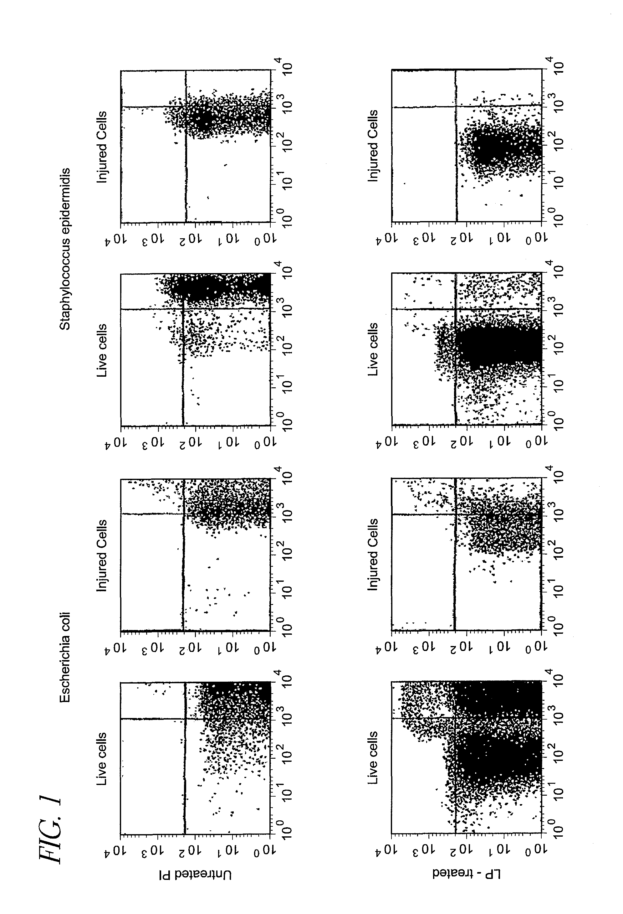
Detection of Live Cells and Injured Cells of Escherichia coli and Staphylococcus epidermidis Suspended in Physiological Saline (LP-Treated or Untreated) with Flow Cytometer

(1) Preparation of Samples

1-1. Preparation of Suspensions of Live Cells and Injured Cells

1-1-1. Escherichia coli Suspensions

[0106]Escherichia coli DH5α was inoculated into L broth, and cultured at 10° C. for 12 hours as stationary culture, and then the culture was subjected to refrigerated centrifugation at 4° C. and 3,000×g for 10 minutes to harvest the cells. The harvested cells were suspended in physiological saline (Otsuka Pharmaceutical, the same shall apply to the following description), and the suspension was centrifuged to harvest the cells again. The same procedure was repeated once more to wash the cells.

[0107]The washed cells were suspended in physiological saline, the suspension was appropriately diluted, and 0.1 ml of the suspension was applied on L agar medium and cultured to measure live cells count...

control example 2

Detection of Escherichia coli Live Cells (LP-Treated) Suspended in Homogenized Milk Subjected to Ultra High Temperature Pasteurization with Flow Cytometer

(1) Preparation of Samples

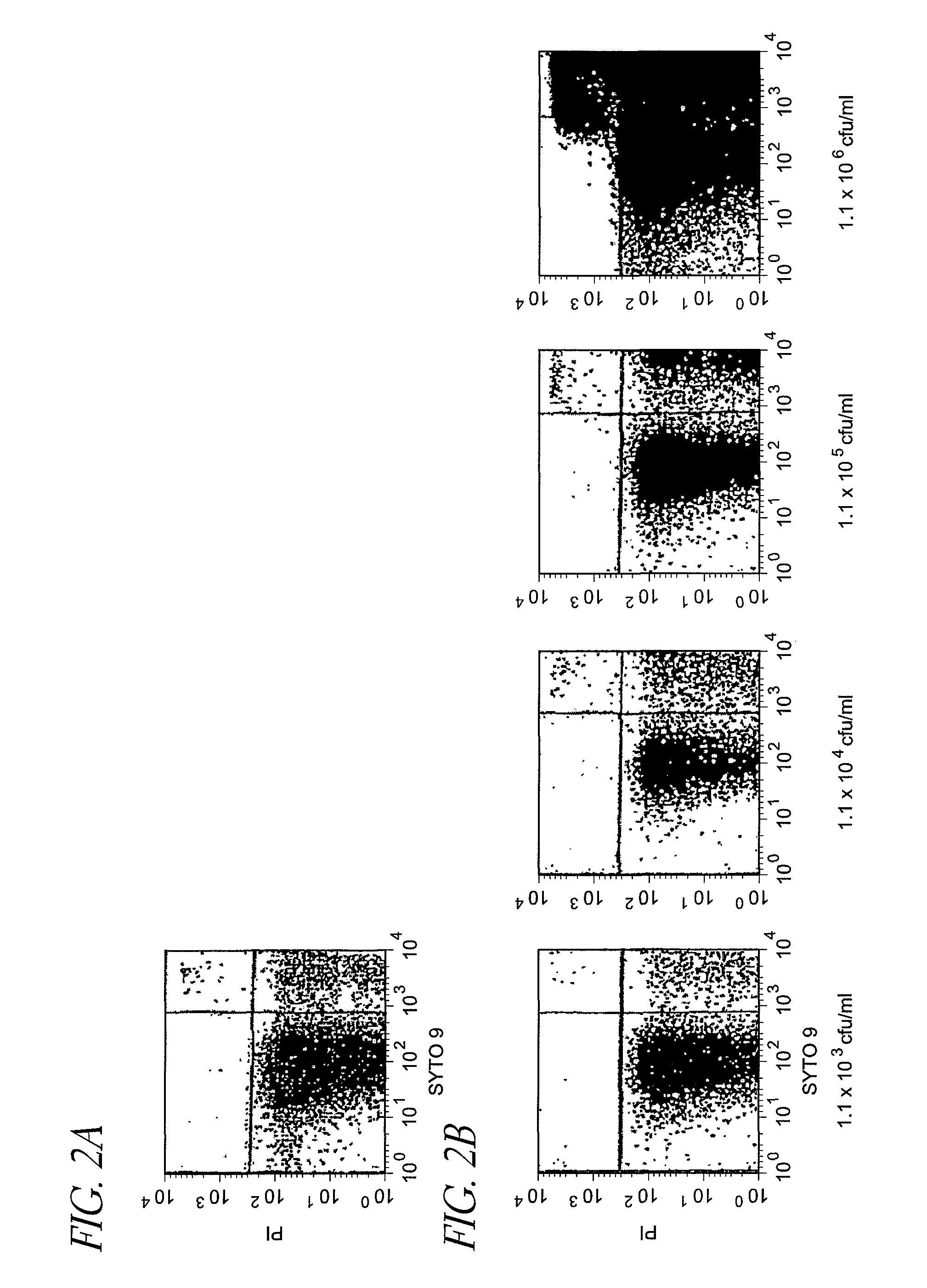
[0121]To 1 ml of 1.1×10⁷ cfu / ml Escherichia coli suspension (live cells) prepared in the same manner as that in Control Example 1, 9 ml of commercial homogenized cow's milk (subjected to ultra high temperature pasteurization (130° C., 2 seconds) henceforth also referred to as the “UHT homogenized milk”) was added to dilute the suspension 10 times, and the diluted suspension was serially diluted in the same manner to prepare UHT homogenized milk inoculated with 1.1×10² to 1.1×10⁶ cfu / ml Escherichia coli (live cells). As the aforementioned UHT homogenized milk, one confirmed not to form colonies when it was incubated at 37° C. for 48 hours and at 25° C. for 72 hours on an agar medium was used.

[0122]Then, the UHT homogenized milk of various dilution rates inoculated with Escherichia coli (live cells) and the ...

control example 3

Detection of Live Cells and Injured Cells of Escherichia coli and Staphylococcus epidermidis (LP-Treated) Suspended in Non-Homogenized Milk Subjected to Low Temperature Long Time Pasteurization (LTLT) with Flow Cytometer

(1) Preparation of Samples

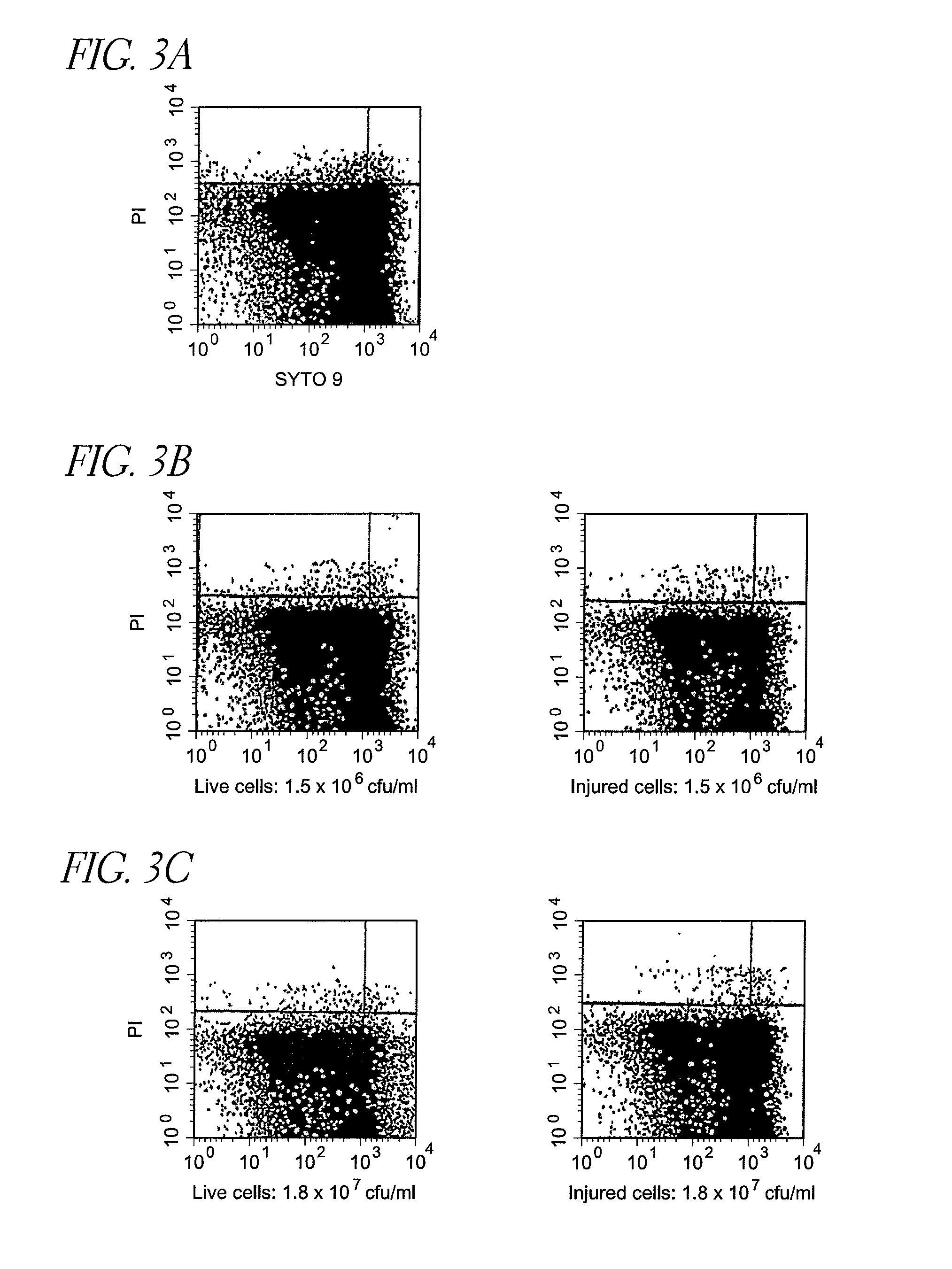
[0129]To 1 ml each of 1.5×10⁷ cfu / ml Escherichia coli suspensions (live cells and injured cells) prepared in the same manner as that in Control Example 1, 9 ml of commercial cow's milk not subjected to a homogenization treatment (non-homogenized, subjected to low temperature long time pasteurization (63° C., 30 minutes), henceforth also referred to as the “LTLT non-homogenized milk”) was added to dilute the suspensions 10 times, and diluted suspensions were serially diluted in the same manner to prepare LTLT non-homogenized milk inoculated with 1.5×10² to 1.5×10⁶ cfu / ml Escherichia coli (live cells and injured cells). As the aforementioned LTLT non-homogenized milk, one confirmed not to form colonies when it was incubated at 37° C. for 48 ho...


PUM


Abstract

According to the following steps, live cells, injured cells, VNC cells and dead cells of a microorganism in a test sample are detected by flow cytometry:a) the step of treating the test sample with an enzyme having an activity of decomposing cells other than those of the microorganism, colloidal particles of proteins or lipids existing in the test sample,b) the step of treating the test sample with a topoisomerase poison and / or a DNA gyrase poison.c) the step of treating the test sample treated in the steps a) and b) with a nuclear stain agent, andd) the step of detecting the microorganism in the test sample treated with the nuclear stain agent by flow cytometry.

Description

CROSS-REFERENCE TO RELATED APPLICATIONS[0001]This application is the U.S. National Phase under 35 U.S.C. §371 of International Application PCT / JP2006 / 302889, filed Feb. 17, 2006, which was published in a non-English language, which claims priority to JP 2005-211190, filed Jul. 21, 2005.TECHNICAL FIELD[0002]The present invention relates to a method for detecting a microorganism contained in foodstuffs or clinical samples, a method for preparing a sample used for the method, and a kit for detecting a microorganism. More precisely, the present invention relates to a method and a kit for detection of a microorganism that enable even distinction of live cells, injured cells and dead cells of a microorganism contained in foodstuffs or clinical samples.BACKGROUND ART[0003]The plate culture method has been conventionally used for measurement of general live bacterial counts in foodstuffs, clinical samples or environments. However, the plate culture method requires time of about two days to ...

Claims


Application Information

Patent Timeline
Patent Type & Authority Patents(United States)
IPC IPC(8): C12Q1/02C12Q1/18
CPCC12Q1/04C12Q1/34C12Q1/533C12Q2304/10C12Q1/37
Inventor YOSHIDA, SHINICHISOEJIMA, TAKASHI
Owner MORINAGA MILK IND CO LTD
Learn More