

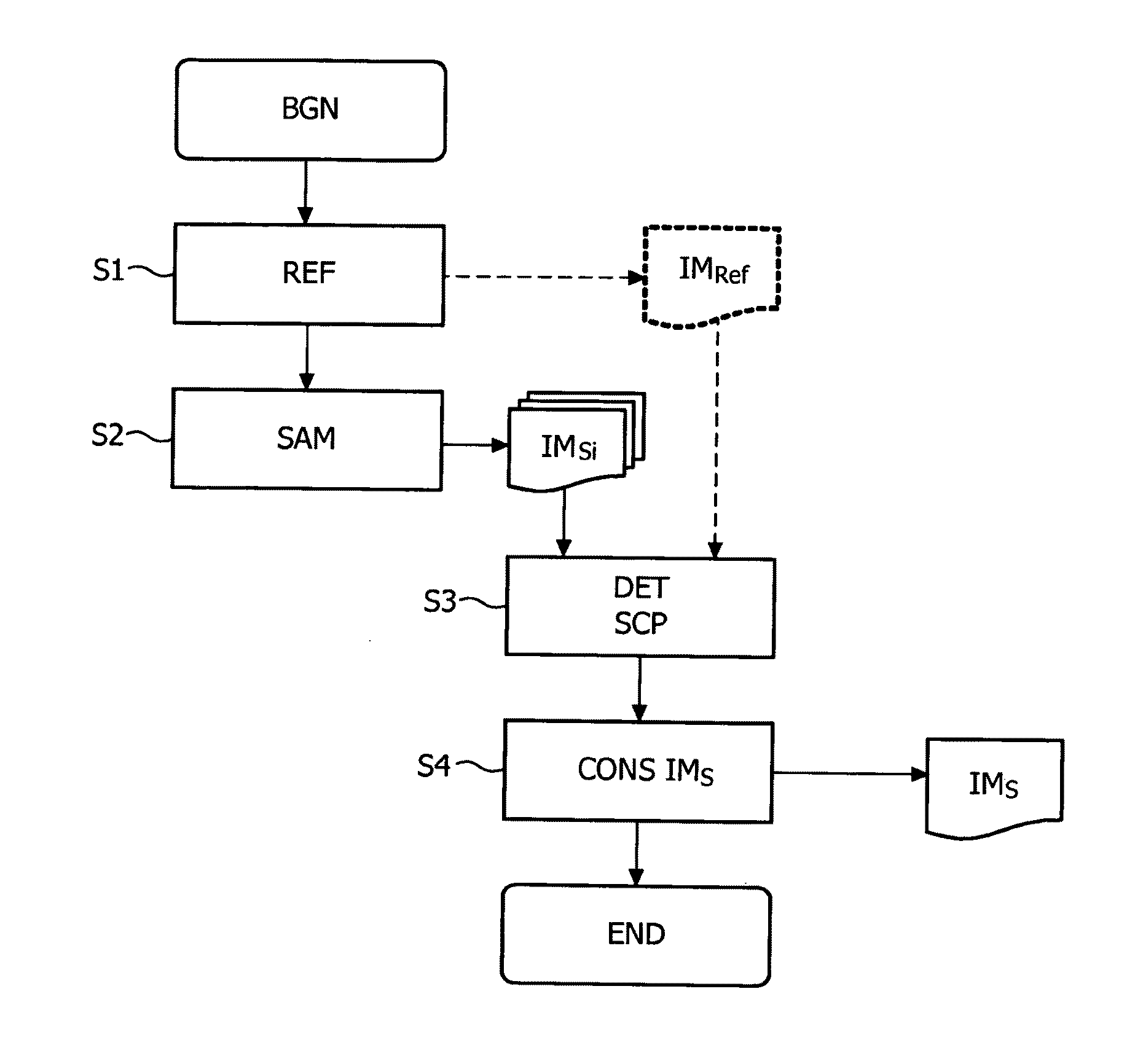
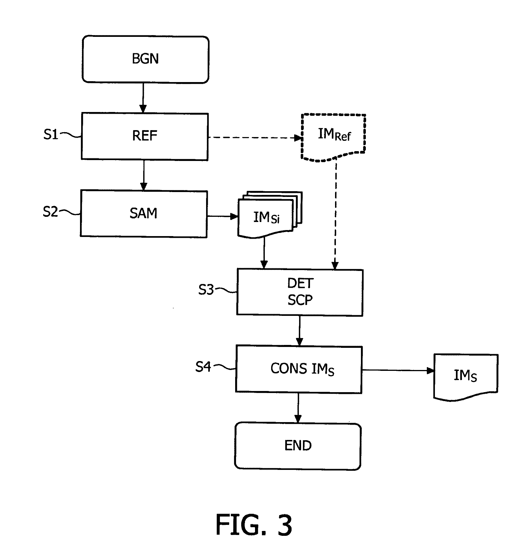
Method of imaging a sample
a sample and processing method technology, applied in the field of image processing methods, can solve the problems of difficult imaging of samples like unstained samples or biological samples, and achieve the effect of enhancing the image contrast of samples
- Summary
- Abstract
- Description
- Claims
- Application Information
AI Technical Summary
Benefits of technology
Problems solved by technology
Method used
Image

Examples
first embodiment
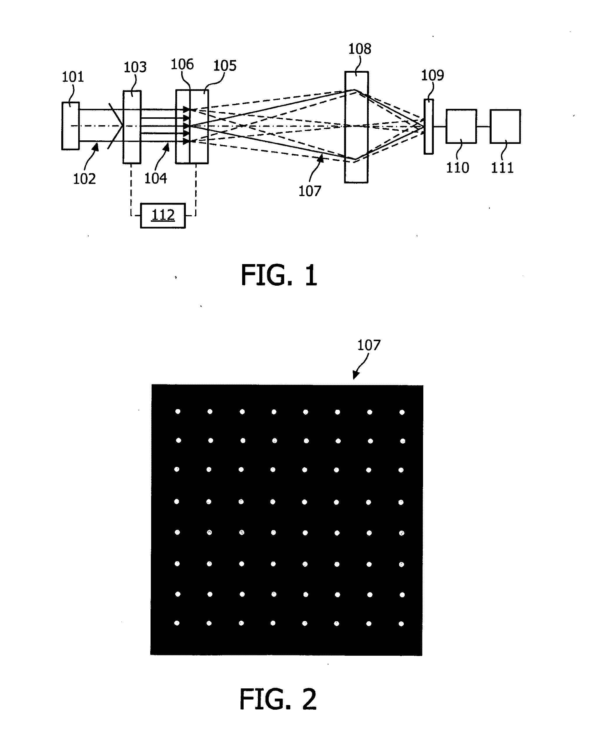
[0088] the spot characterizing parameter determination for the plurality of spot comprises determining the position shift between a reference position and a sample position. FIG. 7 schematically illustrates the position shift of a spot on a portion of a pixelated detector between a nominal position NP and a sample position SP. More precisely, for the plurality of spot a displacement vector DV from the reference array of spots to the sample related array of spots is calculated. The reference position for the plurality of identified spot within the reference array of spots and the sample position for the plurality of identified spot within the imaged sample related array of spots are determined. Then, the displacement vector for a plurality of spot is determined by calculating the difference between the reference position and the sample position of the plurality of associated spot.
[0089]According to a first alternative, the image construction step depends on the magnitude DV of the di...
second embodiment
[0093] the spot characterizing parameter determination for the plurality of spots comprises determining for the plurality of spots an alteration of the spot shape due to the reference array of spots interacting with the sample. The alteration may be for example the deviation from the circular symmetry of the spot shape. The alteration of the spot shape may be measured by determining the height and / or the width in at least one direction of the spot. FIG. 8 schematically depicts a spot on a portion of a pixelated detector and illustrates the alteration (e.g. longitudinal elongation) of a spot between a nominal position NP and a sample position SP.
third embodiment
[0094] the spot characterizing parameter determination for the plurality of spots comprises determining for the plurality of spots an alteration of the polarization due to the reference array of spots interacting with the sample. The alteration may be for example due to birefringence in the sample. The alteration of the polarization may be measured by adding a polarization filter to the detection light path.
[0095]According to a fourth embodiment, the spot characterizing parameter determination for the plurality of spots comprises summing the pixels intensity of areas associated with the plurality of spots. More precisely, an area of grouped pixels of the pixelated detector is associated with the plurality of sample spots. The areas are defined such that the pixels within the area are the closest to the identified reference spot corresponding to the identified sample spot. The spot characterizing parameters are determined by summing pixel intensities of each area. For example, the in...
PUM
 Abstract
Description
Claims
Application Information