Methods for the characterization of microorganisms on solid or semi-solid media

Active Publication Date: 2011-02-10
BIOMERIEUX INC
View PDF21 Cites 19 Cited by
  • Summary
  • Abstract
  • Description
  • Claims
  • Application Information

AI Technical Summary

Benefits of technology

[0006]The present invention provides methods for detecting, monitoring, characterizing, and/or identifying microorganisms on solid and/or semi-solid media. Characterization encompasses the broad categorization or classification of microorganisms as well as the actual identification of a single species. As used herein “identification” means determining to which family, genus, species, and/or strain a previously unknown microorganism belongs to. For example, identifying a previously unknown microorganism to the family, genus, species, and/or strain level. The methods disclosed herein allow the detection, characterization and/or identification of microorganisms more quickly than prior techniques, resulting in faster diagnoses (e.g., in a subject having or suspected of having an infection) and identification of contaminated materials (e.g., foodstuffs and pharmaceuticals). The steps involved in the methods of the invention can be carried out i

Problems solved by technology

Direct examination of microorganisms on growth medium has been considered problematic d

Method used


Image

  • Methods for the characterization of microorganisms on solid or semi-solid media
  • Methods for the characterization of microorganisms on solid or semi-solid media
  • Methods for the characterization of microorganisms on solid or semi-solid media

Examples


example 1

Obtaining Spectra from Colonies on Plates and Membranes

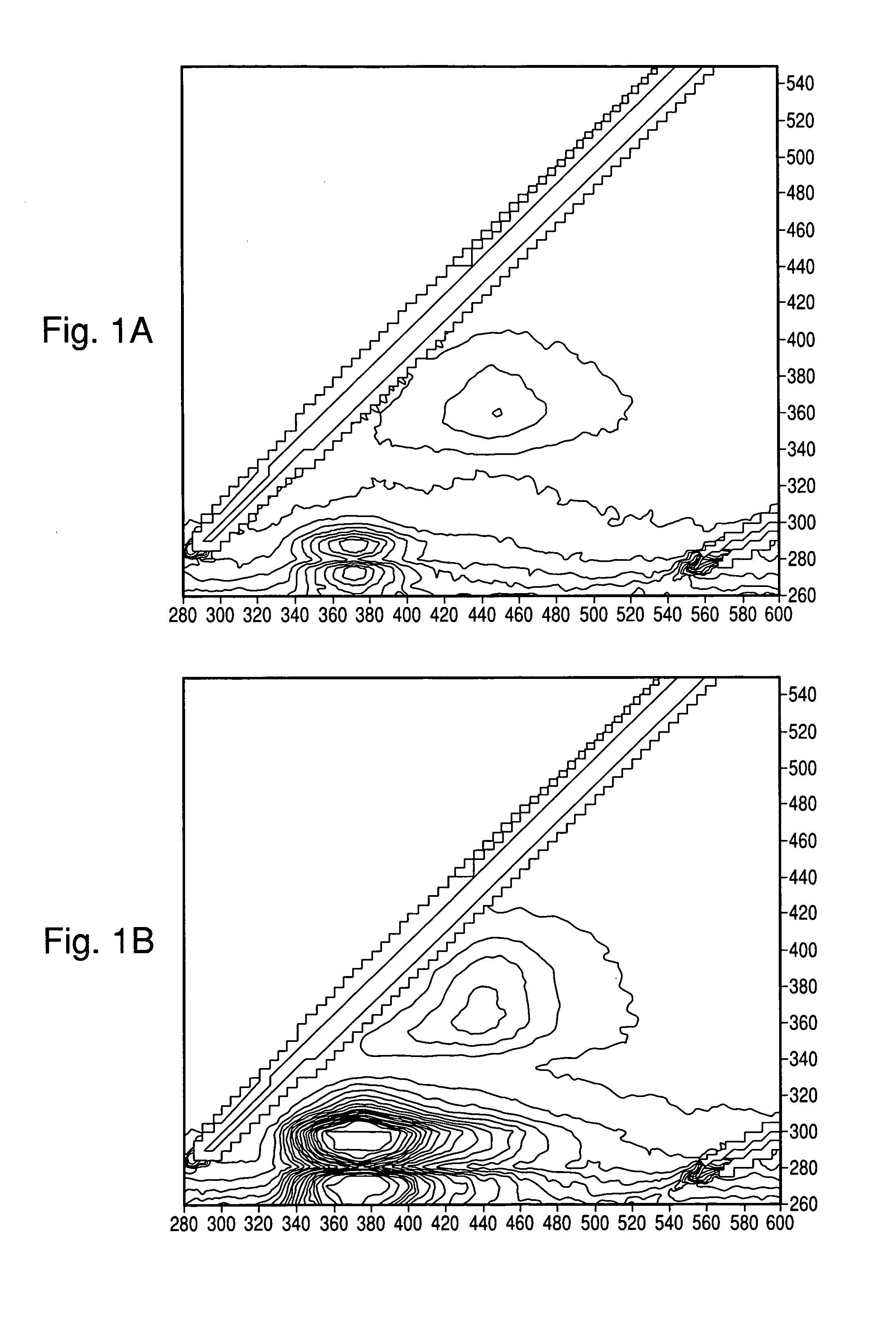
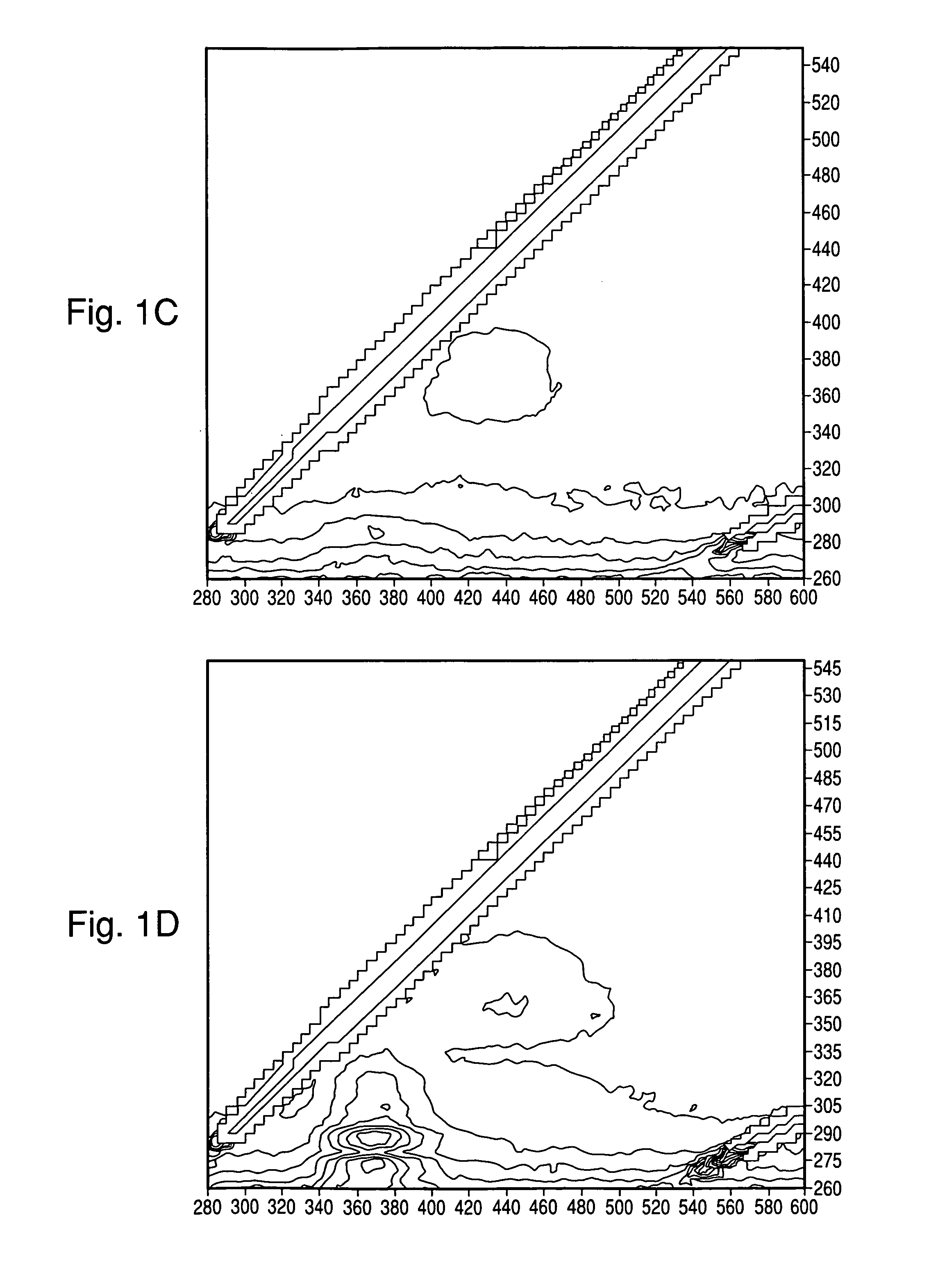
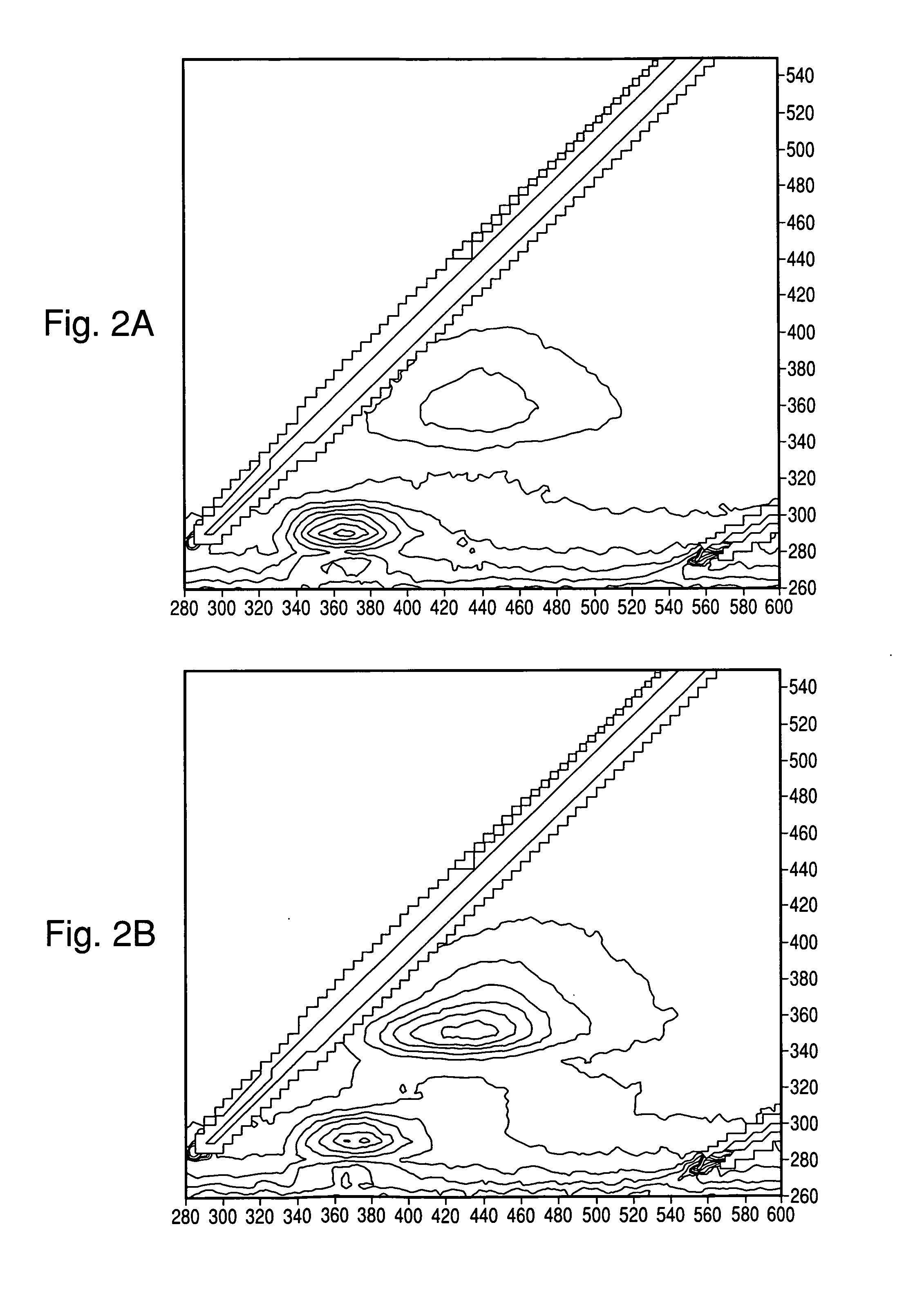
[0095]Tests were conducted to determine whether useful spectra could be obtained of colonies directly on blood agar plates (BAP; tryptic soy agar with 5% sheep blood), with and without black membranes (Table 1). Colonies of E. coli (EC), S. aureus (SA), E. faecalis (EF), and P. aeruginosa (PA) were grown as indicated in Table 2 and spectra were taken through the UV microscope (10× Objective) coupled with a fiber optic adaptor to a Fluorolog3 spectrometer (Horiba Jobin Yvon, Edison N.J.) and a PMT detector. The EEM was acquired through a wavelength range of Excitation (Ex)=260-550 nm, and Emission (Em)=280-600 nm, every 5 nm with a slit width=5 nm. Where indicated, the interrogation area was narrowed by placing a 1 mm pinhole in the emission path which resulted in an observed area of approximately 0.1 mm. Without the pinhole, the excitation and emission circles as projected on the colonies were equal at approximately 1 mm diamete...

example 2

Scanning for Microcolonies Through the Microscope

[0100]Tests were carried out to determine whether colonies growing under the microscope on a motorized stage could be located by using point-by-point IF measurements, and have IF spectra automatically collected of each colony detected. A UV microscope was coupled to a Fluorolog3 (Horiba Jobin Yvon, Edison N.J.) spectrometer, which served as the fluorescence excitation source and the emission measurement device, via fiber optic cables. The microscope's motorized stage was fitted with a compact plate incubator constructed with coils of tubing fed by a circulating waterbath set to 36° C. The incubator was also equipped with a UV transparent window made from a quartz coverslip. Various agar media were inoculated by spread method with E. coli ATCC 25922 (EC) and / or S. aureus ATCC 25923 (SA) as indicated in Table 3. Some runs used a light blocking material, either a black Whatman Mixed Ester (WME) membrane or charcoal, to reduce the fluores...

example 3

Classification of Microorganism Colonies on Agar Plates

[0112]Tests were carried out to determine whether microbial colonies could be classified from IF spectra taken directly on the agar plate where they were grown.

[0113]The spectral acquisition was done across an excitation (Ex) and emission (Em) matrix of wavelengths 260-580 nm and 260-680 nm, respectively, in a subset of 300 EEM points selected to reduce the acquisition time required. Additionally, all reflectance wavelengths (where Ex=Em) were also read. For fluorescence, the slit widths were set to 5 nm bandpass and the integration time was 1000 ms. Each 300 point acquisition took approximately 8.1 min to complete.

[0114]Table 4 lists the microorganisms tested, which comprised 6 isolates each of 20 species for a total of 120 tests. Where used to indicate groupings other than by species, the term Clinical Gram (ClinGram) refers to the classification level possible by a highly skilled observer reading a Gram stain, not just positi...


PUM

PropertyMeasurementUnit
Diameteraaaaaaaaaa
Temperatureaaaaaaaaaa

Abstract

The present invention relates to methods and systems for scanning, detecting, and monitoring microorganisms on solid or semi-solid media using intrinsic fluorescence (IF) measurements. The methods are further directed to detection, characterization and/or identification of microorganisms on a solid or semi-solid media using intrinsic fluorescence (IF) measurements that are characteristic of said microorganisms.

Description

CROSS REFERENCE TO RELATED APPLICATION[0001]This application claims the benefit of U.S. Provisional Patent Application No. 61 / 122,925, entitled, “Methods for Characterization of Microorganisms on Solid or Semi-Solid Media”, filed Dec. 16, 2008, which is incorporated herein.FIELD OF THE INVENTION[0002]The present invention relates to methods and systems for detecting, monitoring, characterizing, and / or identifying microorganisms on solid or semi-solid media.BACKGROUND OF THE INVENTION[0003]Microorganisms isolated for the purpose of clinical diagnostics, as well as those isolated to monitor contamination of foodstuffs, medical tissues, or the environment, often need to be characterized in order to determine the appropriate response to the presence of the organisms found. Traditional automated phenotypic identification assays, such as the Vitek®, Phoenix™, and Microscan® systems, or manual phenotypic tests such as API, require that microorganisms be in an appropriate growth phase and f...

Claims


Application Information

Patent Timeline
IPC IPC(8): C12Q1/04C12Q1/68G01N33/566
CPCC12Q1/04G01N21/31G01N21/76G01N33/483
Inventor WALSH, JOHNHYMAN, JONES M.THORPE, THURMAN
Owner BIOMERIEUX INC
Why Eureka
Try Eureka