Medical image diagnostic apparatus and method using a liver function angiographic image, and computer readable recording medium on which is recorded a program therefor

a technology of angiographic image and diagnostic apparatus, which is applied in the field of medical image diagnostic, can solve the problems of inability to accurately meet the needs of conventional technologies, difficult to understand the detail of a time series variation of the signal value of a region of interest, and unrealistic to make a judgment based only on a parameter image in an actual image diagnosis site, etc., to achieve easier and more appropriate image diagnosis and improve image reading efficiency.

Inactive Publication Date: 2011-03-03
FUJIFILM CORP
View PDF24 Cites 49 Cited by
  • Summary
  • Abstract
  • Description
  • Claims
  • Application Information

AI Technical Summary

Benefits of technology

[0061]More specifically, according to the first embodiment of the present invention, segment functional level information representing a functional level of at least one segment of a liver of diagnostic target is obtained from a liver function angiographic image obtained with a contrast agent that produces a contrast effect according to a functional level of a liver of diagnostic target and presented. This allows easier and more appropriate image diagnosis to be made using the functional level and segments of the liver. For example, when a target region of a liver for resection surgery is determined through image diagnosis of the liver, the functional level of the liver may be confirmed on a segment-by-segment basis, so that a determination may be made easily and appropriately as to which segment is to be a resection target.
[0062]According to the second embodiment of the present invention, with respect to each of liver angiographic images of two or more time phases of interest obtained in a contrast examination using a contrast agent that reflects liver blood flow and liver function in an image, a region of interest positionally corresponding to each other between each of the time phases of interest is set, a local image representing the region of interest is generated with respect to each of the time phases of interest based on each of the liver angiographic images of the time phases of interest, and the generated local image with respect to each of the time phases of interest is displayed in a manner that allows comparative reading. This allows comparative reading of the displayed local images of the respective time phases of interest to be made easily, and a time series variation in signal value...

Problems solved by technology

In the mean time, the technology described in Patent Document 4 performs liver segment identification but does not intend to the evaluation of liver function.
Further, the technology described in Non-patent Document 4 performs the evaluation of liver cell function using an EOB contrast agent but does not pay attention to liver segments.
These conventional technologies, therefore, can not be said to appropriately meet the needs in image diagnosis of liver described above.
It is, therefore, difficult to understand a time series variation of signal value of a region of interest in detail.
Further, it is unrealistic to make a judgment based only on a parameter image in an actual image diagnosis site, and the judgment is made by returning to the original image.
In t...

Method used


Image

  • Medical image diagnostic apparatus and method using a liver function angiographic image, and computer readable recording medium on which is recorded a program therefor
  • Medical image diagnostic apparatus and method using a liver function angiographic image, and computer readable recording medium on which is recorded a program therefor
  • Medical image diagnostic apparatus and method using a liver function angiographic image, and computer readable recording medium on which is recorded a program therefor

Examples


first embodiment

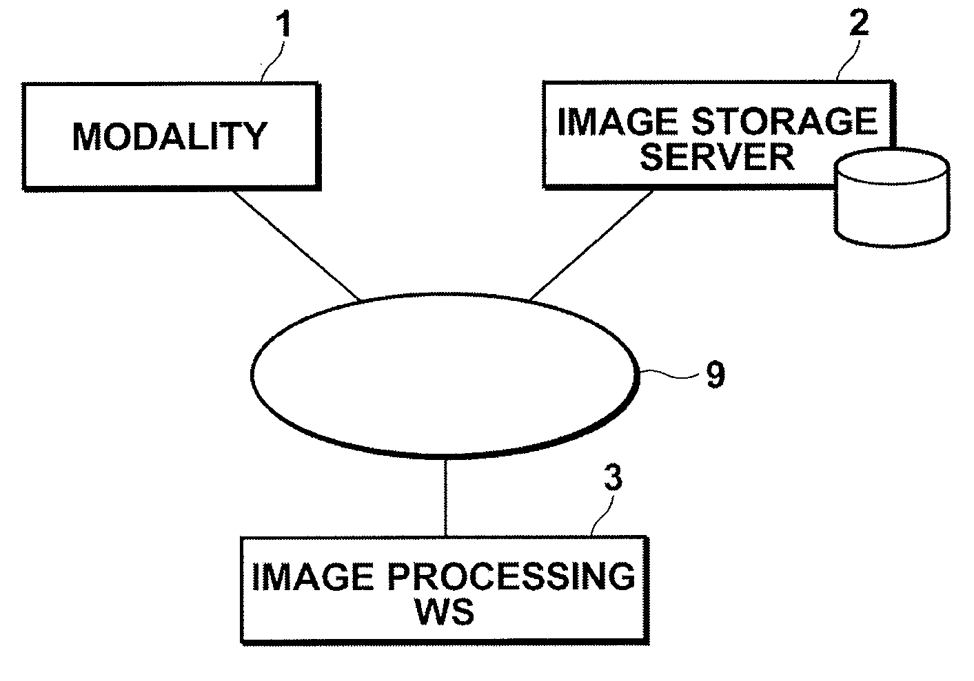
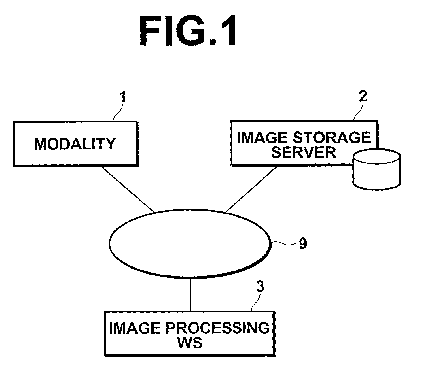
[0091]In the invention, modality 1 includes an MRI system. The MRI system is capable of performing dynamic imaging in which three-dimensional medical images (voxel data) Vt0, Vt1, - - - , Vt6 (hereinafter, collectively referred to as three-dimensional dynamic images Vtn) of an abdomen (including liver) are obtained before administering a contrast agent that includes gadoxetate sodium (Gd-EOB-DTPA) as an active ingredient (EOB contrast agent) and, for example, 20 sec, 1 min, 2 min, 5 min, 10 min, and 20 min after administration of the EOB contrast agent. Here, as described above, the EOB contrast agent has a property to be selectively absorbed in normal liver cells, so that three-dimensional dynamic images Vtn obtained by such imaging correspond to liver function angiographic images.

[0092]Image storage server 2 is a computer for storing image data of three-dimensional dynamic images Vtn obtained by modality 1 and of medical images generated through image processing in image processin...

second embodiment

[0139]As described above, in the present invention, liver segments R3S1, R3S2, - - - in a three-dimensional morphological image V3 representing an anatomical structure of a liver are identified and then liver segments R2S1, R2S2 - - - in three-dimensional dynamic images V2tn, in which liver function is imaged, corresponding to liver segments R3S1, R3S2 - - - identified in the three-dimensional morphological image V3 are identified using a moving direction / amount DR of each pixel. Thus, even when three-dimensional dynamic images V2tn do not clearly represent the anatomical structure of a liver, this allows evaluation values F2S1, F2S2, - - - respectively representing functional levels of liver segments R2S1, R2S2, - - - to be obtained from the three-dimensional dynamic images V2tn by supplementing information of the anatomical structure of the liver by way of the three-dimensional morphological image V3.

[0140]In the embodiment described above, the combination of segment identificatio...

third embodiment

[0162]As described above, according to the present invention, region of interest setting unit 132 respectively sets liver regions of interest VOI[1], VOI[2], - - - , VOI[8], which are positionally correspond to each other between each time phase, in liver angiographic images V[1], V[2], - - - V[8] obtained in a contrast examination using an EOB contrast agent that reflects blood flow and function of a liver in an image. Then, based on the liver angiographic images V[1], V[2], - - - V[8] in the respective time phases, local image generation unit 134 generates, with respect to each time phase, local sectional images LIA[1]·LIC[1]·LIS[1], LIA[2]·LIC[2]·LIS[2], - - - , LIA[8]·LIC[8]·LIS[8] representing regions of interest VOI[1], VOI[2], - - - , VOI[8], and display control unit 136 causes the generated local sectional images with respect to each time phase LIA[1]·LIC[1]·LIS[1], LIA[2]·LIC[2]·LIS[2], - - - , LIA[8]·LIC[8]·LIS[8] to be displayed side-by-side in one display screen. This al...


PUM


Abstract

Performing image diagnosis in an easier and more appropriate manner by focusing on functional levels and segments of a liver. A segment function level calculation unit obtains, from liver function angiographic images obtained using a contrast agent that produces a contrast effect according to a functional level of a liver of diagnostic target, evaluation values representing functional levels of a plurality of liver segments in liver regions, a display image generation unit generates an image based on the evaluation values, and the image so generated is displayed on a display unit.

Description

BACKGROUND OF THE INVENTION[0001]1. Field of the Invention[0002]The present invention relates to a medical image diagnostic apparatus and method for performing image diagnosis based on a liver angiographic image obtained through the use of a contrast agent that reflects at least liver function in the image. The invention also relates to a computer readable recording medium on which is recorded a program for causing a computer to perform the method described above.[0003]2. Description of the Related Art[0004]Various types of apparatuses for performing image diagnosis using an image of a liver administered with a contrast agent have been proposed.[0005]For example, an ultrasonic diagnostic system developed by focusing attention on the fact that, when a liver injected with a contrast agent is examined by ultrasonography, a serious liver tumor, such as primary hepatoma, liver cell carcinoma, or the like, can be detected relatively easily, in comparison with normal tissues around it or a...

Claims


Application Information

Patent Timeline
IPC IPC(8): A61B5/05G06K9/62
CPCA61B5/055A61B6/507A61B5/4244A61B6/481A61B6/504A61K49/105G01R33/5601G01R33/5608G01R33/5635G06T7/0016G06T15/08G06T19/20G06T2200/24G06T2207/10072G06T2207/20104G06T2207/20216G06T2207/30056G06T2210/41G06T2219/2012A61B5/416
Inventor MASUMOTO, JUNKOBAYASHI, RYOICHI
Owner FUJIFILM CORP
Why Eureka
Try Eureka