Biological Imaging Device

a bioimaging device and optical technology, applied in the direction of optical radiation measurement, spectral modifiers, radiation measurement, etc., can solve the problems of inability to obtain reproducible data, long time period, inability to monitor the growth of cancer cells in the same animal body, etc., and achieve the effect of reducing the astigmatism of a spherical mirror

Active Publication Date: 2011-04-28
SHIMADZU CORP
View PDF7 Cites 9 Cited by
  • Summary
  • Abstract
  • Description
  • Claims
  • Application Information

AI Technical Summary

Benefits of technology

[0063]Further, according to the present invention, a cheap spherical concave lens is unexpectedly suitable for obtaining such an advantage and astigmatism of a spherical mirror can be reduced by taking some measures. These two additional two remarkable points of the present invention will be supplementarily described below.
[0064]As described above to some extent with reference to the second principle, the first remarkable point is that a spherical concave mirror is particularly suitable for 1:1 imaging. It is well known that light emitted from one focal point of an ellipsoidal mirror converges to the other focal point. A spherical mirror can be regarded as a variation of an ellipsoidal mirror where the two focal points are at the same position, and therefore, it is natural that light emitted from the center of curvature C of a spherical mirror is focused on the point C without aberration. It can be expected from the fact that a clearer image can be obtained when the imaging magnification is closer to 1 and the distance between the center of curvature C and each of a pair of the point P on the sample and the point Q on the image of the sample is smaller.
[0065]FIG. 12 shows a comparison of spot diagrams obtained by calculating aberration at magnifications of 1:0.5 (top), 1:1 (middle), and 1:2 (bottom), assuming that an image of the point P is formed by a concave mirror on the point Q. As can be seen from FIG. 12, 1:1 imaging using a spherical concave mirror is particularly advantageous. In the five spot diagrams in each case, the central spot diagram (marked with a circle) is at the best imaging position and the other four spot diagrams are at positions shifted back and forth from the focal point by a certain amount (mm). It is to be noted that aberration was calculated under the conditions where the R (radius of curvature) of the concave mirror was 150 mm, the diameter of a beam of light impinging on the concave mirror was 24 mm, and the angle ∠PGR was 2°. The scale bar on the left side equals 0.05 mm and represents the spot size.
[0066]As can be seen easily from a comparison of the central spot diagrams marked with a circle, 1:1 imaging is advantageous as expected.
[0067]The second remarkable point relates to astigmatic aberration. Astigmatic aberration is usually explained using such a diagram as shown in FIG. 13. Assuming that the point G is the center of a circular beam of light impinging on the concave mirror and the point PGR are present in a horizontal plane, light emitted from the point Q located at a distance of R (radius of curvature) from the point G is reflected by the concave mirror and the reflected light traveling in a horizontal plane is focused on the point P1 slightly in front of the target point P on which light should be focused, and therefore, light intensity at the point P1 is distributed along a vertical line as shown in FIG. 13. On the other hand, the reflected light traveling in a vertical plane is focused on the point P2 slightly behind the point P, and therefore, light intensity at the point P2 is distributed along a horizontal line. The point P intermediate between the point P1 and the point P2 is at a distance of R from the point G. Light intensity at the point P, on which light should be focused, is distributed in a circular pattern, but the diameter of a circular beam at the point P increases as the distance between the point P1 and the point P2 increases. Therefore, the distance between the point P1 and the point P2 (axial astigmatism) is preferably smaller.
[0068]Axial astigmatism can be determined using the following formulas.

Problems solved by technology

In this case, however, it is impossible to monitor the growth of cancer cells in the same animal body over a long period of time.
However, according to a conventional observation technique, a specimen is usually observed only from above, and cannot be simultaneously observed from various directions.
However, in this case, reproducible data cannot be obtained, and the mouse cannot be simultaneously observed from various directions.
On the other hand, the intensity of luminescence changes with time, and therefore, when image pickup is performed every time the observation direction is changed, image pickup conditions are different among image pickup directions, and thus, resulting images are useless.
However, the method disclosed in Patent Document 1 uses a rotating reflection mirror and therefore has the following drawbacks: a specimen is measured from various directions in a time-sharing manner, that is, multi-directional simultaneous measurement cannot be performed, and therefore, it takes a long time to complete measurement; images observed from different directions are picked up at different times, and therefore, measurement conditions vary according to time in the case of, for example, luminescence measurement because the intensity of luminescence changes with time; and a device using a rotating reflection mirror has a complicated structure.
Therefore, the imaging lens cannot simultaneously focus both direct light from the sample and light reflected by the back-side mirror toward the lens.

Method used


Image

  • Biological Imaging Device
  • Biological Imaging Device
  • Biological Imaging Device

Examples


first embodiment

[0135](Optical System for Simultaneous Observation from Five Directions)

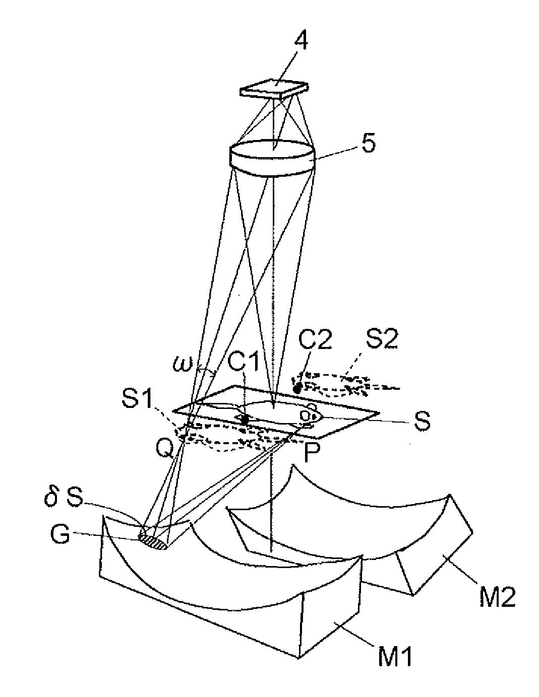
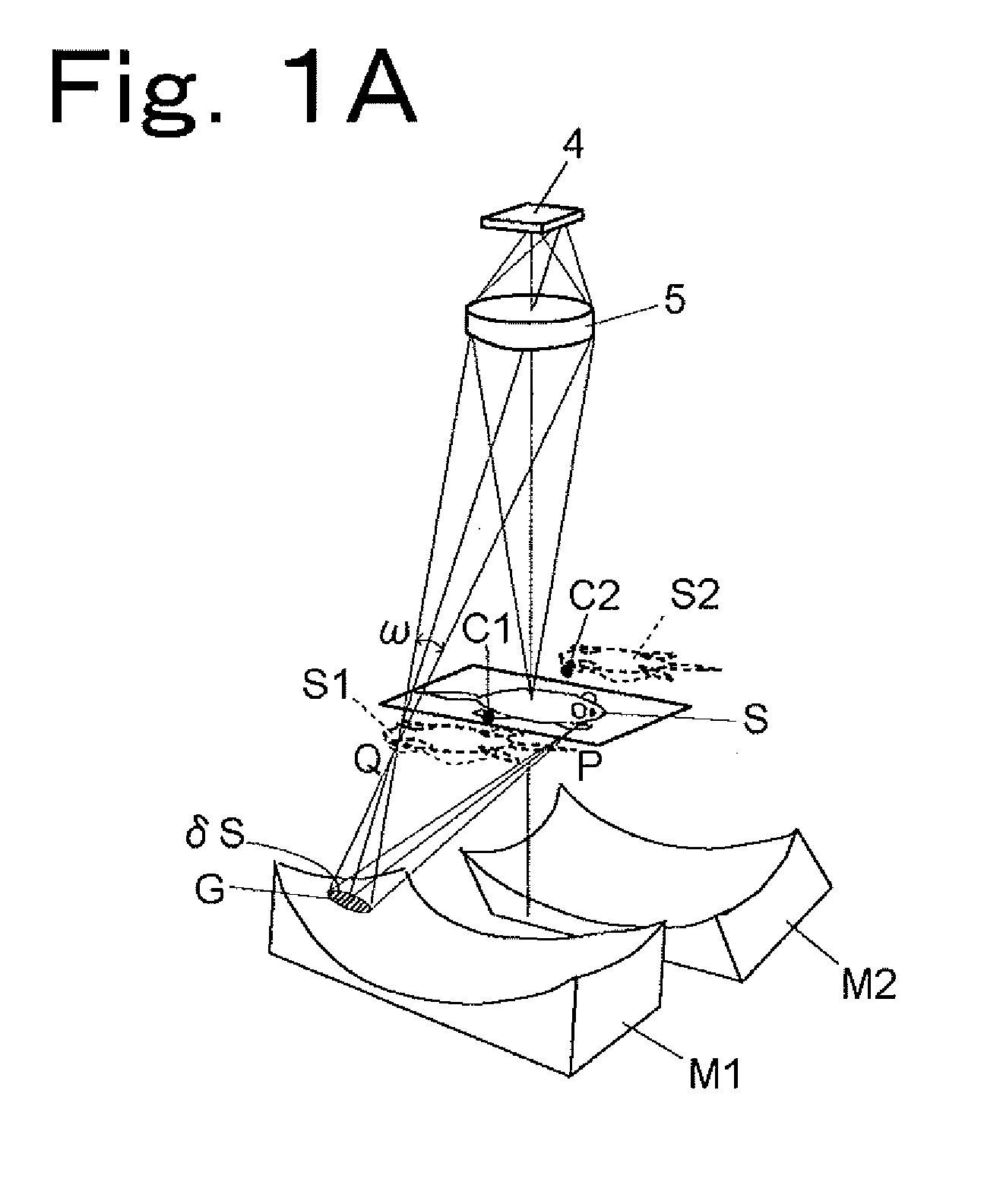
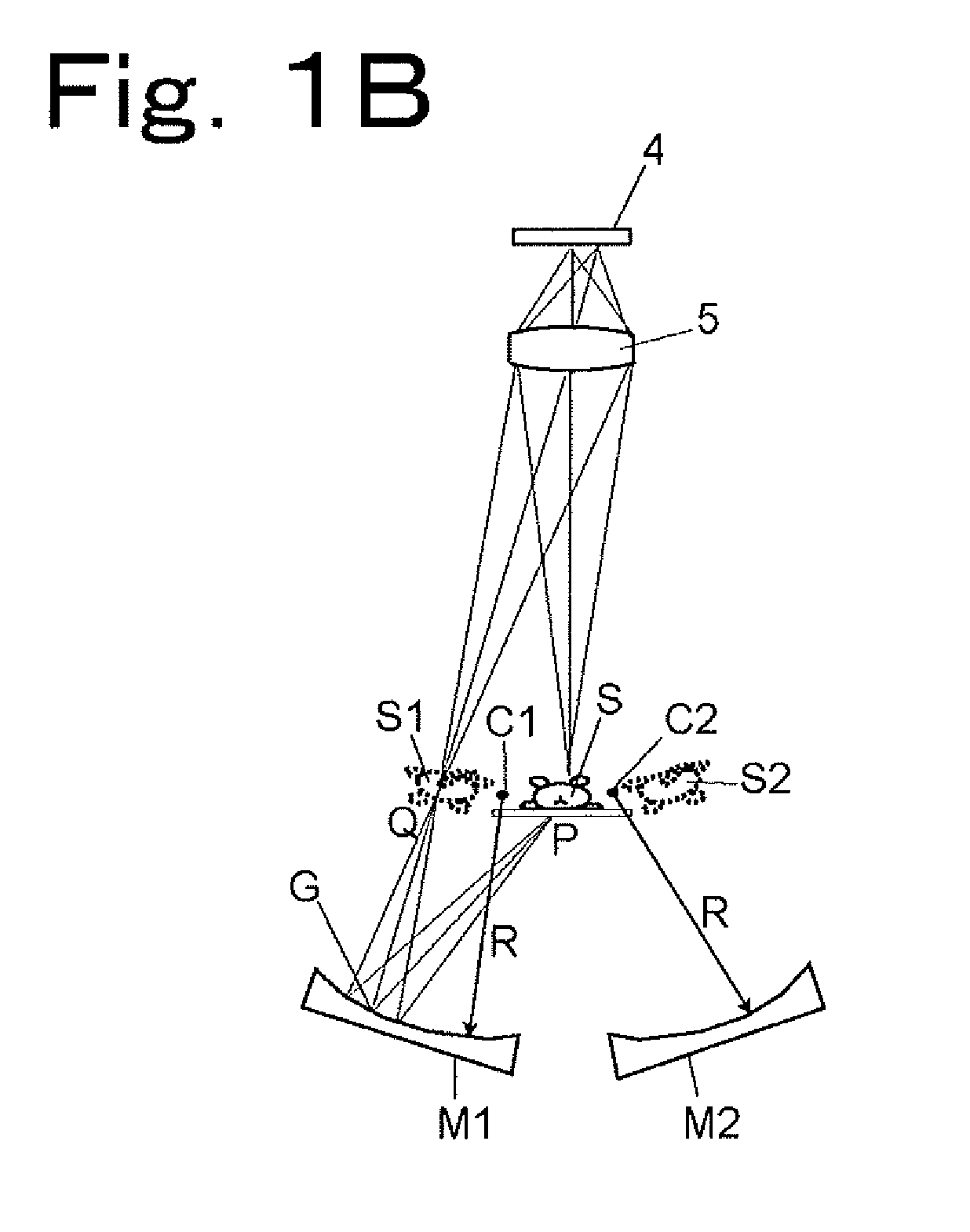
[0136]Hereinbelow, an optical system for simultaneous observation from five directions will be described by way of example with reference to FIGS. 4A and 4B. As shown in FIG. 4A, a sample S is placed in the center of the optical system, and a main imaging lens 5 and a CCD 4 are arranged directly above the sample S (in a direction corresponding to an observation angle of 0°), and four concave mirrors M3, M1, M2, and M4 are arranged in directions corresponding to observation angles of 60°, 135°, 205°, and 300°, respectively. That is, an optical waveguide path in the direction of 0° provides a region occupied by light traveling directly from the sample S to the lens 5, an optical waveguide path in the direction of 60° provides a region occupied by light traveling from the sample S through the concave mirror M3 and a plane mirror M5 to the lens 5, an optical waveguide path in the direction of 135° provides a region ...

second embodiment

[0152]A second embodiment is the same as the first embodiment for five-directional observation except that the two concave mirrors as means for acquiring “images viewed from obliquely above” are changed to plane mirrors. The second embodiment will be described with reference to FIG. 6. As shown in FIG. 6, plane mirrors M5 and M6 are arranged obliquely above the sample S instead of the concave mirrors for acquiring “images viewed from obliquely above” (M3 and M4 shown in FIG. 4A) to form virtual images S3 and S4, respectively. That is, among the five optical waveguide paths, the two optical waveguide paths in the directions in which the sample S is observed from obliquely above are changed to an optical waveguide path in which light travels from the sample S through the plane mirror M5 to the lens 5 and an optical waveguide path in which light travels from the sample S through the plane mirror M6 to the lens 5, but the other three optical waveguide paths in three different directions...

third embodiment

[0157]A third embodiment uses reflection mirrors in combination to measure not the entire body but part of a sample, for example, the head (brain) of a mouse as a sample from four directions (i.e., from above, below, right, and left). The third embodiment will be described with reference to FIGS. 7A to 7C.

[0158]As in the cases of the first and second embodiments, the third embodiment also has the main imaging lens 5 and the CCD 4 arranged above the sample S. However, the third embodiment is different from the first and second embodiments in that the left-side image of the sample S is received by the concave mirror M1 and then reflected by the plane mirror M3 toward the lens 5 and the right-side image of the sample S is also received by the concave mirror M2 and is reflected by the plane mirror M4 toward the lens 5. This technique is a direct reflection of the second principle described above with reference to FIG. 2, and therefore, lateral images S2 and S3 of the sample S are formed...


PUM

PropertyMeasurementUnit
focal lengthaaaaaaaaaa

Abstract

Optical waveguide paths to observe a sample on a sample holder from a plurality of directions while guiding an image of light in each direction which is emitted out of the sample toward a direction of a two dimensional detector via a main imaging lens include an optical waveguide path which never receives the light directly from the sample. The optical waveguide path which never receives the light directly from the sample forms an image of the sample within a substantial focus range of the main imaging lens, and includes optical elements arranged such that a light beam after formation of the image proceeds toward a direction of the main imaging lens. Optical elements on at least one optical waveguide path are those for forming real images. Therefore, the main imaging lens images the sample and those real images in block on the two dimensional detector.

Description

BACKGROUND OF THE INVENTION[0001]1. Field of the Invention[0002]The present invention relates to an optical bioimaging technology for biological samples such as small animals.[0003]2. Description of the Related Art[0004]In medical and biological fields, imaging of the distribution of molecular species in a living body is an important research technique. Heretofore, cellular-level imaging of molecular species has been widely performed using a microscope and a molecular probe labeled with fluorochrome or a chemiluminescent molecular probe. Hereafter, there is a demand for a device capable of observing the distribution of molecular species of interest in a living body not at the cellular level but at the level of organ or the entire animal body larger than cells. Such a device makes it possible to acquire images of a living body, such as a mouse, whose cancer cells are labeled with fluorescent probes every day or every week to monitor the growth of cancer cells of interest with the lap...

Claims


Application Information

Patent Timeline
Patent Type & Authority Applications(United States)
IPC IPC(8): G01N21/64G01J1/58G06K9/00
CPCG01N21/6456G01N21/6486H04N13/0217H04N5/2254G01N21/763H04N13/218
Inventor ODA, ICHIROYAJIMA, ATSUSHIHIZUMI, KENTAROMEGA, AKIMASAKAMIKAKE, TADAFUMIKURATANI, YUTAKAFUJITA, TAKESHITSUNAZAWA, YOSHIO
Owner SHIMADZU CORP
Why Eureka
Try Eureka