Methods of obtaining geometry from images

Active Publication Date: 2011-05-05
BIO TREE SYST
View PDF8 Cites 82 Cited by
  • Summary
  • Abstract
  • Description
  • Claims
  • Application Information

AI Technical Summary

Benefits of technology

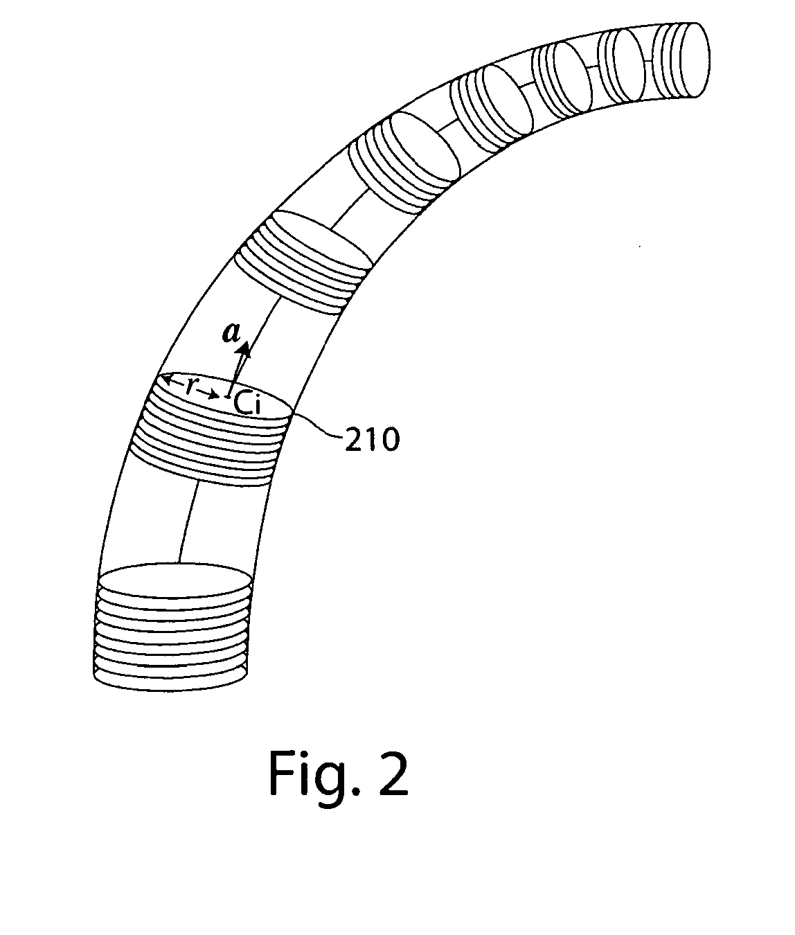
[0016]According to aspects of the invention, a poker chip representation of a vasculature may be mined for physiological, biological, and/or medical purposes. In some embodiments, geometrical information associated with a single poker chip may be mined. In some embodiments, geometrical information associated with a plurality of poker chips, optionally including local linkage information may be mined. Accordingly, aspects of the invention relate to obtaining vessel geometry, determining one or more structural features from the vessel geometry, and/or analyzing the one or more structural features for medical diagnostic, prognostic, and/or research applications.
[0017]Aspects of the invention provide methods for analyzing structures such as blood vessels and evaluating their association with disease, responsiveness to therapeutic treatments, and/or other conditions. Aspects of the invention provide quantitative and analytical methods for evaluating and/or comparing the vessels in different regions of the same body (e.g., a human body) or within ex vivo tissues or between different bodies (e.g., the same regions in different bodies) or different ex vivo tissues. Aspects of the invention can be useful in assisting and/or automating the analysis of vascular patterns and their association with disease diagnosis, prognosis, response to therapy, etc., or any combination thereof. Aspects of the invention can be used in connection with vessel structural information that is obtained from vessel images (e.g., blood vessel images), scan data, vessel representations (e.g., a reconstructed vasculature, a representation that can be viewed as being similar in some ways to a stack of poker chips with varying diameters and is that is referr

Problems solved by technology

However, this process may be difficult and time consuming.
For example, it may be difficult to assess 3D biological structure by attempting to follow 2D structure through a series of stacked 2D images.
In particular, it may be perceptually difficult and time consuming to understand how 2D structure is related to 3D structure as it appears, changes in size and shape, and/or disappears in successive 2D image slices.
To further frustrate this process, when anatomical structure of interest is small, the structure may be difficult to discern or it may be difficult to understand how numerous structures relate to a biological whole.
While the human visual c

Method used


Image

  • Methods of obtaining geometry from images
  • Methods of obtaining geometry from images
  • Methods of obtaining geometry from images

Examples


example 1

Xenotopic Tumor Models

[0261]A tumor model can be generated by inoculating human non-small cell lung tumor cell line (A549 from ATCC, Inc.) subcutaneously in immunodeficient mice (SCID). SCID male mice (6-8 weeks old from Charles River Inc.) are inoculated subcutaneously in the lower back with a suspension of 1×10⁶ human lung tumor cells (A549) in 0.2 ml of PBS. All mice are fed normal chow diet throughout the duration of the experiment. All mice weights are measured throughout the experiment. Tumor size is measured with calipers twice-a-week and tumor volume is calculated using the formula Length2×Width×0.52. All mice are randomized into two treatment groups (approximately 10 mice per group) when the median tumor volume reaches approximately 500 mm3. The treatment groups can be treated according to the following schedule using intraperitoneal (i.p.) administration of either a control composition or an anti-angiogenic compound. For example, different levels of an anti-angiogenic comp...

example 2

Perfusion with Casting Agent

[0267]Perfusion with a casting agent, Mercox (available from Ladd Research, Williston, Vt.) can be performed as follows. An initial anticoagulation step for each animal is performed using an i.v. injection of heparin (10,000 U / ml, 0.3 cc / mouse). After 30 minutes, the animals are anesthetized. Each animal's heart is cannulated and the animal perfused with warm physiological saline at physiological pressure (with an open vein draining the organ or with an open vena cava). Perfusion is continued until the organ or animal is clear of blood. Mercox monomer is filtered through a 0.5 μm filter and a casting resin is prepared by mixing 8 ml Mercox, 2 ml methylmethacrylate, and 0.3 ml catalyst. The resin is infused through the same cannula until the onset of polymerization (the resin changes color to brown and emits heat, ˜10 min). The organ or animal is carefully immersed in a 60° C. water bath for 2 hours (or overnight in a sealed container). The tissue is remov...

example 3

Xenotopic Tumor Models Response to Anti-Angiogenic Therapy

[0268]Xenotopic mouse models obtained as described in Example 1 were treated with either a control solution of saline / PBS or an anti-angiogenic preparation of Avastin® at 0.5 mg / kg / i.p. as described above. At the end-point, vascular casts were prepared as described in Example 2 above and analyzed for two treated mice (both treated with Avastin® at 0.5 mg / kg / i.p.) and one control mouse. The resulting vascular casts were scanned using a micro CT-scanner and the results of the structural analysis are shown in FIGS. 14-17. The analysis was performed by determining the number of blood vessels within bins of different diameter ranges for the xenotopic tumor in the treated and control animals. The bins were each 13.8 μm wide and the smallest bin included blood vessels having a diameter of between 20.7 μm and 34.5 μm. Mean tumor volumes did not differ significantly between the groups at the end of the experiment. However differences ...


PUM


Abstract

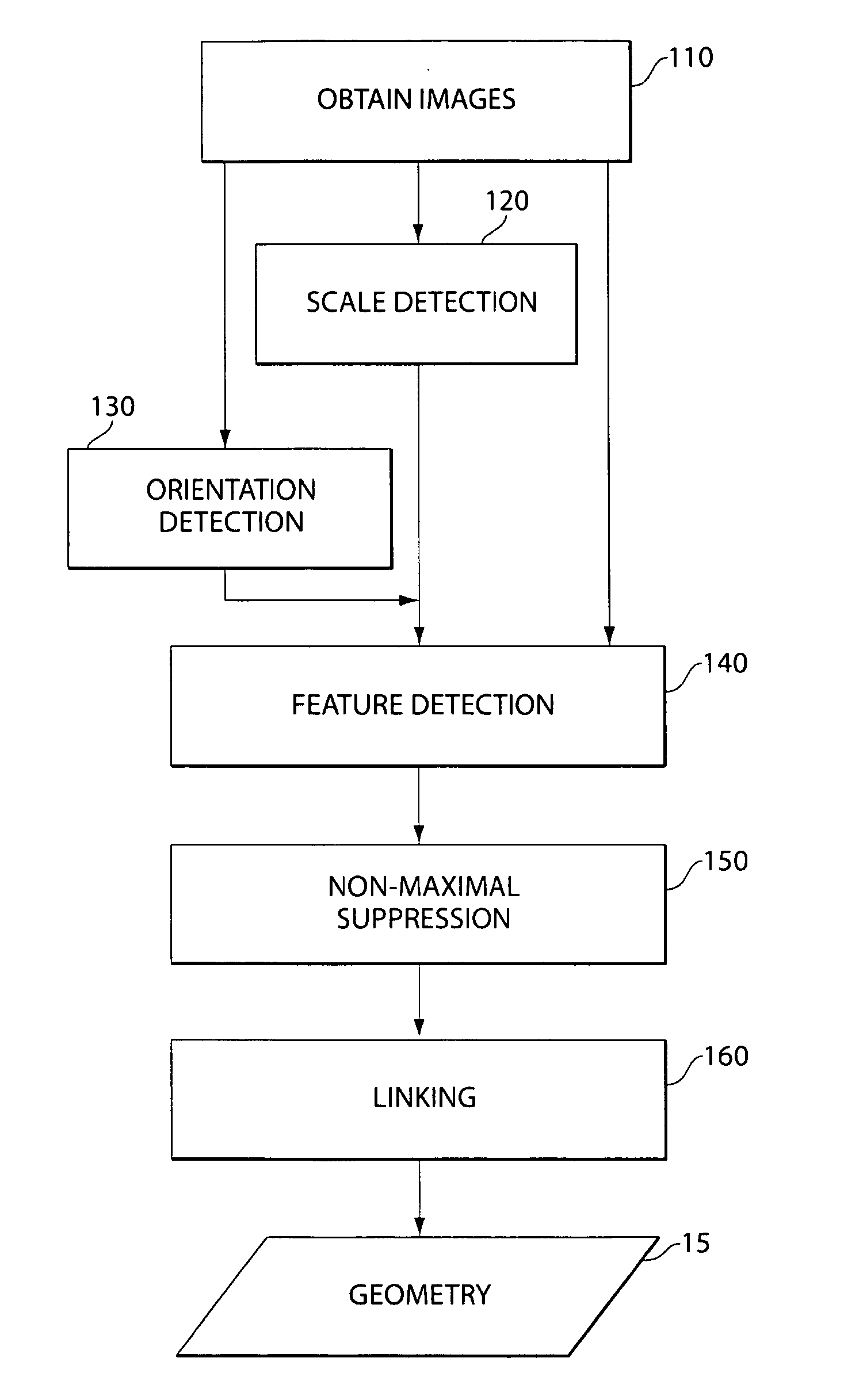
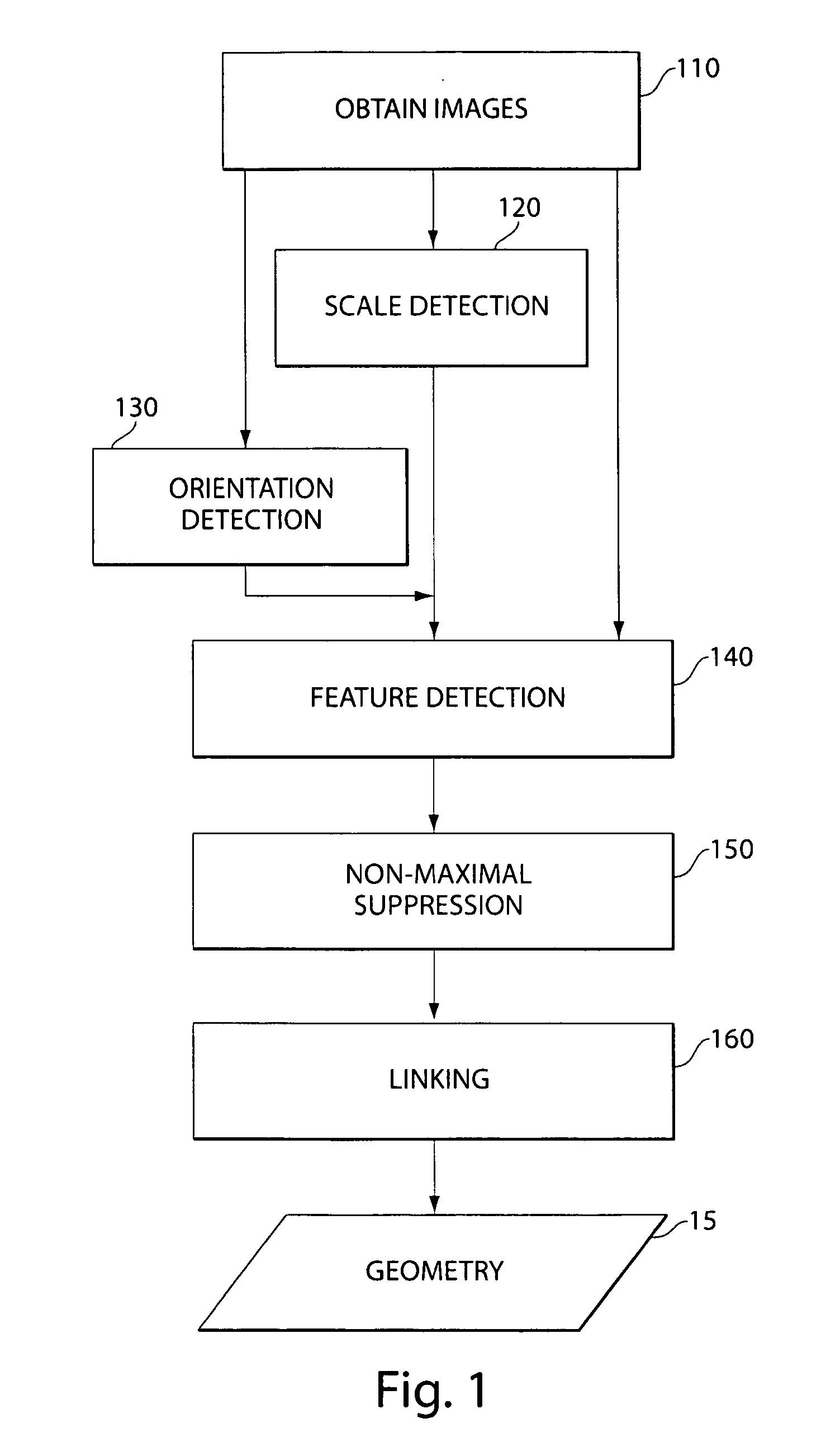
In one aspect, a method of detecting at least on feature associated with a blood vessel in at least one image of at least one blood vessel using a matched filter adapted to respond to the at least one feature is provided. The method comprises applying a scale detection filter to selected voxels in the at least one image to determine a scale for the matched filter at each of the selected voxels, determining an orientation for the matched filter at each of the selected voxels, wherein determining the orientation is assisted by using the scale determined at each of the selected voxels, applying the matched filter at each of the selected voxels at the scale and the orientation determined at each of the selected voxels to obtain a filter response at each of the selected voxels, and analyzing the filter response at each of the selected voxels to determine if the respective voxel corresponds to the at least one feature.

Description

RELATED APPLICATIONS[0001]This application claims priority under 35 U.S.C. §119(e) to U.S. Provisional Application Ser. No. 61 / 009,872 entitled “METHODS OF ANALYZING VESSEL DISTRIBUTIONS AND USES THEREOF,” filed on Jan. 2, 2008, and U.S. Provisional Application Ser. No. 61 / 010,080 entitled “METHODS OF ANALYZING VESSEL DISTRIBUTIONS AND USES THEREOF,” filed on Jan. 3, 2008, both of which are herein incorporated by reference in their entirety.FIELD OF THE INVENTION[0002]Aspects of the present invention relate to extracting geometry from one or more images for use in analyzing biological tubular structures for diagnostic and therapeutic applications in animals. In particular, aspects of the invention relate to extracting geometry from images of blood vessels to identify structural features useful for detecting, monitoring, and / or treating diseases, and / or for evaluating and validating new therapies.BACKGROUND OF THE INVENTION[0003]A wide range of imaging methods and devices are commonl...

Claims


Application Information

Patent Timeline
IPC IPC(8): G06K9/00
CPCG06T7/0044G06T2207/10072A61B6/508G06T2207/20004G06T2207/30101G06T2207/10116G06T7/74G06F18/22G06T7/0012G06T7/20G06T15/08A61B5/055A61B6/504A61B2576/02G06T7/60G06T2207/10028G06T7/62A61B5/02007A61B5/107A61B5/489G06T2207/30172G06T2211/404
Inventor KANG, KONGBINBRAUNER, RAUL A.WU, YANCHUNCRUZ, JOAO
Owner BIO TREE SYST
Why Eureka
Try Eureka