Graft composition for neural tissue regeneration, method of production and uses thereof

a neural tissue and composition technology, applied in the field of neural tissue regeneration, can solve the problems of inability to produce long-distance regeneration on their own, cells face ethical and histopathological challenges, and trophic factors cannot solve the lack of ecm guidance, etc., and achieves a high survival rate and is easy to shap

Inactive Publication Date: 2011-05-26
FINA BIOTECH S L U
View PDF2 Cites 8 Cited by
  • Summary
  • Abstract
  • Description
  • Claims
  • Application Information

AI Technical Summary

Problems solved by technology

However, they do not leave the immediate site of injury and appear incapable of producing any long-distance regeneration on their own, quickly becoming dystrophic (Li, Y. et al., Exp Neurol.
However, these cells face ethical and histo-pathological challenges for their harvesting and use.
However, trophic factors did not solve the lack of ECM guidance.
However, collagen induces a response that leads to the sealing and isolation of the implant, rendered unable of interconnecting with the host tissue.
However, the degradation products of PLGA particles, and the prolonged presence of an exologous material, may exert unpredictable deleterious effects on cell physiology.
However, at the present, there are no studies on PRP scaffolds application in the field of neurological diseases.
In summary, materials and strategies currently used for cell therapy in spinal cord and brain injury repair have not been completely satisfactory.

Method used


Image

  • Graft composition for neural tissue regeneration, method of production and uses thereof
  • Graft composition for neural tissue regeneration, method of production and uses thereof
  • Graft composition for neural tissue regeneration, method of production and uses thereof

Examples


example 1

Comparative Study Between Fibrin Gel Implants and PRP Gel implants in fostering cell multiplication, migration and differentiation.

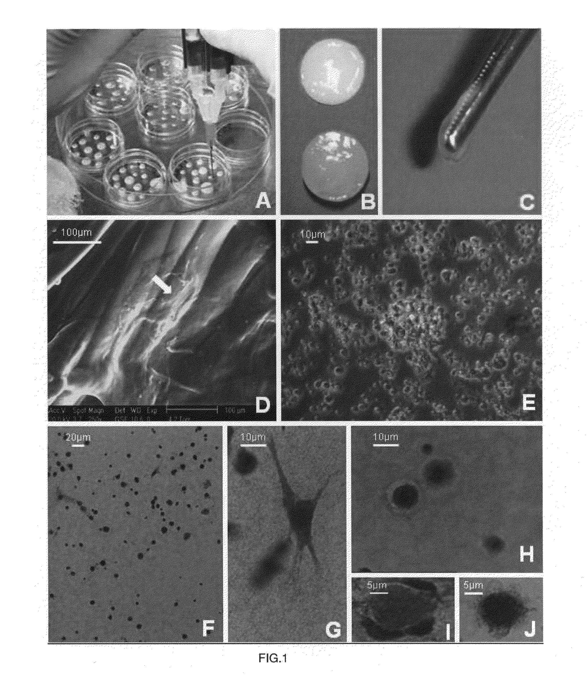
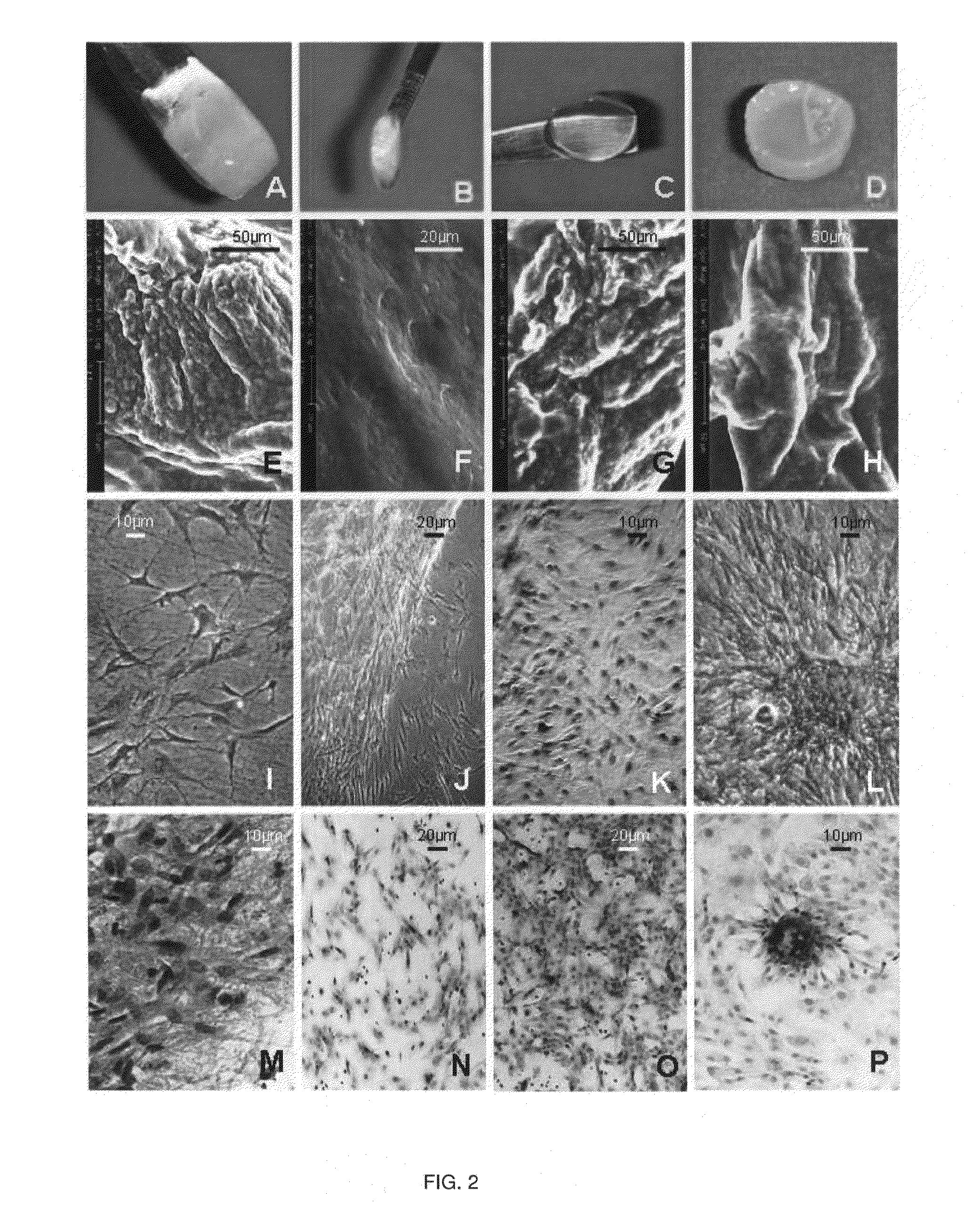
[0063]Cell Morphology after Seven Days in Culture

[0064]In the present study, hBMSC showed the following profile after phenotypic flow cytometry studies: CD29+, CD44+, CD105+, CD166+, CD34−, CD45−.

[0065]FG-scaffolds were consistent and fairly manageable, but hBMSC were not properly adhere to the fibrin mesh and a few hours of culture appeared to form clusters of cells with a rounded morphology and were only occasionally able to display any cell with a typical mesenchymal morphology (Table III).

TABLE IIIMorphological aspects of the seeded hBMSC in the course of theseven days of culture, according to the scale described in themethodology (Whillert et al.).FG-scaffold:5 / 2 scaffold10 / 4 scaffold20 / 100 scaffold100 / 500 scaffoldday 1+++++day 2+++++day 3+++++day 4++++++day 5+++++++day 6+++++++day 7++++++++

[0066]FIG. 1 shows different morphological aspects of FG-sc...

example 2

[0079]A total of 10 adult Wistar rats were used. All of them were submitted to a cortical lesion, through brain cortex resection at the parietal level after craniotomy. Immediately after the lesion, blood was extracted from the animals and gel was prepared according to the protocol:

Component A: Platelet Rich plasma (PRP)+cells (500.000)+BDNF (100 ng / ml)

Component B: Thrombine 5 UI+calcium chloride (34 micromoles / ml).

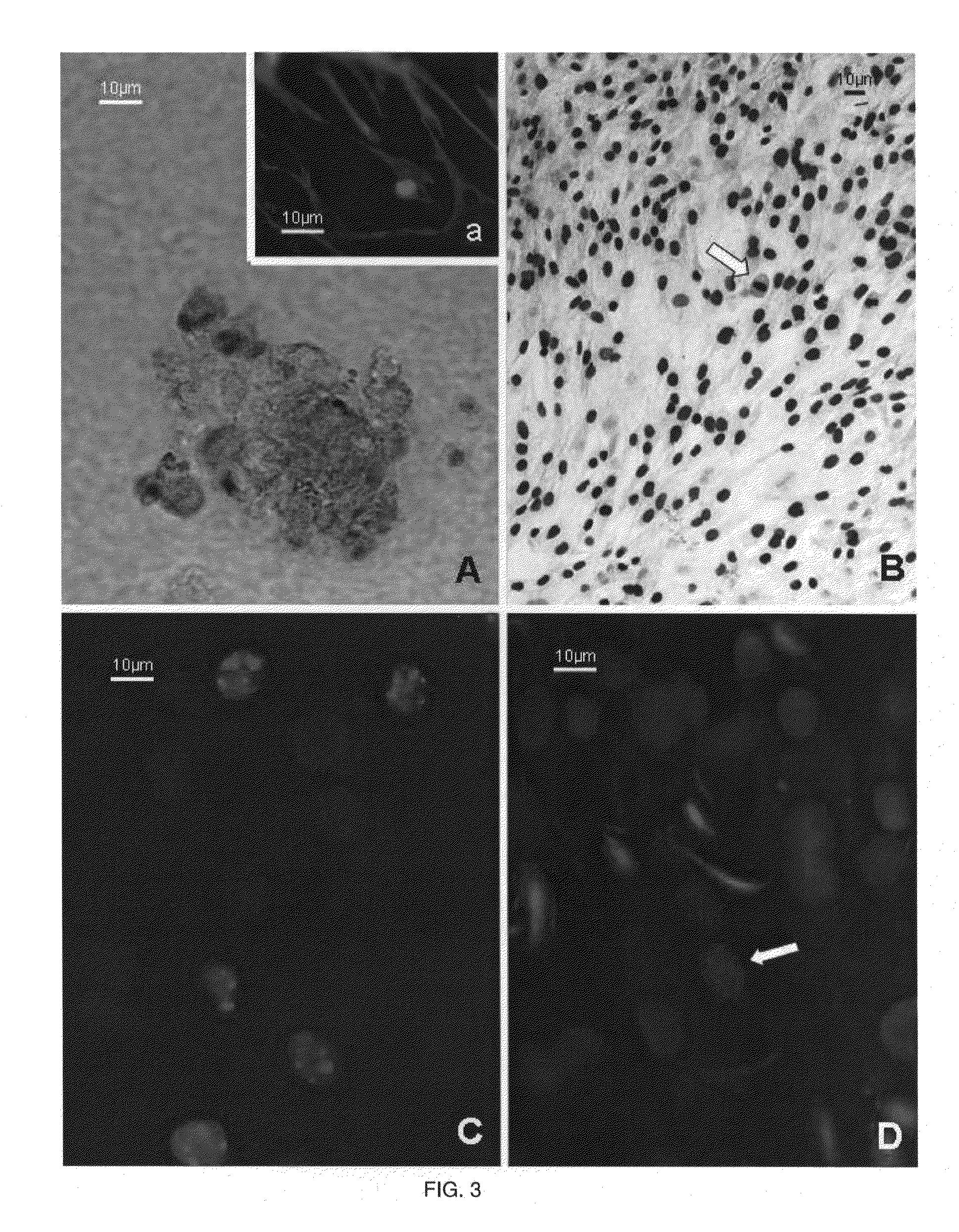
Both components were mixed and the resulting gel was placed on the brain lesion in 6 animals (FIGS. 5A and B). In 4 animals, the same protocol was carried out, but without cells.

[0080]After a week, histological studies were carried out. In FIG. 5C the aspect of grafted cells are shown (study developed through bisbenzimide label analysis). It was observed that grafted cells showed a normal aspect and their viability was higher than 60%. In those animals in which cells encapsulated in the gel were implanted, viability of grafted cells with neural differentiation signals wer...


PUM

PropertyMeasurementUnit
densityaaaaaaaaaa
densityaaaaaaaaaa
temperatureaaaaaaaaaa

Abstract

The present invention provides a biocompatible, biodegradable graft composition for regeneration of neural tissue, comprising the following components: a) a gel scaffold formed from an isolated platelet containing fluid and an activating agent comprising calcium chloride, with or without thrombin, b) a nerve growth factor selected from BDNF, NGF, retinoic acid and combinations thereof, and c) a culture of at least 50,000 progenitor stem cells; the method of production and the uses thereof.

Description

TECHNICAL FIELD OF THE INVENTION[0001]The present invention is related to the field of neural tissue regeneration in the Central Nervous System (CNS). Particularly, the present invention relates to a biocompatible, biodegradable gel graft composition for regeneration of neural tissue and to methods for its manufacture and uses thereof.BACKGROUND OF THE INVENTION[0002]Regeneration after injury to the Central Nervous System (CNS) is limited by inhibitory influences from the extracellular environment counteracting the repair of myelin and neurons. Often, after spinal cord or brain tissue injury, a cavity originates due to loss of native cells. This cavity is void of extracellular matrix (ECM) and usually isolated from the tissue by a glial scar, which was initially thought to block regeneration within the CNS after injury (Windle et al., J Comp Neurol. 1950; 93:241-258). More recently, researchers have demonstrated that in several different experimental models, CNS axons are capable of...

Claims


Application Information

Patent Timeline
Patent Type & Authority Applications(United States)
IPC IPC(8): A61K35/12C12N5/02C12N9/74A61K35/16A61K35/19A61K35/30
CPCA61K35/16C12N2502/115A61K35/30A61K38/185A61K38/4833C07K14/475C07K14/48C12N5/0068C12N5/0663C12N2533/56C12N2533/90A61K35/19A61L2430/38A61L2300/414A61L27/58A61L27/54A61L27/52A61L27/3834A61L27/3616A61K45/06A61K2300/00
Inventor ZURITA CASTILLO, MERCEDESVAQUERO CRESPO, JESUSAGUAYO FERRER, CONCEPCIONOTERO ORTEGA, LAURA
Owner FINA BIOTECH S L U
Why Eureka
Try Eureka