Dynamical visualization of coronary vessels and myocardial perfusion information

a technology of myocardial perfusion and visualization, applied in the field of dynamic visualization of coronary vessels and myocardial perfusion information, can solve the problems of difficult correlation of information acquired during the different coronary stages of blood circulation, and achieve the effect of improving positioning accuracy

Active Publication Date: 2011-06-09
KONINKLIJKE PHILIPS ELECTRONICS NV
View PDF2 Cites 5 Cited by
  • Summary
  • Abstract
  • Description
  • Claims
  • Application Information

AI Technical Summary

Benefits of technology

[0021]By visualizing different cardiac data in a superimposed presentation for example the geometry or topology of the cardiac vessels may be overlaid to the information about the myocardial perfusion. Thus, it may be easier for example for a user like a physician to perform a diagnosis. When, for example information of the cardiac vessel topology is displayed together with perfusion data corresponding to the same phase of the heart motion, it may be easier to assess visually, whether the coronary arteries are properly providing the myocardium with blood—and where a possible perfusion problem is coming from. Furthermore, the method as described above may facilitate the assessment of data by providing useful high-contrasted, low-noise information.
[0040]The selection of a full cardiac cycle with optimal coronary arteries visibility as the first cardiac data may be advantageous. For example, if the topology of the coronary arteries of the heart is to be overlaid to the perfusion data it may be important to have the best possible visibility of the arteries. Furthermore, the acquisition and / or selection of a full cardiac cycle may be important because in a full cycle the full motion of the heart is represented and may be overlaid several times for example as a time loop to the perfusion data of the second cardiac data. It may be advantageous to select the cardiac cycle with the better or best coronary arteries visibility for example in the filled-in state, when the vessels are full of the blood volume with the contrast agent. To approximately select the optimal cycle, the last cycle before the beginning of the perfusion stage may be selected.
[0044]The background subtraction may for example be done with the help of a cardiac digital subtraction angiography (DSA). As described above, mask images may be acquired before the injection of a contrast agent, then after injection of a contrast agent into the vessels to be observed, a contrast image may be acquired. Subtracting the mask images from the contrast images may provide good contrasted vessels filled with the contrast agent. The subtraction of background features may result in images where only the injected coronaries and / or the myocardial perfusion are much better visible.
[0047]The background subtraction may be a great support and important feature of the method, because it helps to significantly improve the visibility of the vessels and processes in the heart, for example by enhancing the relative contrast of the images.
[0053]The computing of spatial correspondence between the first cardiac data and the second cardiac data allows for better accuracy in the positioning of the vessels like the coronary artery during the visualization of the cardiac data sets in a superimposed presentation.

Problems solved by technology

Furthermore, for a physician looking at the images or image sequences acquired during an examination it may be difficult to correlate the information acquired during the different coronary stages of the blood circulation.

Method used


Image

  • Dynamical visualization of coronary vessels and myocardial perfusion information
  • Dynamical visualization of coronary vessels and myocardial perfusion information
  • Dynamical visualization of coronary vessels and myocardial perfusion information

Examples


Embodiment Construction

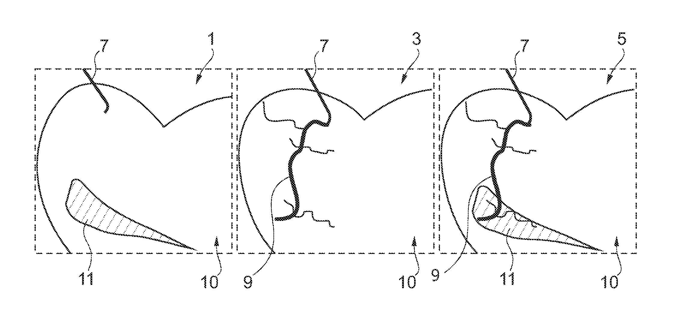
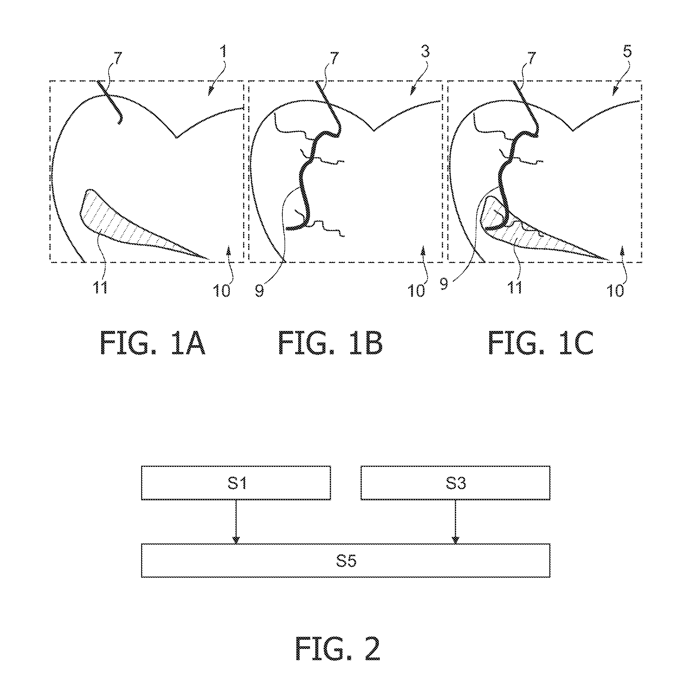
[0066]In FIGS. 1A to 1C exemplary snap shots of a continuous presentation, i.e. “movie”, of data acquired and visualized with the method according to the invention are presented. In FIG. 1A an image based on data acquired during a perfusion stage of the heart is shown. The image shown in FIG. 1B is based on data acquired during an arterial inflow of contrast agent into the vessels of the heart and the image shown in FIG. 1C shows a snap shot of a continuous overlay of the images shown in FIG. 1A and FIG. 1B.

[0067]In the FIG. 1A to 1C a catheter tip 7 is visible. The catheter tip 7 can be used to inject a contrast agent into the vessels 9 of the heart. Furthermore, the catheter tip 7 can be used for medical possibly surgical treatments. The location of the catheter tip 7 is the same in all the images 1A to 1C. Thus, the catheter tip 7 may be used as a helpful orientation to compute spatial correspondence between two images, as for example the image 3 acquired during an arterial stage...


PUM


Abstract

A method for dynamically visualizing coronary information and an apparatus adapted to implement such method is described. In a preferred embodiment of the method, first dynamic cardiac data is acquired during a first cardiac stage and second dynamic cardiac data is acquired during a second cardiac stage. Then, the two data sets are visualized continuously the in a superimposed presentation, wherein the first cardiac data and the second cardiac data corresponding to a same phase within the cardiac cycle are visualized simultaneously. In this way for example information about the vessel geometry may be immediately linked with information about the muscle irrigation or perfusion. Furthermore, this useful information may be displayed in a high-contrasted and low-noise presentation.

Description

FIELD OF THE INVENTION[0001]The present invention relates to the field of dynamically visualizing coronary vessels and myocardial perfusion information. Particularly, the present invention relates to a method and an apparatus for acquiring and dynamically visualizing coronary information. Furthermore, the present invention also relates to a computer program element adapted for performing such method when executed on a computer and to a computer-readable medium on which such computer program element is stored.BACKGROUND OF THE INVENTION[0002]During an examination of the heart it is important to supply accurate images to the physician to facilitate the procedure. A well established method for providing images of the lumen of a vessel is angiography, where after the injection of a contrast agent (CA) a series of images is acquired for example with the help of X-rays.[0003]There are several approaches to provide images with high image quality. An example for such an approach is a proced...

Claims


Application Information

Patent Timeline
Patent Type & Authority Applications(United States)
IPC IPC(8): H05G1/64
CPCA61B6/463A61B6/464A61B6/481A61B6/503A61B6/507A61B6/5217A61B6/5235A61B6/5264A61B6/541A61B6/504G16H50/30
Inventor BUIJNS, ANTONIUS JOHANNES CORNELIUSRONGEN, PETER MARIA JOHANNESAUVRAY, VINCENT MAURICE ANDREBONNEFOUS, ODILEFLORENT, RAOUL
Owner KONINKLIJKE PHILIPS ELECTRONICS NV
Why Eureka
Try Eureka