
Means and Methods for Detecting Bacteria in an Aerosol Sample

a technology of aerosol sample and bacteria, applied in the field of spectroscopic medical diagnostics, can solve the problems of inconvenience for patients, poor sensitivity, and sore throat of patients with infectious respiratory diseases

Inactive Publication Date: 2011-07-28
OPTICUL DIAGNOSTICS LTD
View PDF4 Cites 24 Cited by
  • Summary
  • Abstract
  • Description
  • Claims
  • Application Information

AI Technical Summary

Benefits of technology

"The present invention provides a method for detecting and identifying specific bacteria within an uncultured sample. The method involves obtaining an absorption spectrum of the sample, dividing it into segments based on the absorption spectrum, and calculating features from each segment. These features can include correlation, peak's wavelength, peak's height, peak's width, peak's cross section, peak's area, and the coefficients of a fitted polynomial curve. The features are then used to assign the bacteria to a specific genus or species using algorithms such as Sequential Backward Selection, Sequential Forward Selection, or Support Vector Machine. The method can be applied to a variety of samples, such as cough, sneeze, saliva, mucus, bile, urine, vaginal secretions, middle ear aspirate, pus, pleural effusions, synovial fluid, abscesses, cavity swabs, serum, blood, and spinal fluid."

Problems solved by technology

A patient who suffers from infectious respiratory diseases will usually endure sore throat and have trouble swallowing.
Moreover, the test causes some inconvenience to the patient.
Although these test give fast results (10 to 30 minutes) their sensitivity is very poor and they are not user friendly.
None of the prior art literature discloses means and method that can quickly (without culturing) and accurately detect bacteria from a sample, and none demonstrates identification within a wet sample.
Furthermore, none of the prior art literature discloses means and method that can eliminate the water influence from the sample so as to better detect the bacteria.
Moreover all of the above require a skilled operator and / or the use of reagents or a complicated sample preparation for the detection of bacteria.
Furthermore, none of the above distinguishes among different bacteria in a mixture or within a sample.

Method used

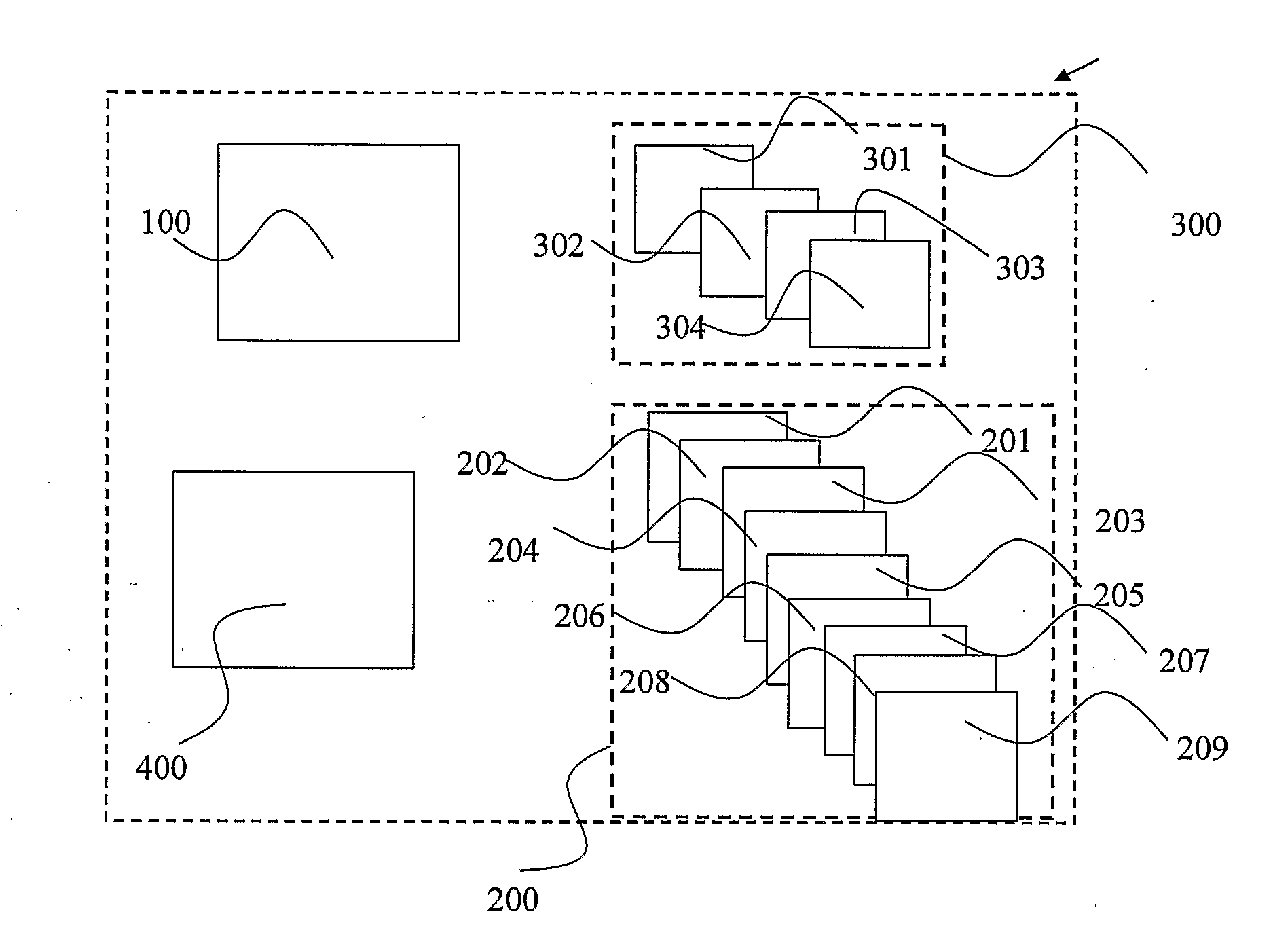
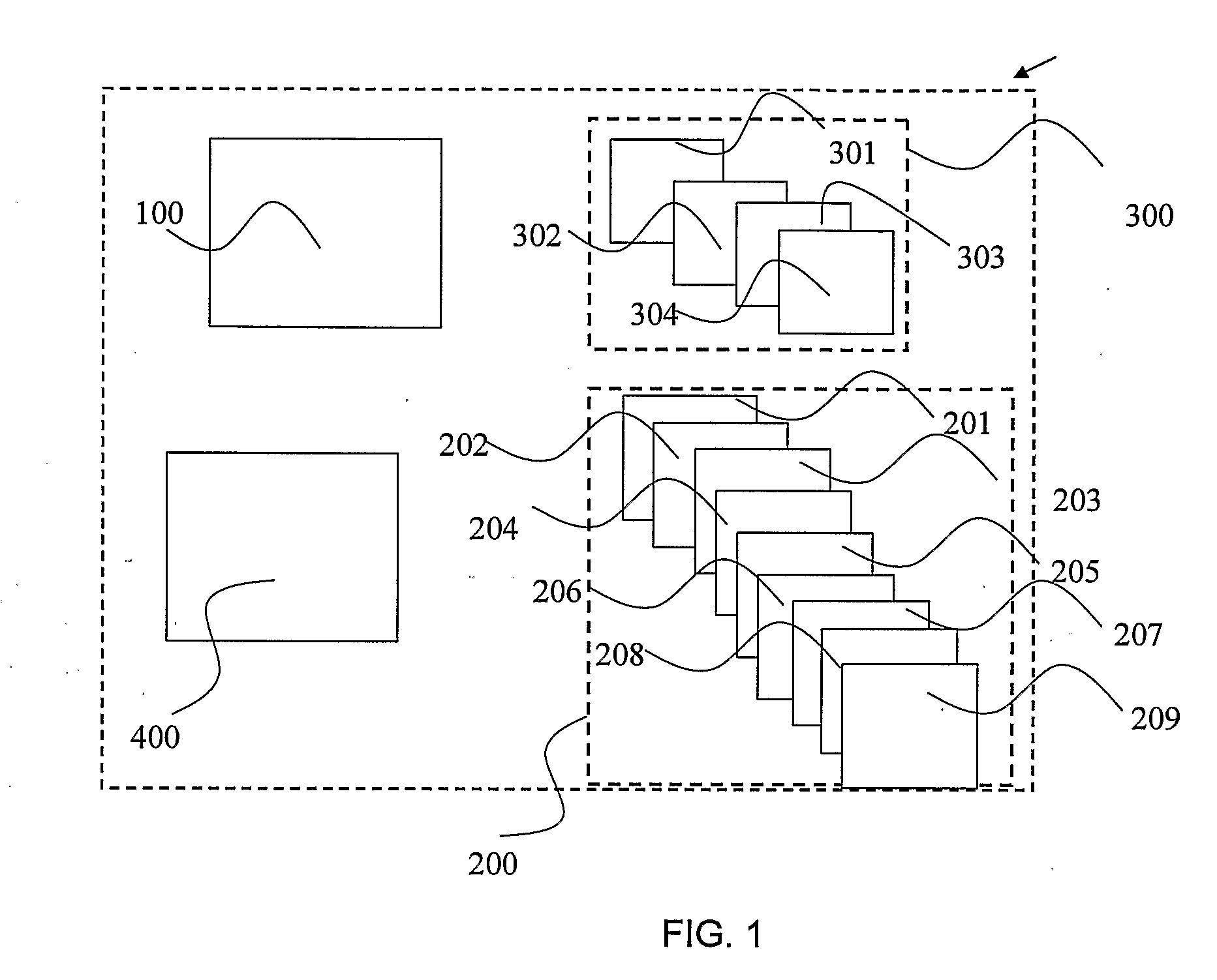
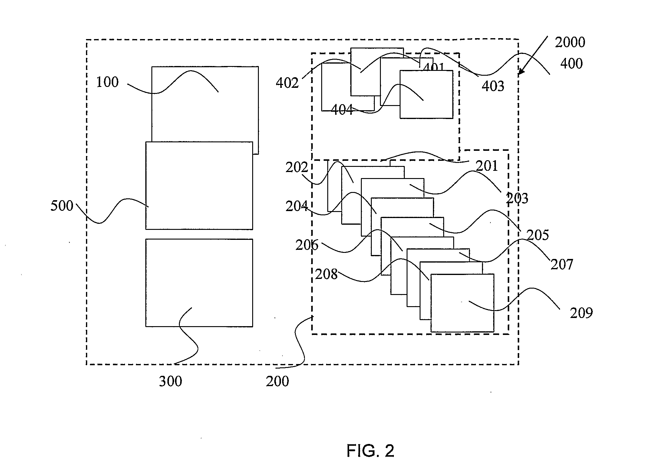

Image

  • Means and Methods for Detecting Bacteria in an Aerosol Sample
  • Means and Methods for Detecting Bacteria in an Aerosol Sample
  • Means and Methods for Detecting Bacteria in an Aerosol Sample

Examples


example 1

Water Influence

[0322]One of the major problems in identifying bacteria from a fluid sample's spectrum (and especially an aerosol spectrum) is the water influence (i.e., the water noise which masks the desired spectrum by the water spectrum).

[0323]The water molecule may vibrate in a number of ways. In the gas state, the vibrations involve combinations of symmetric stretch (v1), asymmetric stretch (v3) and bending (v2) of the covalent bonds. The water molecule has a very small moment of inertia on rotation which gives rise to rich combined vibrational-rotational spectra in the vapor containing tens of thousands to millions of absorption lines. The water molecule has three vibrational modes x, y and z. The following table (table 1) illustrates the water vibrations, wavelength and the assignment of each vibration:

TABLE 1water vibrations, wavelength and the assignment of each vibrationWavelengthcm−1Assignment0.2mm50intermolecular bend55μm183.4intermolecular stretch25μm395.5L1, librations...

example 2

Bacteria's Absorption Spectrum

[0483]Each type of bacteria has a unique spectral signature. Although many types of bacteria have similar spectral signatures there are still some spectral differences that are due to different proteins on the cell membrane and differences in the DNA / RNA structure. The following protocol was used:[0484]1. Strep. β hemolytic (ATCC 19615) were purchased from HY labs.[0485]2. The content of one full plate that was grass seeded with Strep. Pyo by adding 800 μL, of ddH2O to the plate and collecting the content into 1 eppendorf tube 500 μl.[0486]3. Centrifuge the tube for 5 minX14000 rpm[0487]4. Discarding the supernatant[0488]5. Adding 30 μL of ddW solution.[0489]6. Mixing the content;[0490]7. Reference reading of the empty optical cell[0491]8. Putting 500 μL of the tube in a 3 mL spray bottle[0492]9. Spraying one practice squeeze into an eppendorf tube and discarding the tube[0493]10. Spraying 2 squeezes: one on one side, and the other in the other side of ...

example 3

Distinguishing Between Two Bacteria in an Aerosol Sample

[0497]The following examples illustrate in-vitro examples to provide a method to distinguish between two bacteria within an aerosol mixture of—Streptococcus payogenes and Streptococcus Bovis and to identify and / or determine whether Streptococcus payogenes is present within the aerosol sample.

[0498]The following protocol was used:[0499]1. Strep. β hemolytic (ATCC 19615) and Streptococcus bovis (ATCC 9809) were purchased from HY labs.[0500]2. The content of two full plates of Strep pyo. is added with 800 μL of ddH2O to each plate and the content is placed into eppendorf tube. The procedure is repeated twice (collecting the content of 6 full plates to 3 eppendorf tubes).[0501]3. Step 2 is repeated for S. bovis, collecting the content of 8 full plates to 4 eppendorf tubes.[0502]4. Centrifuging the 4 tubes 3 min×9,000 rpm.[0503]5. Discarding the supernatant.[0504]6. Weighting the four eppendorf tubes.[0505]7. Transferring with 1 ml ...


PUM


Abstract

This disclosure provides a method for detecting and / or identifying uncultured bacteria. The sample is an aerosol sample selected from a group consisting of cough, sneeze, saliva, mucus, bile, urine, vaginal secretions, middle ear aspirate, pus, pleural effusions, synovial fluid, abscesses, cavity swabs, serum, blood and spinal fluid. The method comprises obtaining absorption spectra (AS) of the sample, extracting and processing the acquired data, thereby detecting and / or identifying the bacteria.

Description

FIELD OF THE INVENTION[0001]The present invention relates to the field of spectroscopic medical diagnostics of specific bacteria within a sample. More particularly, the present invention provides means and methods for detecting different kinds of bacteria in an aerosol sample by using spectroscopic measurements. The detection can be used for both medical and non-medical applications, such as detecting bacteria in water, beverages, food production lines, sensing for hazardous materials in crowded places, bio-defense etc.BACKGROUND OF THE INVENTION[0002]The identification of microorganisms is clearly of great importance in the medical fields. Furthermore, in recent years the need for efficient and relatively rapid identification techniques has become even more pressing owing to the remarkable expansion of environmental and industrial microbiology. One field in which it there is an urgent need for a rapid and accurate identification of bacteria in an aerosol environment.[0003]Respirato...

Claims


Application Information

Patent Timeline
Patent Type & Authority Applications(United States)
IPC IPC(8): G06F19/00
CPCG06K9/00496G06F18/00G06F2218/00
Inventor BEN-DAVID, MOSHEGANNOT, GALLYAERUV, TOMERMARKOWITZ, ZVI
Owner OPTICUL DIAGNOSTICS LTD
Learn More