Method and apparatus of determining a radiation dose quality index in medical imaging

Active Publication Date: 2011-11-03
REINER BRUCE
View PDF14 Cites 28 Cited by
  • Summary
  • Abstract
  • Description
  • Claims
  • Application Information

AI Technical Summary

Benefits of technology

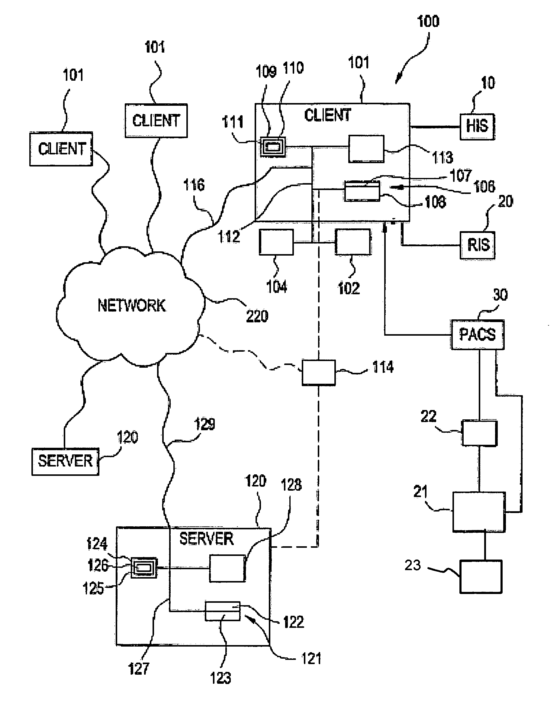
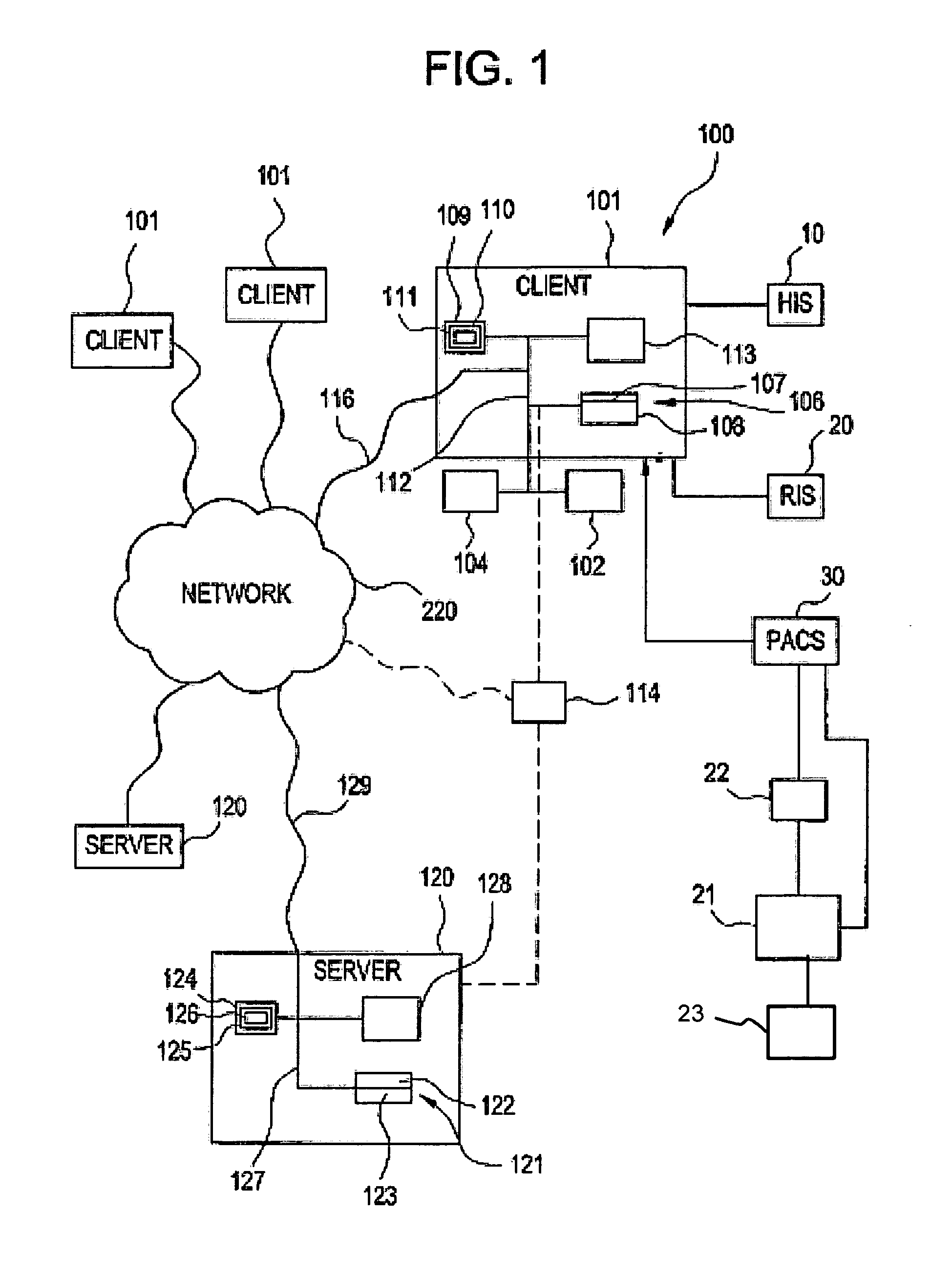
[0009]Thus, the present invention relates to a method and apparatus to accomplish the goal of providing reforms to improve patient safety, quality and accountability, by creating a technology which simultaneously records and tracks objective data referable to radiation safety, medical imaging quality, and accountability among the various stakeholders and technologies in use. This quantifiable data is referred to as the Radiation Dose Quality Index (RDQI), and creates an objective, data-driven measure of quality and safety as it relates to medical practice.
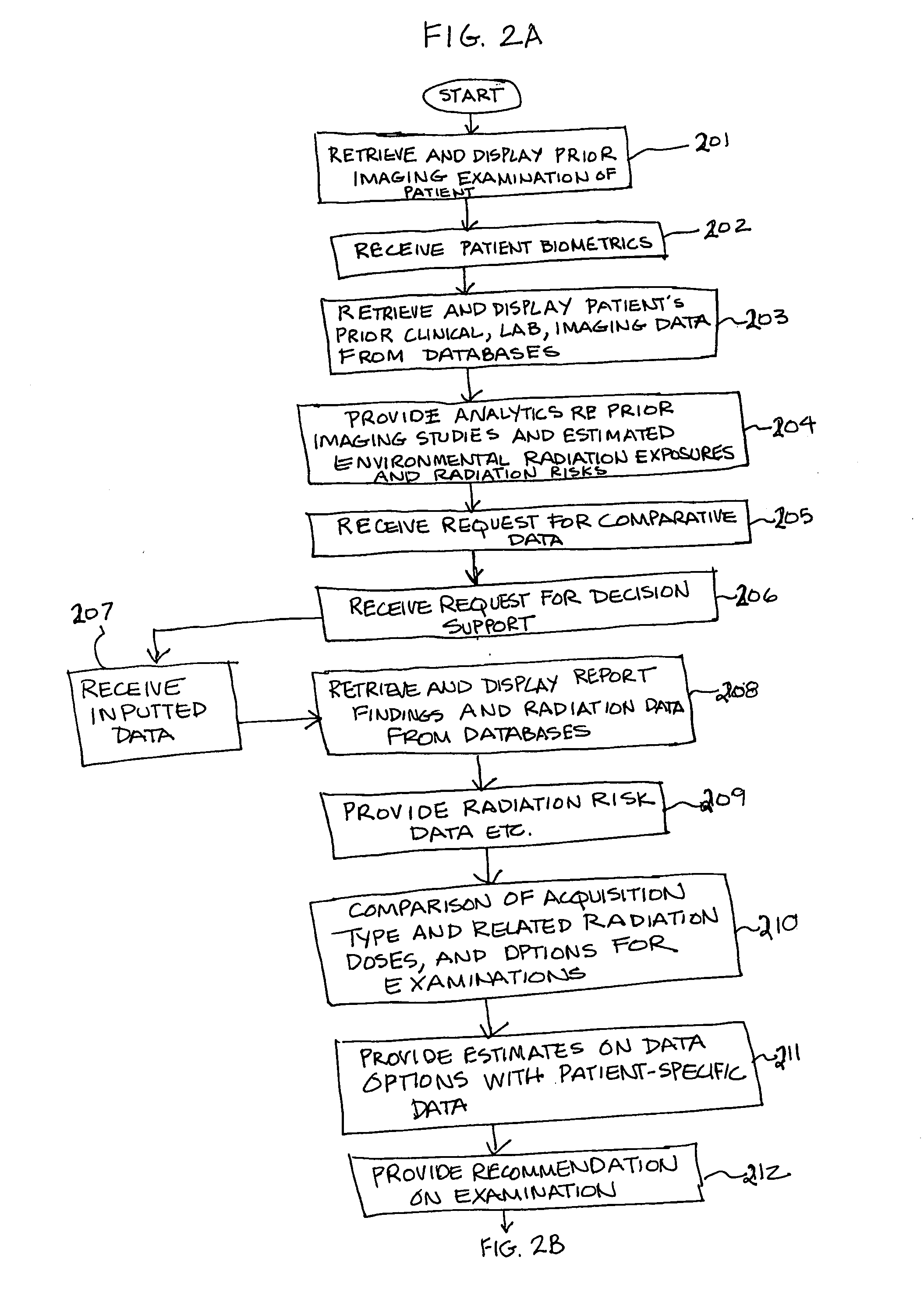
[0011]In one embodiment, the program retrieves and displays the patient's prior clinical, laboratory, and imaging data from the various databases, for clinical review. Included in the imaging data are a series of examination and patient-specific data related to individual exam radiation dose, cumulative radiation dose, and radiation iatrogenic risk. The program provides several analytics to the user, including the carcinogenesis risk associated with prior imaging studies, along with estimated environmental radiation exposures. Other iatrogenic risks of radiation (distinct from carcinogenesis) may be compiled by the program. In addition, the program provides additional comparative data, to assist the user in the examination selection and ordering process.
[0013]In one embodiment, the program provides a detailed analysis to assist in the exam selection process. When the user selects an option, for data on, for example, the estimated dose for conventional CT, an estimated dose for low dose, and estimated dose for ultra-low dose CT, are provided. After selecting one option, the program may provide the decision support option of “protocol optimization”. The program processes the clinical, imaging, and historical data to create the optimal CT protocol to maximize radiation dose reduction, enhance image quality, and provide accurate diagnostic accuracy. The protocol optimization schema would include acquisition parameters, CT kernel, collimation, image processing, and noise reduction filters.
[0023]In one embodiment, a number of technical resources can be utilized by the program to enhance radiologist performance (e.g., image processing, CAD), but the RDQI program still accurately records, tracks, and analyzes potential differences in radiologist performance, and utilizes this data proactively to maximize clinical outcomes.
[0025]In one embodiment, in addition to administrative analysis, the RDQI plays a major role in comparative technology assessment, to effectively use the program to compare different technology providers, after accounting for institutional, staff, and patient differences. The program can quantitatively compare mean exam radiation dose, mean quality, and MQT scores. A prospective customer could make an informed and educated decision as to the most cost effective technology to purchase, after review by the program and factoring in its own institutional, patient, and staff variables. At the same time, a digital mammography vendor can utilize the program to accurately compare radiation and quality data of its customers, to determine the net effect the new and old detector technologies had on technical and clinical performance.

Problems solved by technology

One of the long recognized perils of medical imaging is the use of ionizing radiation, which has been shown to be associated with carcinogenesis.
Collectively, these diagnostic and therapeutic medical applications pose significant iatrogenic risk to the patient, and must be justified through risk-benefit analysis to substantiate the medical efficacy of use.
If one were to attempt to adjust the acquisition parameters in an attempt to reduce radiation dose, a concomitant effect would take place on overall image quality, largely due to increased noise.
As a result, attempts to modify radiation dose (to improve radiation safety) without determining the resultant impact on image quality are somewhat misguided.
While these initiatives are arguably long overdue, minimal technology development and few industry-wide standards are in place to provide the requisite infrastructure to maximize the likelihood of success.

Method used


Image

  • Method and apparatus of determining a radiation dose quality index in medical imaging
  • Method and apparatus of determining a radiation dose quality index in medical imaging
  • Method and apparatus of determining a radiation dose quality index in medical imaging

Examples


example

[0142]In one representative example, a patient presents for the first time to a new primary care physician with the complaint of chronic cough. The various stakeholders included in the analysis will include the patient, primary care physician (i.e., clinician), technologists, radiologist, hospital administrator, radiation oncologist, and technology provider (i.e., vendor).

[0143]At the time of clinical presentation, the patient (Mrs. Jones) reports a chronic, unrelenting cough for the past 3 months, along with an unexplained 10 pound weight loss. In taking the history, the clinician (Dr. Smith) identifies several additional variables which may be of clinical significance, including a 50 pack / year smoking history, prior diagnosis of COPD, surgical history of partial lung resection (for a benign neoplasm), and discontinuation of COPD and heart medications due to financial reasons. One of those medications Mrs. Jones has been taking for several years is used for the treatment of cardiac...


PUM


Abstract

The present invention relates to a method and apparatus which quantifies radiation safety in medical imaging through the creation and analysis of objective data for each individual imaging procedure with respect to acquisition parameters, calculated radiation dose, and clinical data related to imaging examination selection and performance. The present invention also calculates a cumulative radiation dose based upon an individual patient's entire medical record, along with occupational and environmental exposures, to calculate a dynamic cumulative radiation-induced carcinogenesis risk. The present invention provides reforms to improve patient safety, quality and accountability, by creating a technology which simultaneously records and tracks objective data referable to radiation safety, medical imaging quality, and accountability among the various stakeholders and technologies in use. This quantifiable data is referred to as the Radiation Dose Quality Index (RDQI), and creates an objective, data-driven measure of quality and safety as it relates to medical practice.

Description

CROSS-REFERENCE TO RELATED APPLICATIONS[0001]The present application claims priority from U.S. Provisional Patent Application No. 61 / 282,770 filed Mar. 30, 2010, and is a continuation-in-part of pending U.S. patent application Ser. No. 11 / 976,518, filed Oct. 25, 2007, the contents of both of which are herein incorporated by reference in their entirety.BACKGROUND OF THE INVENTION[0002]One of the long recognized perils of medical imaging is the use of ionizing radiation, which has been shown to be associated with carcinogenesis. A number of diagnostic medical imaging modalities and applications utilize ionizing radiation including radiography, mammography, computed tomography (CT), nuclear medicine, and other forms of molecular imaging. In addition, therapeutic radiology utilizes ionizing radiation for treatment in patients with various forms of cancer. Collectively, these diagnostic and therapeutic medical applications pose significant iatrogenic risk to the patient, and must be just...

Claims


Application Information

Patent Timeline
IPC IPC(8): G06Q50/00G16H30/40
CPCG06F19/321G06F19/3431G06Q50/22G06F19/3481G06F19/345G16H50/30G16H50/20G16H30/40
Inventor REINER, BRUCE
Owner REINER BRUCE
Why Eureka
Try Eureka