Method and Device for Enhanced Blood Flow

a technology of enhanced blood flow and localized change, applied in the field of enhanced blood flow, can solve the problems of insufficiently standardized prescription forms, extremely limited improvement in blood circulation, and modest increase in blood circulation, and achieve the effect of promoting a localized change in blood flow and promoting a localized increase in flow

Active Publication Date: 2011-11-24
FLOWAID MEDICAL TECH CORP
View PDF5 Cites 22 Cited by
  • Summary
  • Abstract
  • Description
  • Claims
  • Application Information

AI Technical Summary

Benefits of technology

[0031]According to still further features in the described preferred embodiments, the control unit is designed and configured such that when the plurality of electrodes is disposed on the limb segment, the first, second and third voltage differentials induce at least one substantially radial contraction of a first portion of the muscular tissue in the limb segment, at least partially followed by substantially longitudinal contraction of a second portion of the muscular tissue in the limb segment, so as to effect the localized change in the flow of blood through the blood vessel.
[0032]According to yet another aspect of the present invention there is provided a non-invasive device for promoting a localized change in a flow of blood through a blood vessel in a limb segment of a body, the device including: (a) a plurality of electrodes including at least a first electrode, a second electrode, and a third electrode, each of the electrodes for operatively contacting the limb segment of the body; (b) a signal generator, operatively connected to each electrode, for providing a series of electrical impulses to the limb segment via the plurality of electrodes, the signal generator for connecting to a power supply; (c) a control unit, associated with the signal generator, for controlling the signal generator to produce the series of electrical stimulation impulses, the impulses being of pre-determined voltage differential, form, and duration, wherein the control unit is designed and configured whereby, when the plurality of electrodes is disposed on the limb segment, the series of electrical stimulation impulses induces at least one substantially radial contraction of a first portion of the muscular tissue in the limb segment, the radial contraction at least partially followed by a substantially longitudinal contraction of a second portion of the muscular tissue in the limb segment, so as to effect the localized change in the flow of blood through the limb segment.
[0033]According to yet another aspect of the present invention there is provided a non-invasive method for promoting a localized increase in a flow of blood through a blood vessel in a limb segment on a lower leg of a body of a subject by a series of electrically stimulated contractions of muscle tissue in the limb segment, the method including the steps of: (a) providing a device including: (i) at least a first electrode, a second electrode, and a third electrode, each of the electrodes adapted to operatively contact the limb segment; (ii) a signal generator, operatively connected to each electrode, adapted to produce a series of electrical impulses to the limb segment via the plurality of electrodes, the signal generator connecting to a power supply, and (iii) a control unit, associated with the signal generator, adapted to control the signal generator to produce the series of electrical impulses, the impulses being of pre-determined voltage differential, form, and duration; (b) positioning the plurality of electrodes on the limb segment, wherein the first electrode is positioned on a lower end of the lower leg, the second electrode is positioned on the lower leg, and the third electrode is positioned on an upper end of the lower leg, whereby the first electrode and the third electrode are disposed on opposite ends of the lower leg, and the second electrode and one of the first and third electrodes are disposed on a same end of the lower leg; (c) effecting a sequence of muscular contractions of the lower leg, by operations including: (i) applying at least a first electrical impulse of the electrical impulses between the electrodes on the same end of the lower leg to induce a first muscular contraction of a first portion of the tissue in the lower leg; and (ii) applying at least a second electrical impulse of the electrical impulses between the first and third electrodes to induce a longitudinal muscular contraction of a second portion of the muscular tissue in the lower leg; and (d) repeating operations (i) and (ii), to repeatedly induce at least the first muscular contraction and the longitudinal muscular contraction, to effect the localized increase in the flow of blood.

Problems solved by technology

The efficacy of this personnel-intensive art varies with the experience and technique of the individual massage therapist, and therefore cannot be prescribed in an adequately standardized form.
More importantly, the improvement in blood circulation is also of an extremely limited magnitude.
Any increase in blood circulation is so modest that it is often undetectable using conventional flow-measuring equipment.
Hence, EMS methods, like therapeutic massage, hot-water treatments, etc. are incapable of providing a major increase in the localized flow of blood.
Moreover, because the excitation is random, EMS methods are fundamentally incapable of providing a decrease in the localized flow of blood.
These treatments are of further disadvantage in that they require various device elements to be contacted with the skin.
These elements generally cause discomfort to the patient, and require changing and cleaning after each use in order to ensure good sanitary conditions.

Method used


Image

  • Method and Device for Enhanced Blood Flow
  • Method and Device for Enhanced Blood Flow
  • Method and Device for Enhanced Blood Flow

Examples


example 1

[0165]A conventional Transcutaneous Electrical Nerve Stimulation (TENS) device was used to provide electrical stimulation to the lower leg of a patient suffering from poor circulation in the foot. A first surface electrode of the device was positioned underneath the knee, and a second electrode of the device was positioned above the calf Electrical stimulation was administered for over 60 minutes. A FLIR™ ThermaCAM® EX320 was used to thermally monitor the foot and lower calf. In particular, three locations on the leg were monitored:[0166](1) a point on the big toe;[0167](2) a point on the instep;[0168](3) a point on the lower calf.

Thermographs recorded intermittently during the course of the stimulation treatment are provided in FIGS. 6A-6D. Initially, the lower leg and calf regions, including lower calf point 3, are at a temperature of 33-34C; the instep, including instep point 2, is at a temperature of 33-34C; big toe point 1 is at 28.5C. After about 30 minutes, the temperatures i...

example 2

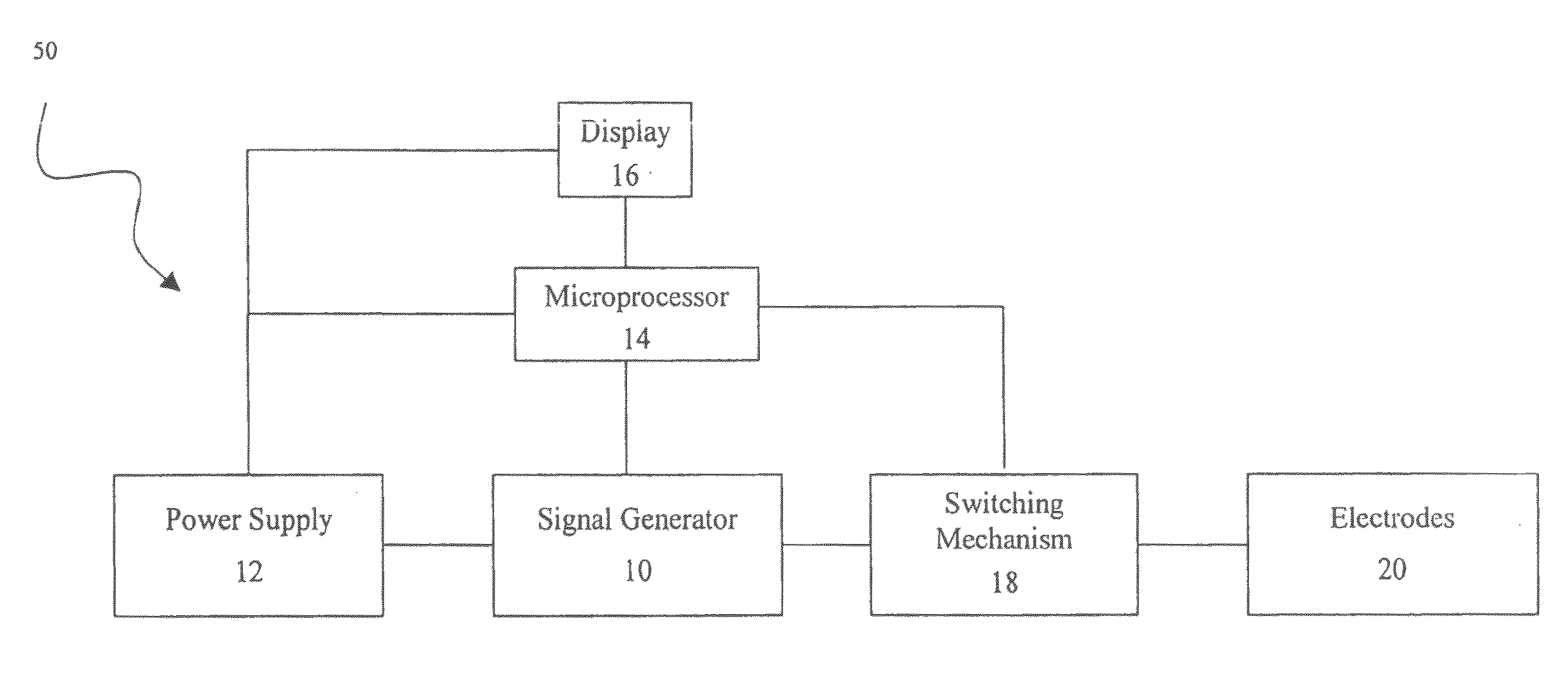
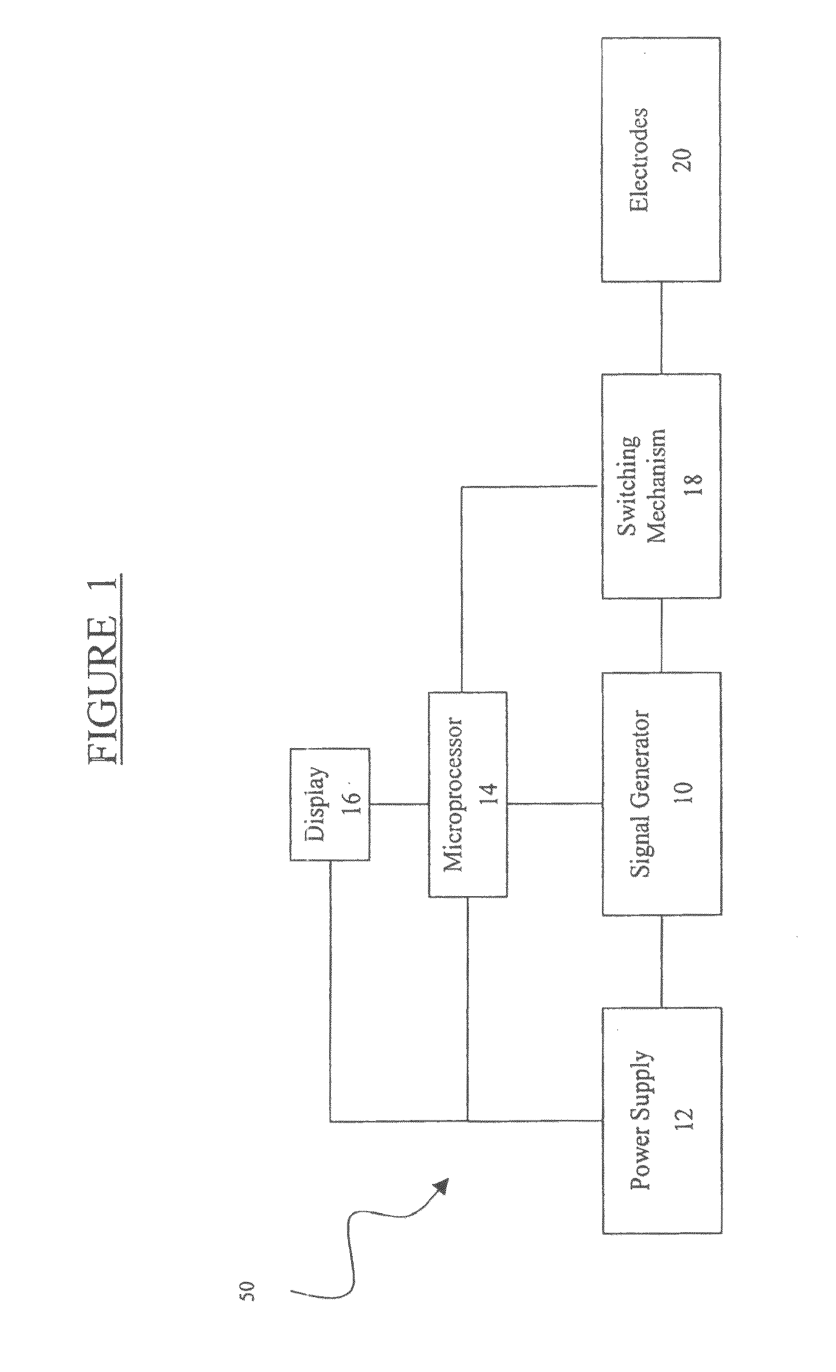
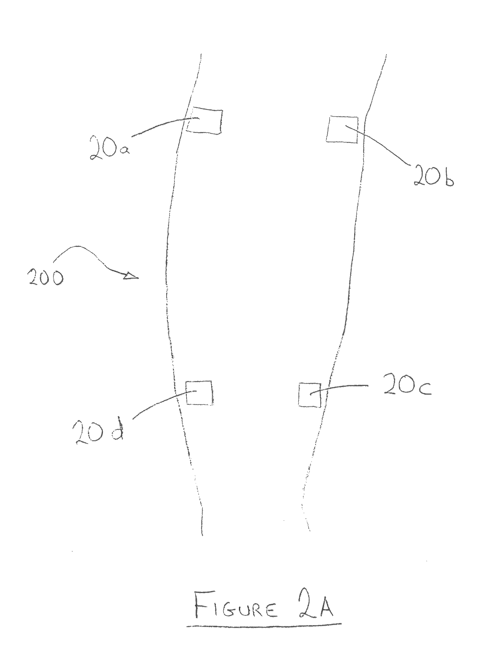
[0170]A device of the present invention was used to provide electrical stimulation to the lower leg of a patient suffering from poor circulation in the foot. Two surface electrodes of the inventive device were positioned underneath the knee, and an additional two surface electrodes of the device were positioned above the calf, substantially as shown in FIG. 2A. The microprocessor used for controlling the device was an ATMEL® 8 bit AVR® microcontroller, model no. ATmega8535, which also contains the signal generator unit.

[0171]Electrical stimulation was administered for over 60 minutes. As in Example 1, a FLIR™ ThermaCAM® EX320 was used to thermally monitor the foot and lower calf. In particular, three locations on the leg were monitored:[0172](1) a point on the big toe;[0173](2) a point at the base of the big toe;[0174](3) a point on the instep.

Thermographs recorded intermittently during the course of the stimulation treatment are provided in FIGS. 7A-7F. In the first thermograph pro...


PUM


Abstract

A non-invasive method and device for promoting a localized change in a flow of blood through a blood vessel in a limb segment of a body by a series of electrically stimulated contractions of muscle tissue in the limb segment, the method including the steps of: (a) providing a device including: (i) first, second and third electrodes, each adapted to operatively contact the limb segment; (ii) a signal generator, operatively connected to the electrodes, adapted to produce a series of electrical impulses to the limb segment via the electrodes, and (iii) a control unit adapted to control the signal generator to produce the series of electrical impulses; (b) positioning the electrodes on the limb segment, wherein the first electrode is positioned on a lower end of the lower leg, the second electrode is positioned on the lower leg, and the third electrode is positioned on an upper end of the lower leg, whereby the first and third electrodes are disposed on opposite ends of the lower leg, and the second electrode and one of the first and third electrodes are disposed on a same end of the lower leg; (c) effecting a sequence of muscular contractions of the lower leg, by operations including: (i) applying a first electrical impulse between the electrodes on the same end of the lower leg to induce a first muscular contraction of a first portion of the tissue; and (ii) applying at least a second electrical impulse between the first and third electrodes to induce a longitudinal muscular contraction of a second portion of the muscular tissue; and (d) repeating operations (i) and (ii), to repeatedly induce the contractions, to effect the increased flow of blood.

Description

[0001]This patent application is a continuation-in-part of: U.S. patent application Ser. No. 11 / 438,070 filed on May 22, 2006; U.S. patent application Ser. No. 12 / 965,883 filed on Dec. 12, 2010, which is a continuation-in-part of PCT / IL2009 / 000584 filed on Jun. 14, 2009; and U.S. patent application Ser. No. 12 / 853,491 filed on Aug. 8, 2010, which is a continuation-in-part of PCT / IL2009 / 000145 filed on Feb. 8, 2009 and which claims priority to U.S. Provisional Patent Application Ser. No. 61 / 027,464, filed on Feb. 10, 2008, all of which are all hereby incorporated in their entirety by reference.FIELD AND BACKGROUND OF THE INVENTION[0002]The present invention relates to a method and device for promoting a localized change in the flow of blood through a blood vessel, and more particularly, to a non-invasive method and device for promoting a localized change in the flow of blood by electrically-induced contractual movement of muscular tissue.[0003]Current treatments for improving blood c...

Claims


Application Information

Patent Timeline
Patent Type & Authority Applications(United States)
IPC IPC(8): A61N1/36A61F13/00A61F13/06
CPCA61N1/36014A61N1/36003A61N1/36034A61N1/36031
Inventor NACHUM, ZVIBREZEL, YAAKOV B.LAMPERT, SHALOM
Owner FLOWAID MEDICAL TECH CORP
Why Eureka
Try Eureka