Validation embedded segmentation method for vascular ultrasound images

a segmentation method and ultrasound technology, applied in image enhancement, instruments, angiography, etc., can solve the problems of posing a further challenge, the way in which speckle reduction is used is very conservative, and the conventional method has certain problems related to accuracy and reliability

Inactive Publication Date: 2011-12-08
ATHEROPOINT
View PDF4 Cites 12 Cited by
  • Summary
  • Abstract
  • Description
  • Claims
  • Application Information

AI Technical Summary

Problems solved by technology

Since the ultrasound vascular scans do not always have the vessel orientation horizontal with respect bottom edge of the image, manual methods can pose a further challenge towards the region of interest estimation.(6) Guiding Method for the calibration System: Since the recognition is followed by the calibration process, the calibration system becomes very robust since the calibration processing is done in the region of interest determined by the automated validation embedded recognition system.
Though speckle reduction is common in ultrasound imaging, but the way speckle reduction is used here is very conservative.
Though conventional methods are generally suitable, conventional methods have certain problems related to accuracy and reliability.
IMT estimation having a value close to 1 mm is a very challenging task in ultrasound images due to large number of variabilities such as: poor contrast, orientation of the vessels, varying thickness, sudden fading of the contrast due to change in tissue density, presence of various plaque components in the intima wall such as lipids, calcium, hemmorage, etc.
Under normal resolutions, a 1 mm thick media thickness is difficult to estimate using stand-alone image processing techniques.
Over and above, the image processing algorithms face an even tighter challenge due to the presence of speckle distribution.
The manual handling of such a repetitive work flow of IMT screenings is tedious, error-prone and subject to lot of variability.
Case (b) is difficult to implement, because it is difficult to identify the LI and MA borders with heavy speckle distribution and the inability of ultrasound physics to generate a clear image where the semi-automated or automated image processing methods are used for IMT estimation.
Besides that

Method used


Image

  • Validation embedded segmentation method for vascular ultrasound images
  • Validation embedded segmentation method for vascular ultrasound images
  • Validation embedded segmentation method for vascular ultrasound images

Examples


Embodiment Construction

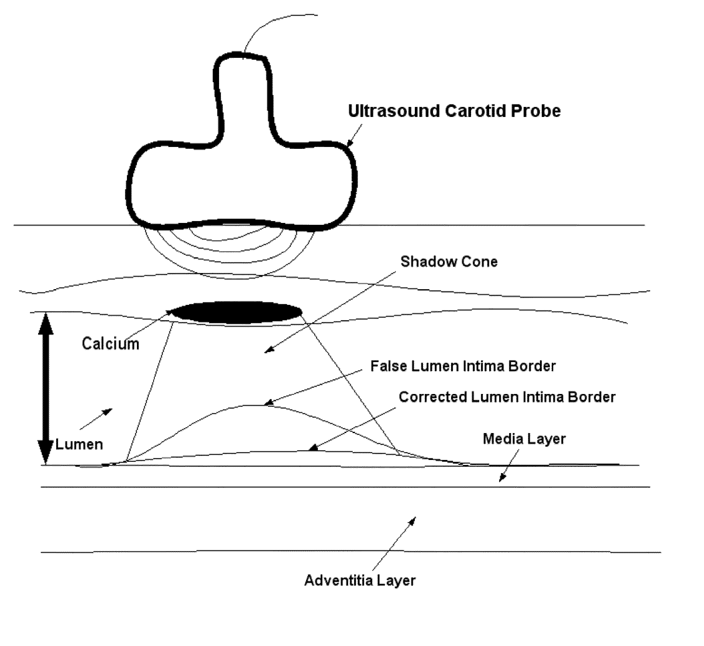
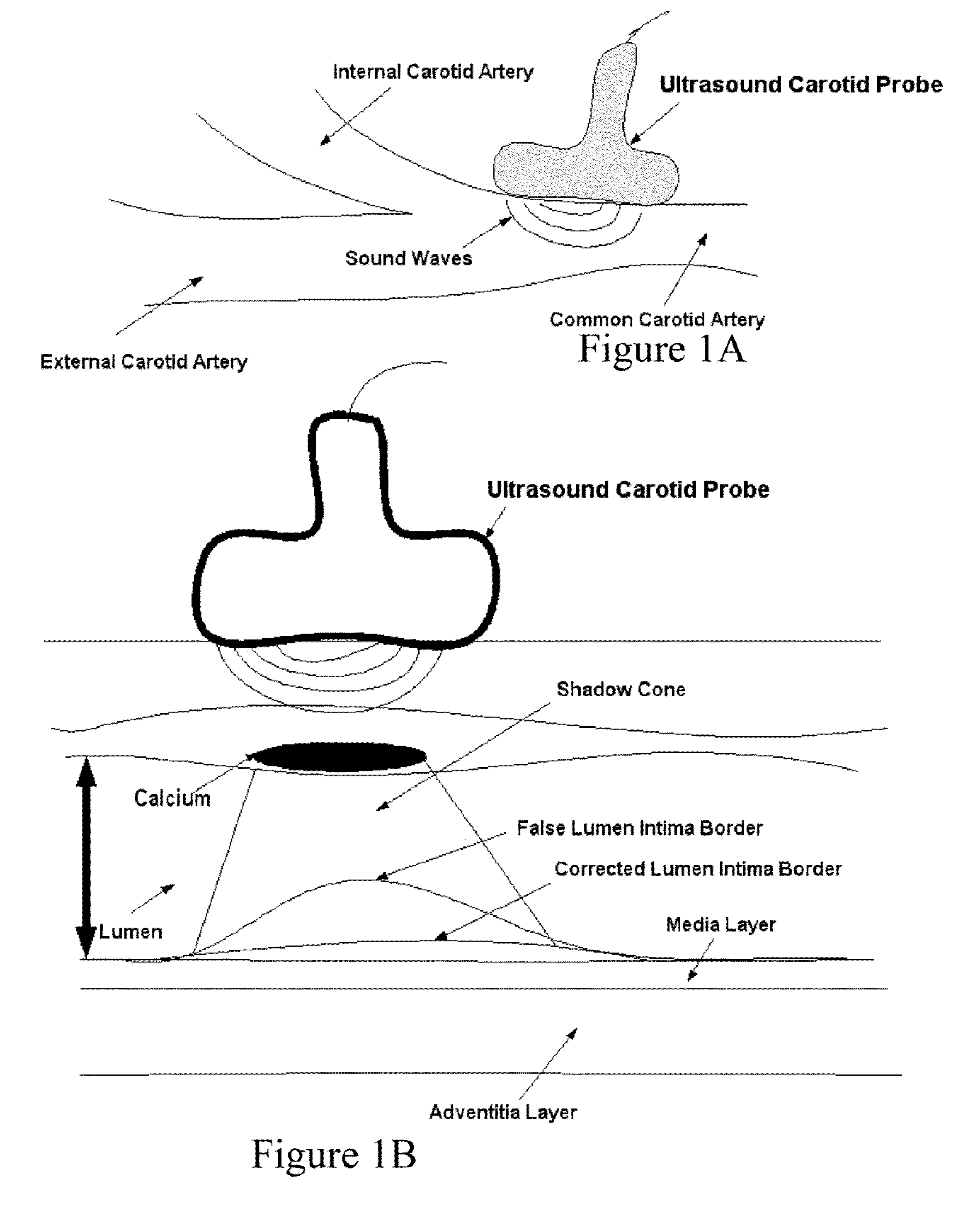
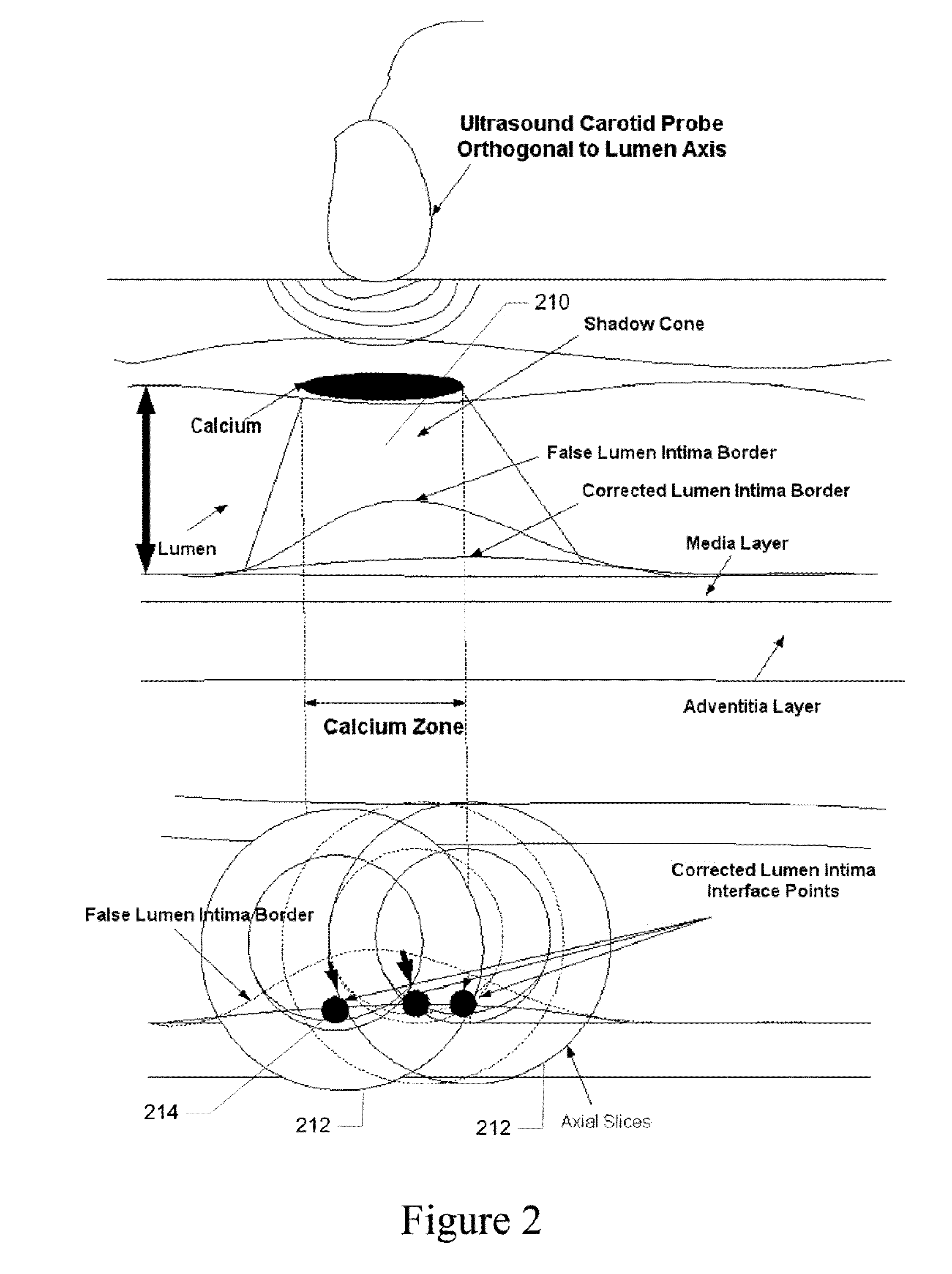
[0041]Recognition of the carotid artery consists of finding a regional layer close to the carotid artery and possibly all along the carotid artery in the image frame. This recognition process must ensure that we are able to distinguish the carotid artery layer from other veins such as jugular vein (JV). We modeled the carotid artery recognition process by taking the hypothesis that carotid artery's far wall adventitia is the brightest in the ultrasound scan frame; hence if we can automatically find this layer, then segmentation process of the far wall would be more systematic and channeled. Since the scanning process of carotid artery yields varying geometries of the carotid artery in the ultrasound scans, one has to ensure that the recognition process is able to handle various geometric shapes of the carotid arteries in the images. The process of location of far adventitia bright layer in the image frame can be supported by the fact that it is very close to lumen region, which carr...


PUM


Abstract

A computer-implemented system and method for intima-media thickness (IMT) measurements using a validation embedded segmentation method. Various embodiments include receiving biomedical imaging data and patient demographic data corresponding to a current scan of a patient; checking the biomedical imaging data in real-time to determine if an artery of the patient has a calcium deposit in a proximal wall of the artery; acquiring arterial data of the patient as a combination of longitudinal B-mode and transverse B-mode data; using a data processor to automatically recognize the artery by embedding anatomic information; using the data processor to calibrate a region of interest around the automatically recognized artery; automatically computing the weak or missing edges of intima-media and media-adventitia walls using edge flow, labeling and connectivity; and determining the intima-media thickness (IMT) of an arterial wall of the automatically recognized artery.

Description

PRIORITY APPLICATION[0001]This is a continuation-in-part patent application of co-pending patent application Ser. No. 12 / 802,431; filed Jun. 7, 2010 by the same applicant. This is also a continuation-in-part patent application of co-pending patent application Ser. No. 12 / 896,875; filed Oct. 2, 2010 by the same applicant. This present patent application draws priority from the referenced co-pending patent applications. The entire disclosures of the referenced co-pending patent applications are considered part of the disclosure of the present application and are hereby incorporated by reference herein in its entirety.COPYRIGHT[0002]A portion of the disclosure of this patent document contains material that is subject to copyright protection. The copyright owner has no objection to the facsimile reproduction of the patent document or the patent disclosure, as it appears in the Patent and Trademark Office patent files or records, but otherwise reserves all copyright rights whatsoever. Th...

Claims


Application Information

Patent Timeline
IPC IPC(8): G06K9/00
CPCG06T7/0012A61B8/0858A61B8/5207G06T2207/20016G06T2207/10132G06T7/0085A61B8/585G06T2207/30101A61B8/14A61B8/0891A61B8/085A61B5/02007A61B8/145A61B8/5223G06T7/13
Inventor SURI, JASJIT S.
Owner ATHEROPOINT
Why Eureka
Try Eureka