Renal cell carcinoma biomarkers

a cancer and biomarker technology, applied in the field of tumor biomarkers, can solve the problems of ms-based cancer biomarker research/discovery, hindering subsequent bioinformatics and statistical analyses, and disappointing translation of proteomic assays to applicable diagnostic and/or prognostic tests in clinical oncology, and achieve the effect of facilitating cancer biomarker discovery

Inactive Publication Date: 2012-01-12
UNITED STATES OF AMERICA
View PDF0 Cites 6 Cited by
  • Summary
  • Abstract
  • Description
  • Claims
  • Application Information

AI Technical Summary

Benefits of technology

[0005]There is an urgent need for methods that can facilitate cancer biomarker discovery from clinically relevant specimens. Described herein is a proteomic approach for tumor biomarker discovery, which represents a major step toward routine detection of tumor-originated proteins that can be utilized to diagnosis a subject with a particular type of cancer, such as renal cell carcinoma (RCC). For example, the disclosed method not only allows low-abundant tumor proteins to be confidentially detected in plasma, but the identification of a panel of proteins that can be specifically linked to a particular tumor type. The generated molecular protein profile identification that includes including posttranslational modifications (PTMs) and corresponding gene profile signature can then be used to diagnosis a subject with the identified tumor type or to identify agents capable of altering the biomarker expression or activity, thereby revealing possible therapeutic agents to treat the specific tumor type.

Problems solved by technology

So far, the translation of proteomic assays to applicable diagnostic and / or prognostic tests in clinical oncology has been disappointing.
A number of factors currently hinder MS-based cancer biomarker research / discovery.
For example, heterogeneity or variations in specific disease / cancer processes as well as the human population (in general) may introduce biases into complex proteomic datasets hindering subsequent bioinformatic and statistical analyses.
Moreover, according to findings reported by the Human Proteome Organization, the dynamic range of the human plasma protein concentration is in the order of 1010 while the dynamic range of contemporary MS instrumentation is 104 at best resulting in a formidable mismatch between the dynamic range of MS instrumentation and the dynamic range of human specimens.
Thus, one type of sample method cannot be used to detect all types of cancer.

Method used


Image

  • Renal cell carcinoma biomarkers
  • Renal cell carcinoma biomarkers
  • Renal cell carcinoma biomarkers

Examples


example 1

Materials and Methods

[0324]Materials and reagents. MARS columns and MARS reagents were purchased from Agilent Technologies (Palo Alto, Calif.). SDS-PAGE materials and reagents were obtained from Invitrogen Life Technologies (Carlsbad, Calif.). Methanol and acetonitrile (HPLC-grade) were purchased from EMD Chemicals (Gibbstown, N.J.). All other chemicals and reagents, except for disclosed ones, were obtained from Sigma-Aldrich (St. Louis, Mo.).

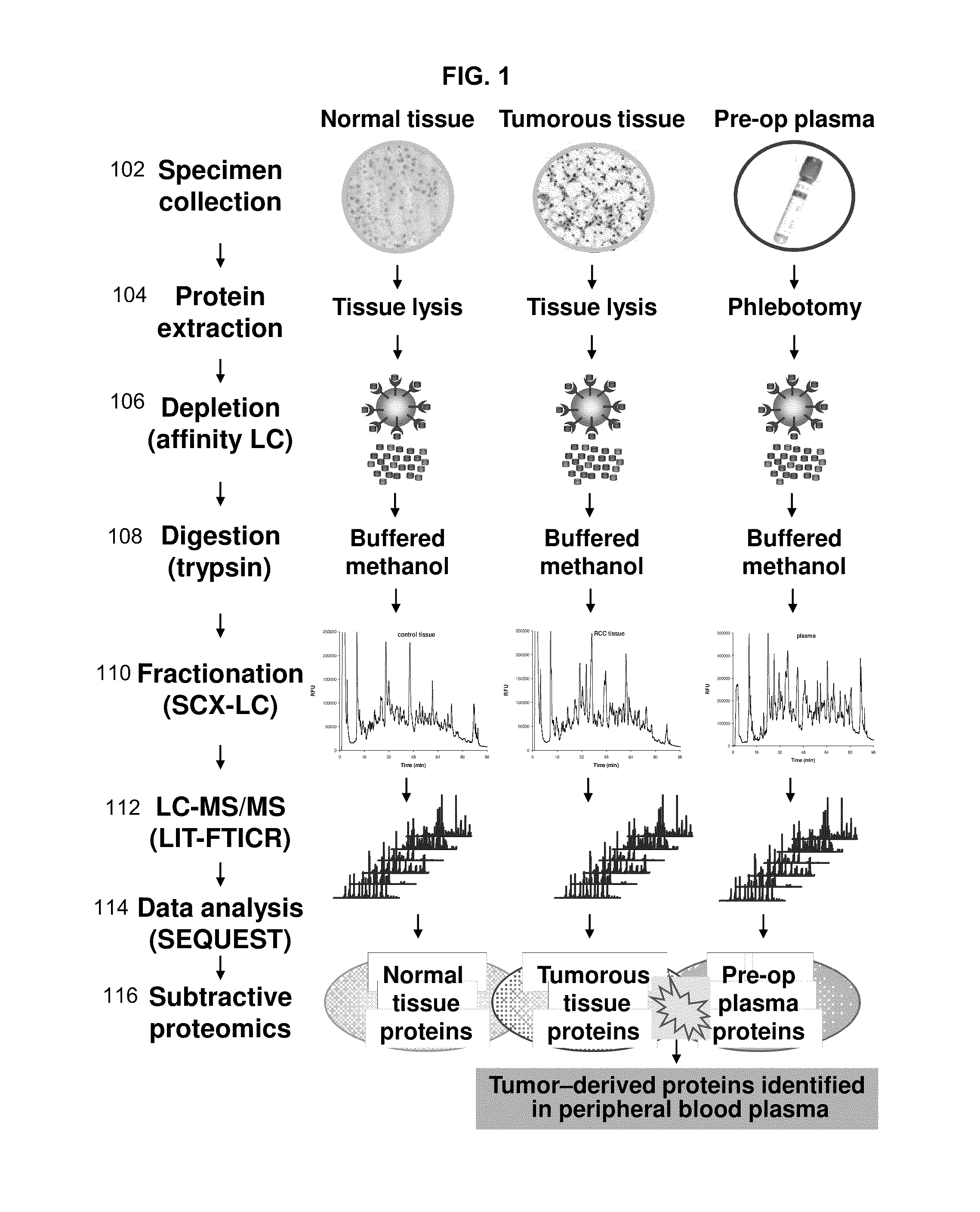
[0325]Specimen collection and processing. Clinical specimens were procured by the National Cancer Institute Cooperative Human Tissue Network in accordance with current regulations and guidance issued by the Office of Human Subjects Review. Tumorous fresh frozen tissue, adjacent non-tumor fresh frozen tissue and plasma specimens were collected prospectively from a single patient diagnosed with the RCC using standard clinical procedures and stored at −80 ° C. Tissue specimens were procured during the surgery while peripheral blood plasma was coll...

example 2

Method of Identifying Tumor Biomarkers including RCC Biomarkers

[0329]This example describes a method of identifying tumor biomarkers using RCC as a model. This example also provides an RCC gene signature that can be used to diagnosis a subject with RCC. One skilled in the art will appreciate that similar methods can be used to identify biomarkers for other cancers, such as breast, lung, colon, or liver cancer.

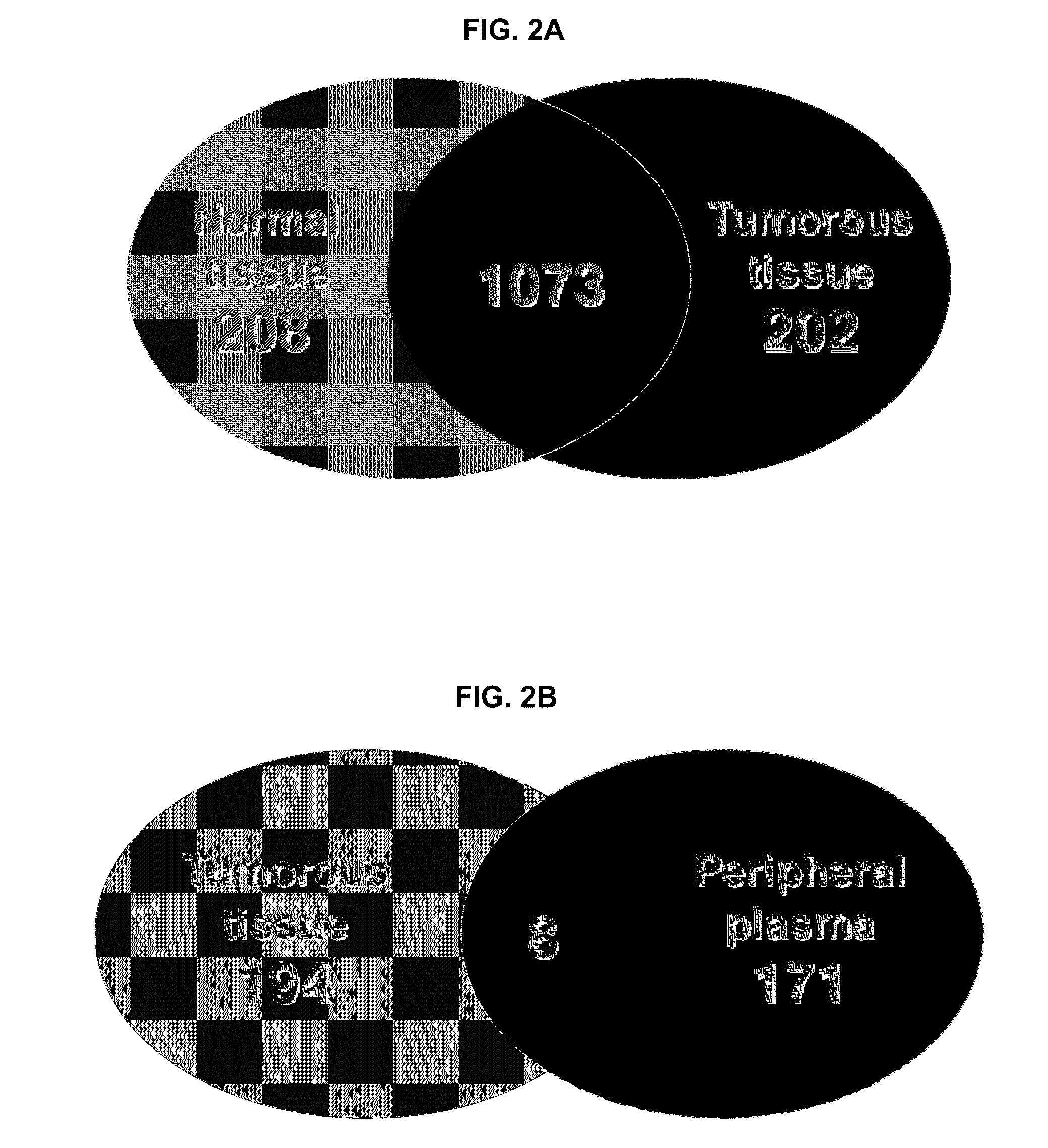
[0330]Tumorous (0.57 grams) and non-tumorous tissue (normal adjacent kidney tissue, 0.56 grams) was obtained from a single patient and freshly frozen in liquid nitrogen along with 0.6 mL of patient's peripheral blood plasma. Since the plasma content of kidney tissue represents up to 22% of its weight the abundant protein depletion was applied to both tissue and plasma specimens. This step significantly contributed to dynamic range reduction and increased overall detection rate of low-abundant proteins. Subsequent tryptic digestion was performed in buffered methanol. The use of ...

example 3

Diagnosing RCC

[0338]This example describes methods that can be used to diagnose a subject with RCC.

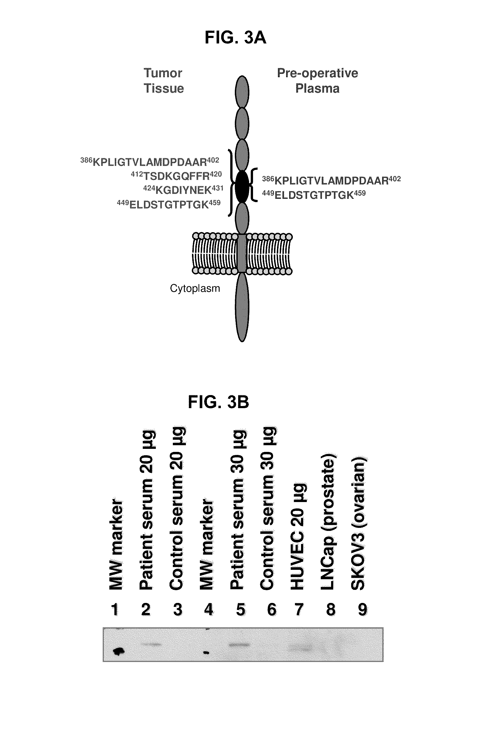
[0339]According to the teachings herein, whether a subject has RCC can be determined by detecting differential expression of at least two RCC biomarkers in a sample obtained from the subject believed to have RCC or at risk of developing RCC. In an example, a peripheral biological sample, such as serum, is obtained from the subject who is believed to have RCC or at risk of developing RCC. The polypeptide levels of two or more of (such as all of) CDH5, CDH11, DDX23, WWC1, CHD4, NCOA6, PKM2, and VCAM1 in the biological sample is then evaluated using a protein array that includes different capture agents, most frequently monoclonal antibodies, each of which is capable of binding to one or more of the disclosed RCC biomarkers and controls, such as positive and negative controls. The amount of RCC biomarker protein measured in the biological sample is then compared to a reference value refle...


PUM

PropertyMeasurementUnit
survival timeaaaaaaaaaa

Abstract

Disclosed herein is a method of identifying a tumor biomarker. In one example, a tumor biomarker is identified by obtaining a peripheral biological fluid sample from a subject with a tumor as well as a tumor sample and an adjacent non-tumor sample from such subject. A protein expression profile is detected in the peripheral biological fluid sample, tumor sample and adjacent non-tumor sample. The protein expression profiles of the peripheral biological fluid sample, tumor sample and adjacent non-tumor sample are then compared, wherein an increase in expression of a specific protein in the tumor sample and peripheral biological fluid sample but not in the adjacent non-tumor sample indicates that the specific protein is a biomarker of the tumor. Also disclosed herein is a gene profiling signature that can be used to diagnosis a subject with renal cell carcinoma (RCC) or to identify agents with therapeutic potential to treat RCC. Thus, methods of diagnosing a subject with RCC are disclosed. Methods are also provided for identifying agents that alter an activity of a RCC biomarker.

Description

FIELD[0001]This disclosure relates to the field of tumor biomarkers and in particular, to methods of identifying (e.g., diagnostic, prognostic, therapeutic and follow up / monitoring) tumor biomarkers for diagnosing specific tumors, such as renal cell carcinoma (RCC), identifying treatment modalities, making prognosis and follow-up for subjects with such conditions.BACKGROUND[0002]Cancer is the second leading cause of death in the United States. There is an acute need for cancer biomarkers that can be detected from clinically relevant samples and used for early diagnosis, therapeutic follow-up, and prognosis of malignant diseases. Proteins are principal regulators and effectors of physiologic and pathophysiologic processes. As such, proteomics is expected to play a role in clinical biomarker discovery.[0003]Clinical proteomics is an emerging area of proteome research. For example, to identify cancer biomarkers by analyzing clinically relevant specimens routinely procured from an indiv...

Claims


Application Information

Patent Timeline
Patent Type & Authority Applications(United States)
IPC IPC(8): A61K39/395C12Q1/68C12Q1/37G01N33/53G01N33/574C07K16/40C07K16/18C07H21/00A61P35/00C40B30/04A61K31/713
CPCC12Q1/6886G01N33/57438G01N33/57484G01N2500/00C12Q2600/136G01N33/6848G01N33/50G01N33/574G01N33/68G01N33/6842C12Q2600/16A61P35/00
Inventor BLONDER, JOSIPWEI, BIH-RONGJOHANN, DONALD J.VEENSTRA, TIMOTHY D.
Owner UNITED STATES OF AMERICA
Why Eureka
Try Eureka