
Biological Components Within the Cerebrospinal Fluid

a technology of cerebrospinal fluid and biological components, applied in the field of biological components within the cerebrospinal fluid, can solve the problems of virtually impossible detection of csf-borne mps, unable to validate mps, and a considerable challenge in the development of disease-modifying therapies

Inactive Publication Date: 2012-07-12
GENFIT SA
View PDF0 Cites 7 Cited by
  • Summary
  • Abstract
  • Description
  • Claims
  • Application Information

AI Technical Summary

Benefits of technology

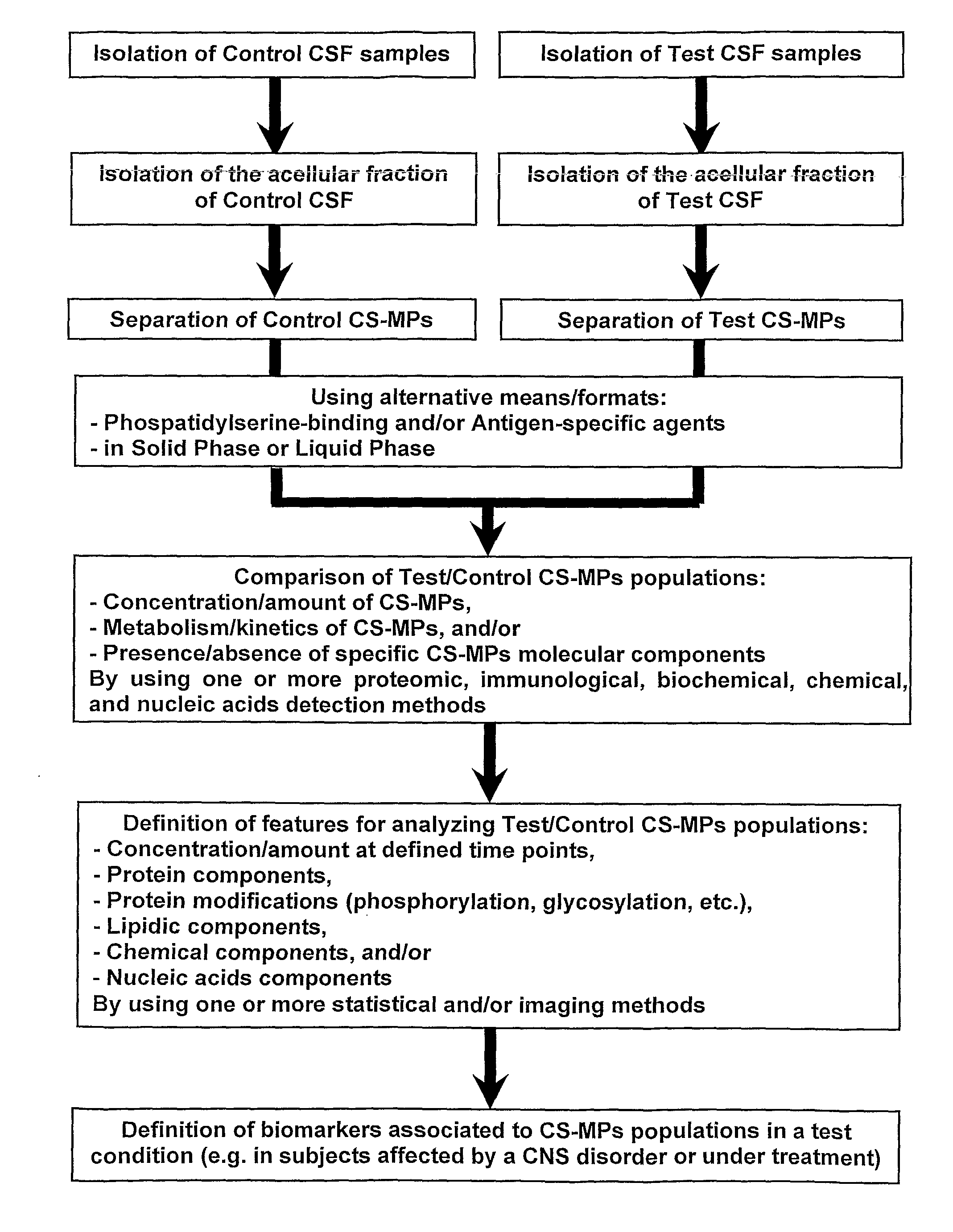
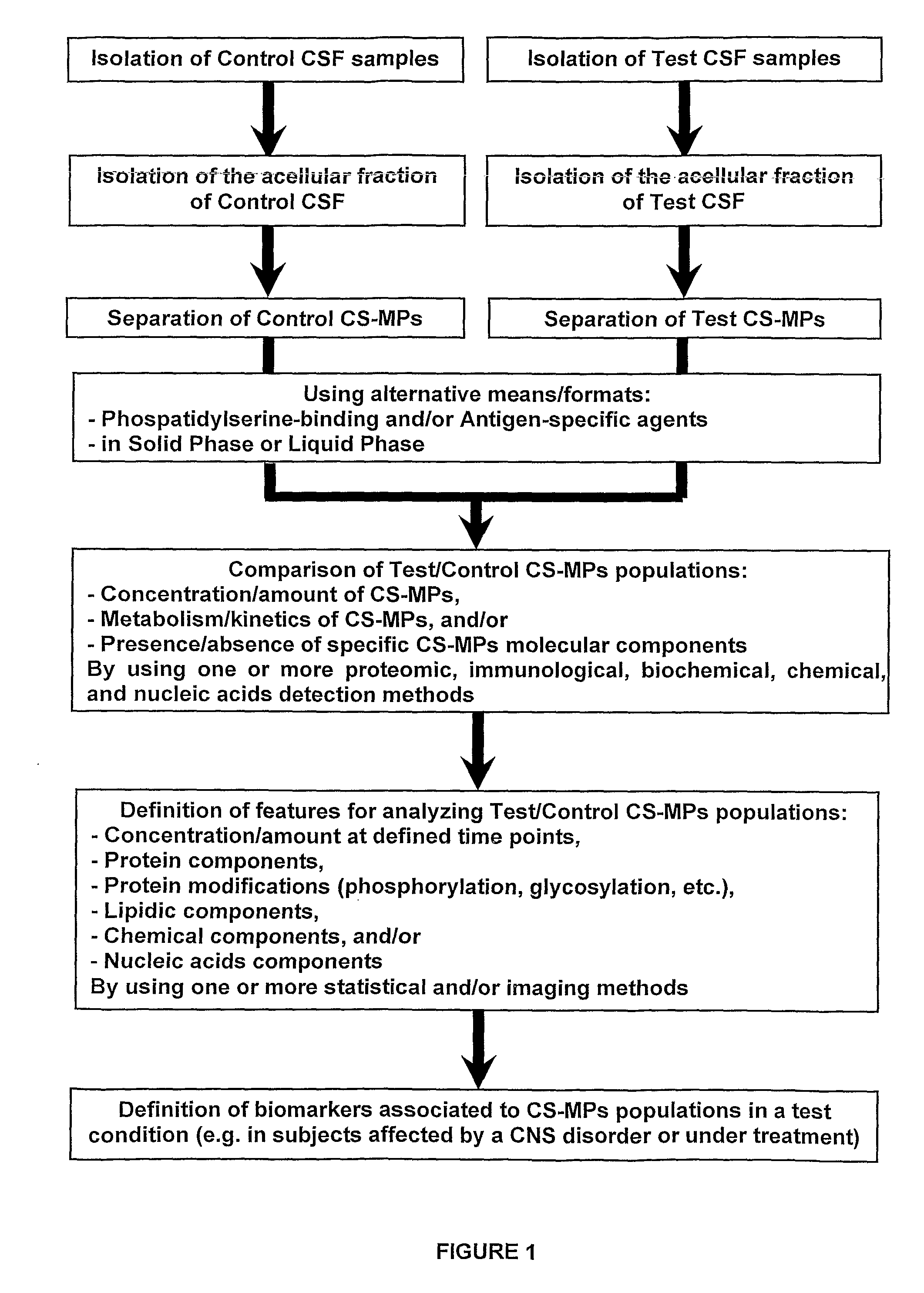
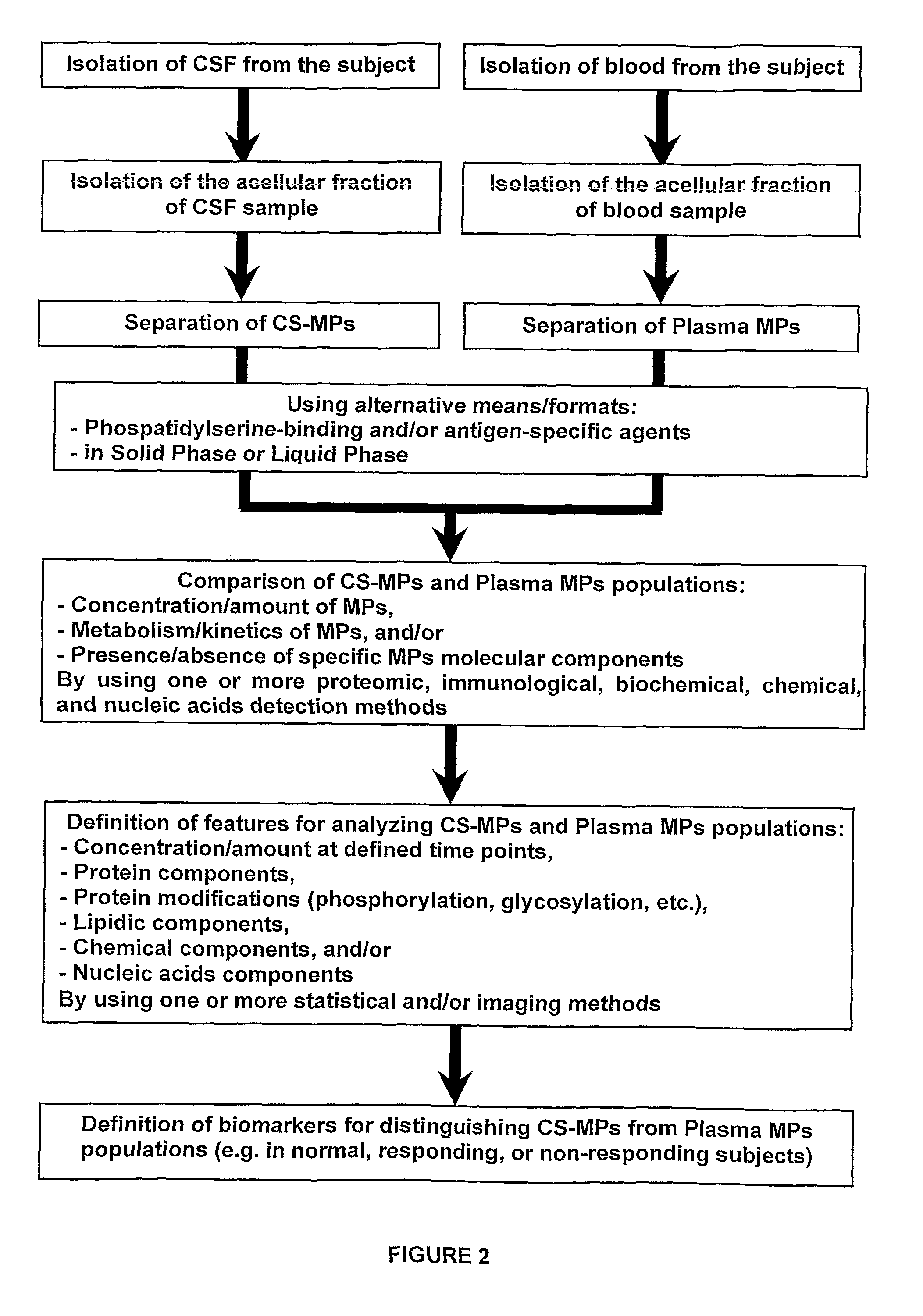
[0017]The CS-MPs that are obtained by this method can be used to establish the concentration and/or components (such as cell type-specific antigens, phospholipids, or glycosylated groups) of CS-MPs that differ between control and test subjects (e.g., normal or at risk of a CN...

Problems solved by technology

However, the development of disease-modifying therapies remains a considerable challenge due to the absence of robust biomarkers for drug development, diagnosis, prognosis, and therapy.
However, all those studies encountered a common problem due to the presence of extremely abundant proteins coming from the blood (such as albumin, transferrin, acute phase proteins and various antibodies) that mask the more significant sub-proteome for identifying the biomarkers of medical interest that are generated within CNS or by other tissues in direct contact with the CSF (such as the cells forming BCSFB, BBB, or the brain-CSF interface).
However, MPs have not been validated yet as biomarkers for any indication in clinical settings and improved means for characterizing MPs of medical interest are needed.
Thus, endothelial or blood-cells derived MPs can enter into CSF and, given the much lower concentration of proteins and particulate into CSF when compared to blood, can make the detection of CSF-borne MPs virtually impossible.
In absence of details such as the dimension, the presence of phosphatidylserine and the protocol for applying centrifugation to CSF samples (Horstman L et al., 2007), the findings in the literature on MPs-like elements that may be produced and shed in the CSF do not provide sufficient evidences in support of such a hypothesis (Smalheiser N, 2009).

Method used


Image

  • Biological Components Within the Cerebrospinal Fluid
  • Biological Components Within the Cerebrospinal Fluid
  • Biological Components Within the Cerebrospinal Fluid

Examples


example 1

Quantification of Rat CS-MPs Compared to Plasma MPs

Materials & Methods

Isolation of Rat CSF

[0066]Male Sprague-Dawley rats (225-250 g, CERJ, France) were anesthetized with pentobarbital (55 mg / kg) and positioned in a stereotaxic frame. The rat head was flexed downward at approximately 45 degrees, a depressible surface with the appearance of a rhomb between occipital protuberances and the spine of the atlas becomes visible. The 25 G needle was punctured into the cisterna magna for CSF collection without making any incision at this region. The blunt end of the needle was inserted into a 10 in. length of PE-50 tubing and other end of the tubing was connected to a collection syringe (Hamilton, 100 μl). The non-blood contaminated sample (100 μl) was drawn into the syringe by simple aspiration. Samples with blood cell contaminations were discarded. A sample was centrifuged at 13,000 g for 2 minutes and the resulting supernatant was subsequently snap-frozen in polypropylene tubes. Samples we...

example 2

Quantification of MPs Produced in Cell Culture Conditions

Materials & Methods

Cell Culture Protocols

[0077]The human neuronal cell line SH-SY5Y (ATCC CRL-2266) was cultured in MEM / Ham's F12 medium (1:1) supplemented with 10% foetal bovine serum. Cells were seeded at 55,000 cells / cm2. Cell differentiation was induced by adding 9-cis retinoic acid (5 μM) for 5 days directly to complete culture medium. After 5 days, medium was replaced with complete medium supplemented with BDNF (50 ng / ml) for additional 5 days. At the end of the differentiation protocol, cells were rinsed with serum-free medium and then treated for 24 hours with complete medium containing Staurosporine (100 and 500 nM; Sigma, France) or vehicle (DMSO 0.1%).

MPs Quantification

[0078]At the end of the treatment, cell supernatants were collected and spun down for 15 minutes at 1,500 g at room temperature. MPs were then isolated by ultracentrifugation at 30,000 g for 45 minutes at room temperature. At this speed, both exosomes...

example 3

Analysis of MP Signature in Human CSF Samples

Materials & Methods

Isolation and Quantification of Human CS-MPs

[0091]The CSF samples were taken for diagnostic purpose from adult patients in medical institution by a lumbar puncture according to a standard procedure. Scientific use of CSF samples was approved by the local Ethics Committee and all patients gave a written informed consent for the diagnostic procedure. No traumatic signs were detected in all the patients.

[0092]50 to 300 μL of CSF was collected from each subject. CS-MPs were obtained after two sequential centrifugations. first at 1,500 g for 15 minutes at room temperature. The supernatant was then carefully removed and transferred to a new tube. The second centrifugation was performed at 13,000 g for 2 minutes at room temperature. Again, the supernatant was carefully transferred into a new tube and snap-frozen using liquid nitrogen. CS-MPs were stored at −80° C. until use.

[0093]CS-MPs quantifications were performed using flu...


PUM

PropertyMeasurementUnit
Diameteraaaaaaaaaa
Diameteraaaaaaaaaa
Nanoscale particle sizeaaaaaaaaaa

Abstract

The invention provides novel methods for isolating, characterizing, comparing, and using biological components that are present in the cerebrospinal fluid. Such biological structures, called CS-MPs, can be used for identifying biomarkers that reflect the status (or anticipate the development) of disorders of the Central nervous System (CNS). The novel methods, biological products, and related kits make possible the use of CS-MPs and of their components as biomarkers for the diagnosis, prognosis, or monitoring of CNS disorders. The CS-MPs have a diameter comprised between 100 and 1000 nm and contain phosphatidylserine (PS).

Description

TECHNICAL FIELD[0001]The present invention relates to methods for isolating, characterizing, comparing and using specific biological components in the cerebrospinal fluid. Such biological components can be used for diagnosing or monitoring diseases in a subject, and / or for evaluating the therapeutic efficacy of a medical treatment or a candidate drug.BACKGROUND OF THE INVENTION[0002]In recent years, huge progress has been made in the understanding of pathophysiology of the disorders of the Central Nervous System (CNS). However, the development of disease-modifying therapies remains a considerable challenge due to the absence of robust biomarkers for drug development, diagnosis, prognosis, and therapy. This aspect is particularly important for neurodegenerative disorders (such as multiple sclerosis and Parkinson's or Alzheimer's diseases) where validated biomarkers as surrogate endpoint for (pre)clinical drug development are needed (Shaw L et al., 2007; Dubois B et al., 2007; Pritcha...

Claims


Application Information

Patent Timeline
IPC IPC(8): G01N21/64G01N33/68
CPCG01N33/6896Y10T428/2982G01N2500/20
Inventor DELERIVE, PHILIPPEMAJD, ZOUHER
Owner GENFIT SA
Learn More