Method, system, software and medium for advanced intelligent image analysis and display of medical images and information

a medical image and intelligent technology, applied in image analysis, image enhancement, instruments, etc., can solve the problems of large misclassification of lesions, large invasive methods of assessing biologic features, and insufficient information about the disease,

Active Publication Date: 2012-07-26
QLARITY IMAGING LLC
View PDF13 Cites 83 Cited by
  • Summary
  • Abstract
  • Description
  • Claims
  • Application Information

AI Technical Summary

Benefits of technology

[0040]Moreover, labeled and unlabeled medical data of the other patients can be obtained, disease and non-disease state lesions in the labeled medical image data and lesions in the unlabeled medical image data can be segmented, and features of the segmented labeled and unlabeled medical image data can be ex

Problems solved by technology

Breast cancer is a leading cause of death in women, causing an estimated 46,000 deaths per year.
Although general rules for the differentiation between benign and malignant mammographically identified breast lesions exist, considerable misclassification of lesions occurs with current methods.
It has been shown that in many cases biologic features of the primary tumor can be correlated with outcome, although methods of assessing the biologic features may be invasive, expensive or not widely available.

Method used


Image

  • Method, system, software and medium for advanced intelligent image analysis and display of medical images and information
  • Method, system, software and medium for advanced intelligent image analysis and display of medical images and information
  • Method, system, software and medium for advanced intelligent image analysis and display of medical images and information

Examples


Embodiment Construction

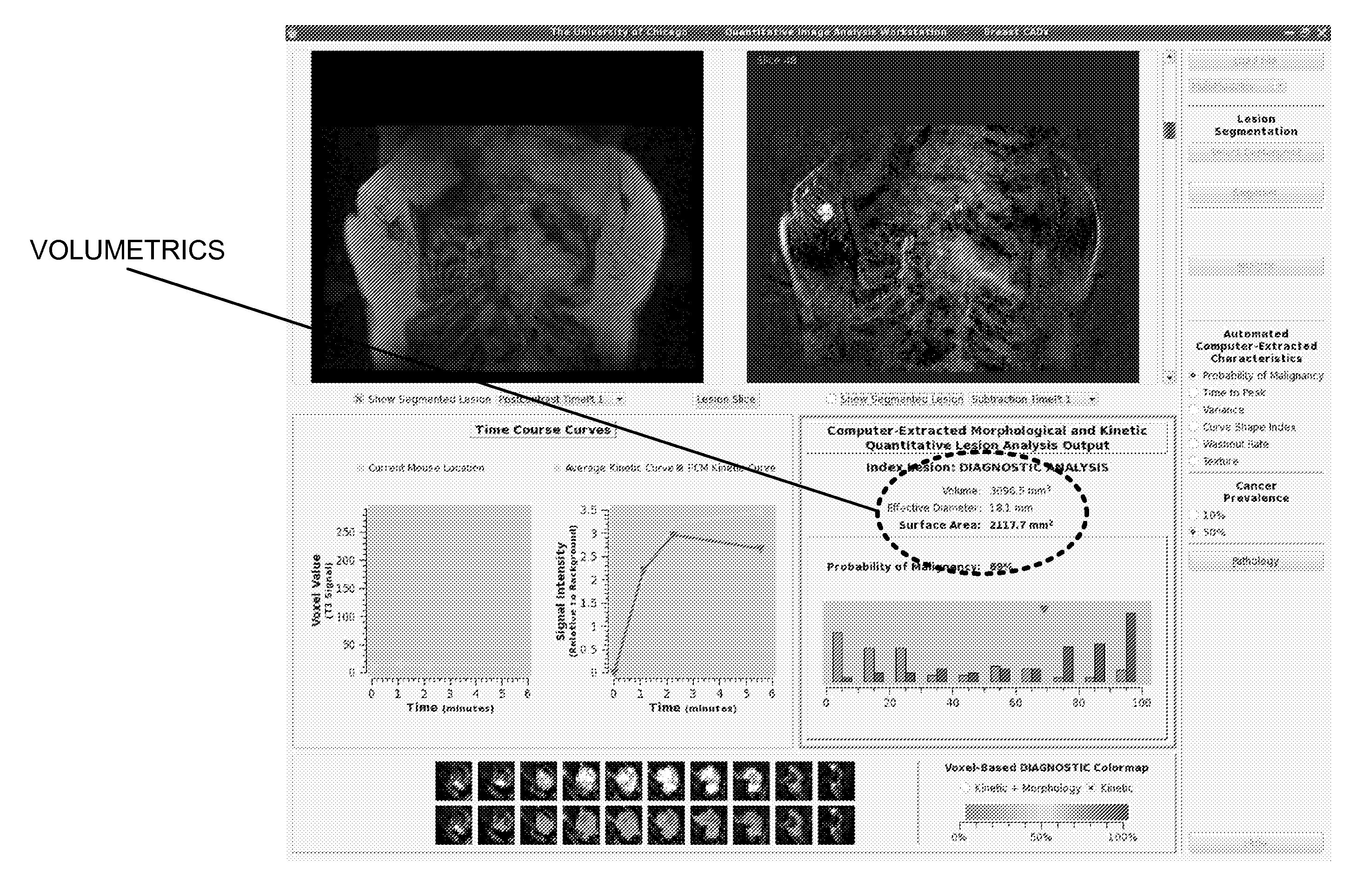
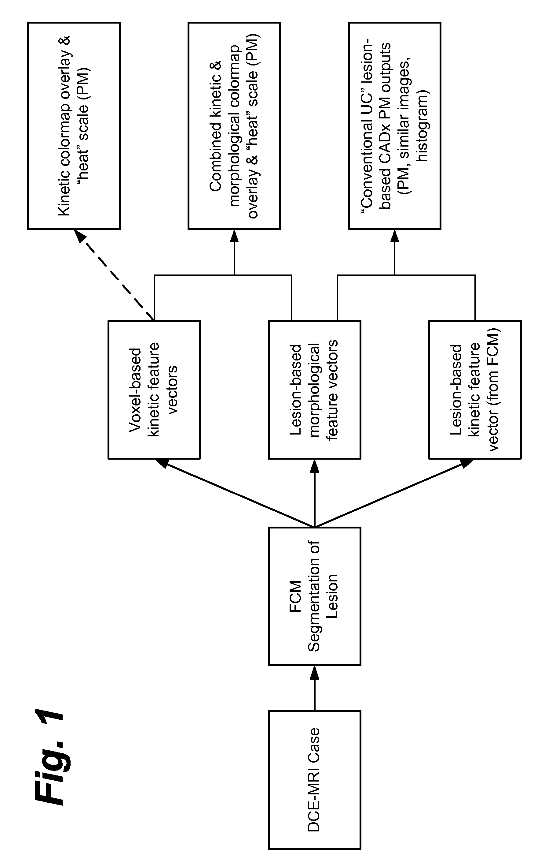
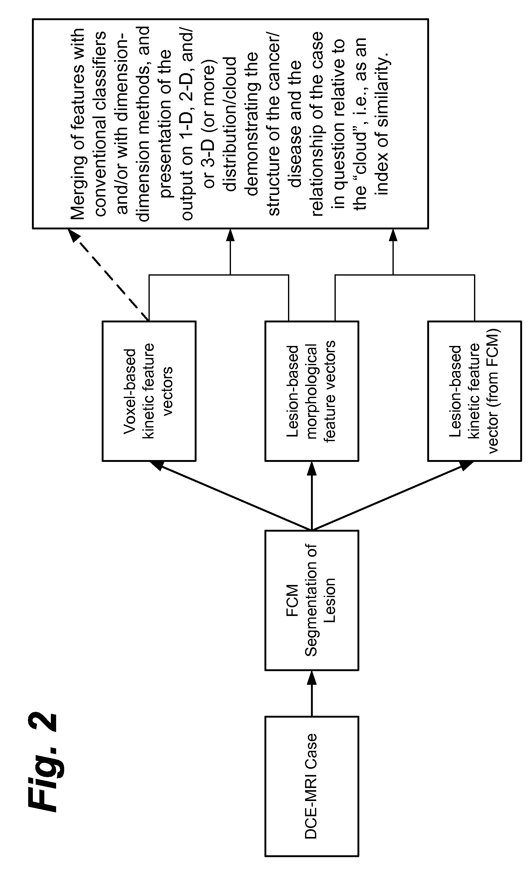
[0071]Embodiments described herein relate to methods and systems for an automatic and / or interactive method, system, software, and / or medium for a workstation for quantitative analysis of multi-modality breast images, which to date includes analysis of full-field digital mammography (FFDM), 2D and 3D ultrasound, and MRI.

[0072]According to one embodiment, a method and a system implementing this method determine and / or employ / incorporate lesion-based analysis, voxel-based analysis, and / or both in the assessment of disease state (e.g., cancer, cancer subtypes, prognosis, and / or response to therapy), and a method for the display of such information including kinetic information, morphological information, and / or both that also may utilize varying the disease state prevalence or prognostic state prevalence within the training or clinical case set.

[0073]According to another embodiment, a method and a system implementing this method determine and / or employ / incorporate, after manual, semi-a...


PUM


Abstract

Computerized interpretation of medical images for quantitative analysis of multi-modality breast images including analysis of FFDM, 2D/3D ultrasound, MRI, or other breast imaging methods. Real-time characterization of tumors and background tissue, and calculation of image-based biomarkers is provided for breast cancer detection, diagnosis, prognosis, risk assessment, and therapy response. Analysis includes lesion segmentation, and extraction of relevant characteristics (textural/morphological/kinetic features) from lesion-based or voxel-based analyses. Combinations of characteristics in several classification tasks using artificial intelligence is provided. Output in terms of 1D, 2D or 3D distributions in which an unknown case is identified relative to calculations on known or unlabeled cases, which can go through a dimension-reduction technique. Output to 3D shows relationships of the unknown case to a cloud of known or unlabeled cases, in which the cloud demonstrates the structure of the population of patients with and without the disease.

Description

CROSS-REFERENCE TO RELATED APPLICATIONS[0001]This application claims the benefit of U.S. Application Ser. No. 61 / 344,951, filed Nov. 26, 2010, the entire contents of which is incorporated herein.BACKGROUND[0002]1. Field of Disclosure[0003]This disclosure relates generally to the fields of computer-aided diagnosis, quantitative image analysis, and image display workstations. Such systems can output a feature value (e.g., characteristic; image-based phenotype) or an estimate of a lesion's probability of disease state (PM) (which can be a probability of malignancy, cancer subtypes, risk, prognostic state, and / or response to treatment), usually determined by training a classifier on datasets.[0004]2. Discussion of the Background[0005]Breast cancer is a leading cause of death in women, causing an estimated 46,000 deaths per year. Mammography is an effective method for the early detection of breast cancer, and it has been shown that periodic screening of asymptomatic women does reduce mor...

Claims


Application Information

Patent Timeline
IPC IPC(8): G06K9/46
CPCG06K9/6253G06T2207/30068G06T7/602G06T2207/10088G06T2207/10116G06T2207/20081G06T7/62G06T15/08G06T15/20G06T17/00G06T19/20G06T2207/10136G06T7/0016G06V10/945G06T7/0012G06T11/003G06T2207/10076G06T2207/10084G06T2207/30004G06F18/40G06T7/40
Inventor GIGER, MARYELLEN L.TOMEK, ROBERTBROWN, JEREMY BANCROFTJAMIESON, ANDREW ROBERTLAN, LICHINANDER, MICHAEL R.DRUKKER, KARENLI, HUIBHOOSHAN, NEHANEWSTEAD, GILLIAN
Owner QLARITY IMAGING LLC
Why Eureka
Try Eureka