
Endoscope system

Inactive Publication Date: 2012-07-26
FUJIFILM CORP
View PDF12 Cites 41 Cited by
  • Summary
  • Abstract
  • Description
  • Claims
  • Application Information

AI Technical Summary

Benefits of technology

[0011]As described above, the special light observation using narrowband light enables the state of the microvessels and microstructure in the superficial layer of the tissue to be more precisely observed. Therefore, the cancer lesion can be more correctly diagnosed.
[0015]An object of the invention is to solve the foregoing prior art problems and to provide an endoscope system with which even a less-experienced doctor can properly diagnose the cancer invasion depth from endoscopic images of microvessels in the superficial layer.
[0029]Therefore, according to the endoscope system of the invention, even a doctor with less experience in the endoscopic cancer diagnosis can properly diagnose the cancer invasion depth from endoscopic observation images.

Problems solved by technology

However, the blood stream in the microvessels of the superficial layer is complicated and varies with the cancer invasion depth and the site of the affected area such as esophagus or stomach.
Therefore, there arises a problem that doctors have difficulty in correctly diagnosing the cancer invasion depth unless they have sufficient experience in the endoscopic diagnosis of cancer.

Method used


Image

  • Endoscope system
  • Endoscope system
  • Endoscope system

Examples


Embodiment Construction

[0038]On the following pages, the endoscope system of the invention is described in detail with reference to the preferred embodiments illustrated in the accompanying drawings.

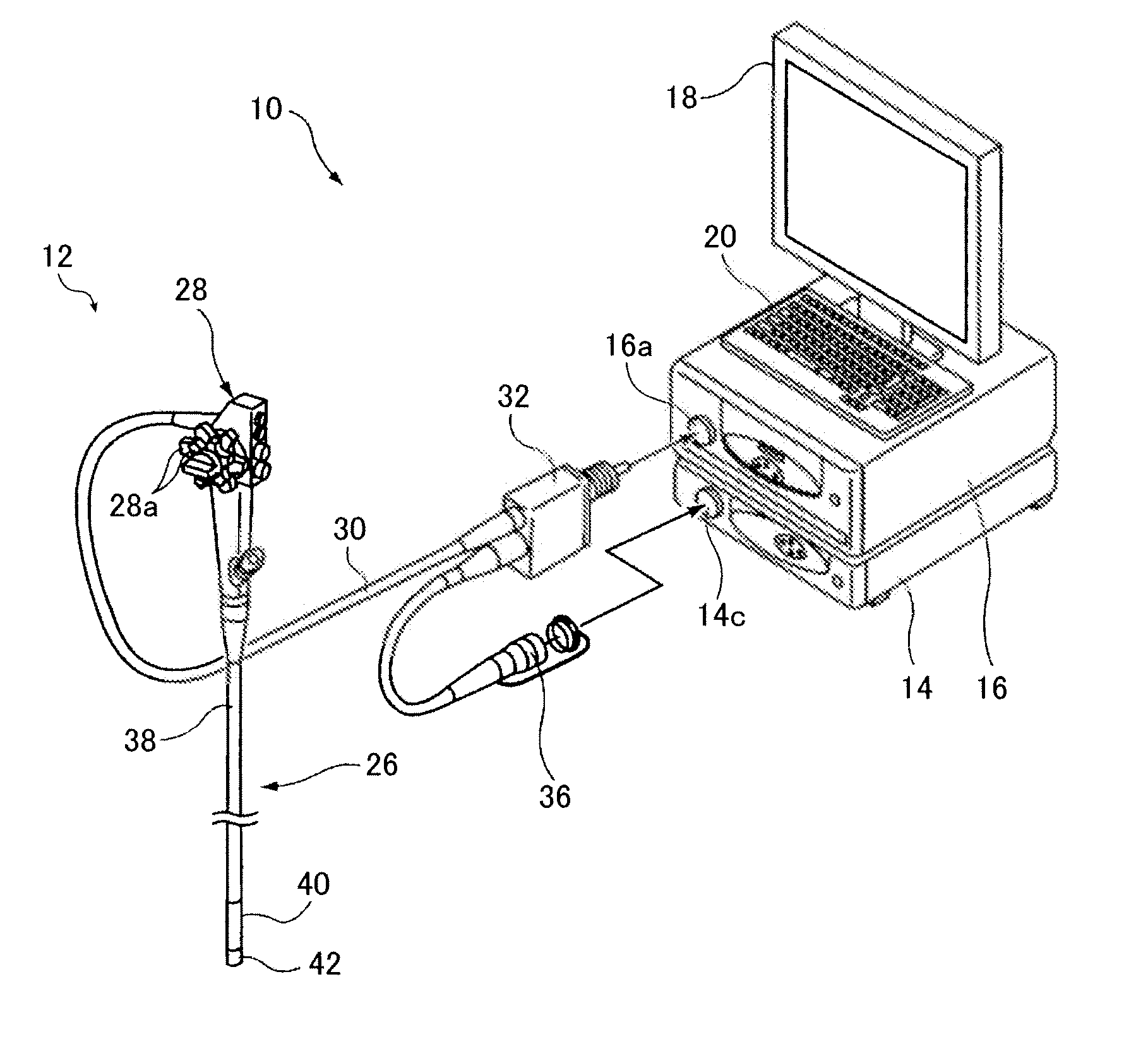
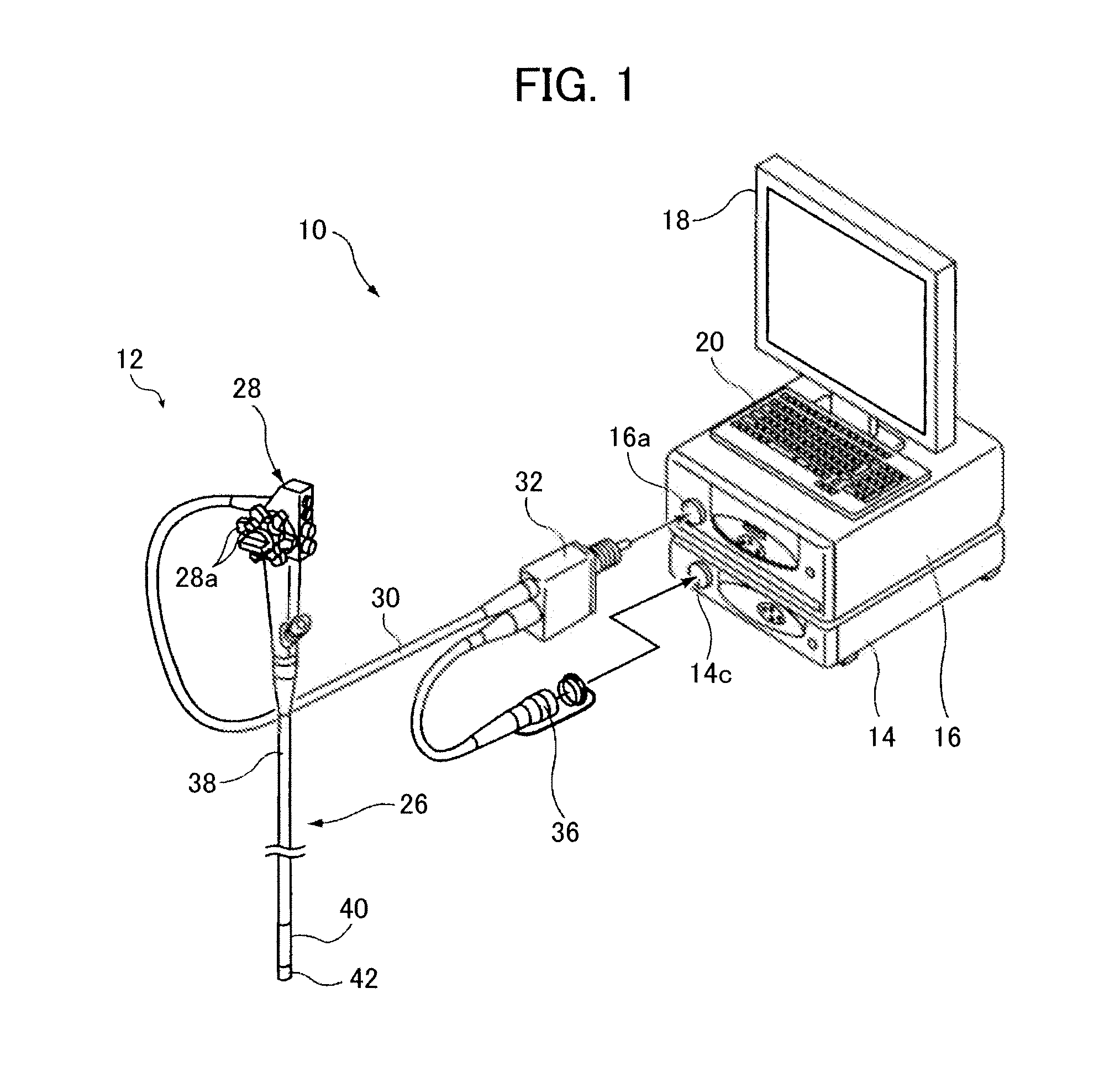
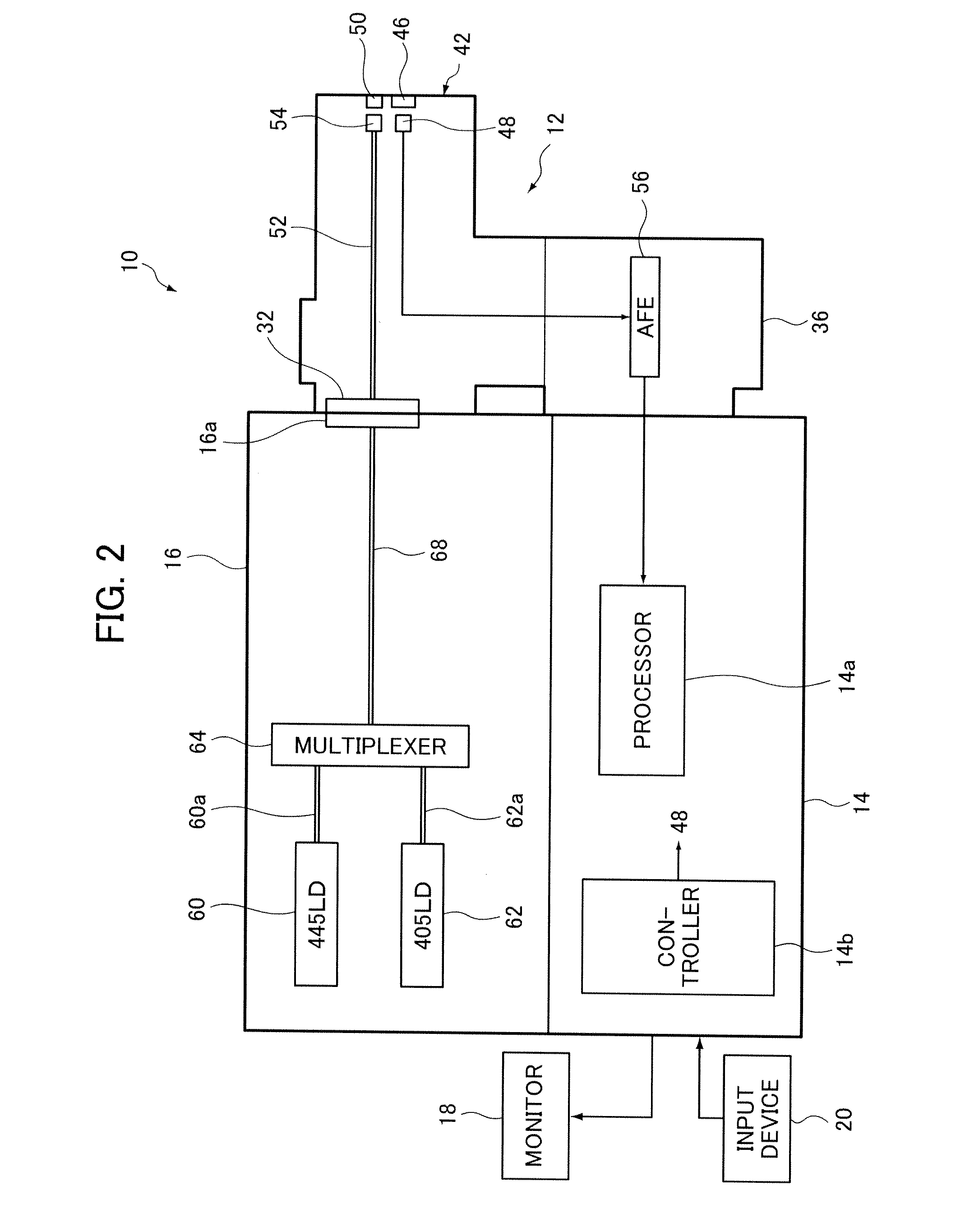
[0039]FIG. 1 is a conceptual perspective view showing an embodiment of the endoscope system of the invention and FIG. 2 conceptually shows the configuration of the endoscope system shown in FIG. 1.

[0040]The illustrated endoscope system 10 includes, for example, an endoscope 12, a processing device 14 for processing an image captured by the endoscope 12 and a light source device 16 for supplying observation light (illumination light) for use in observation and image capture using the endoscope 12.

[0041]The processing device 14 is connected to a monitor 18 for displaying an image captured by the endoscope and an input device 20 for inputting various instructions. The processing device 14 may further be connected to a printer (recording unit) for outputting an image captured by the endoscope as a hard copy.

[0042]...


PUM


Abstract

An endoscope system is provided in which blood vessels (pits) are extracted from an image captured by an endoscope to perform pattern matching with vascular patterns (pit patterns) associated with the cancer invasion depth and operations such as enhanced display of blood vessels, suggestion of preferred imaging conditions and display for diagnosis support are performed based on the matching result. The endoscope system enables even a less-experienced doctor to correctly diagnose the cancer invasion depth.

Description

BACKGROUND OF THE INVENTION[0001]The present invention relates to an endoscope system suitable to the diagnosis using, for example, microvessels in the superficial layer. The invention more specifically relates to an endoscope system with which even a less-experienced doctor can properly diagnose the cancer invasion depth from microvessels in the superficial layer.[0002]In recent years, use is made of an endoscope system capable of so-called special light observation in which a mucosal tissue of a living body is irradiated with specific narrow wavelength band light (narrowband light) as observation light to obtain tissue information at a desired depth of the tissue of the living body.[0003]The special light observation can simply visualize biological information which is not obtainable from the normal observation image, such as the microstructure in the superficial layer of a neovascular vessel formed in a mucosal layer or a submucosal layer, or an enhanced lesion. For example when ...

Claims


Application Information

Patent Timeline
IPC IPC(8): A61B1/04
CPCA61B1/00009A61B1/0005A61B1/0638A61B1/0653A61B1/063G06T2207/10068G06T2207/30096G06T2207/30101G06T7/0014A61B1/000094
Inventor KAKU, TOSHIHIKO
Owner FUJIFILM CORP
Learn More