
Methods and compositions for diagnosis of ectopic pregnancy

a technology of ectopic pregnancy and composition, applied in the field of methods and compositions for diagnosing ectopic pregnancy, can solve the problems of life-threatening condition, no good experimental model system, significant morbidity and mortality, and remains difficult to diagnos

Active Publication Date: 2012-08-23
THE TRUSTEES OF THE UNIV OF PENNSYLVANIA +1
View PDF3 Cites 5 Cited by
  • Summary
  • Abstract
  • Description
  • Claims
  • Application Information

AI Technical Summary

Benefits of technology

[0007]In one aspect, a diagnostic reagent or kit for use in diagnosing an ectopic pregnancy in a mammalian subject includes: (a) one or more ligands, wherein each ligand binds to a different gene expression product or protein selected from Table 2 and/or from FIG. 7, or fragments thereof; or (b) one or more polynucleotides or oligonucleotides, wherein each polynucleotide or oligonucleotide hybridizes to a different gene, gene fragment, gene transcript or expression prod...

Problems solved by technology

As the fetus grows, this condition becomes life-threatening due to potential tubal rupture and internal hemorrhage.
However, EP, for which there is no good experimental model system, remains difficult to diagnose at an early stage.
Approximately 50% of patients with this condition initially are misdiagnosed—resulting in significant morbidity and mortality.
Efforts to diagnose EP at an early point in the pregnancy using blood tests have been hampered because of the lack of useful and reliable serum biomarkers which reliably characterize EP.
Considerable difficulty in determining and identifying biomarkers for EP diagnosis has been attributed to a number of factors such as the high complexity of serum proteomes; a wide protein abundance range spanning more than 10 orders of magnitude; the presence of most clinically useful biomarkers at very low levels; a high patient-to-patient variability; and potential biases due to variations in sample collection and processing.
However, patient-to-patient heterogeneity requires that relatively large numbers of patient samples be analyzed.

Method used


Image

  • Methods and compositions for diagnosis of ectopic pregnancy
  • Methods and compositions for diagnosis of ectopic pregnancy
  • Methods and compositions for diagnosis of ectopic pregnancy

Examples


example 1

Systematic Discovery of Ectopic Pregnancy Serum Biomarkers Using 3-D Protein Profiling Coupled with Label-Free Quantitation

[0122]We used a 3-D method to systematically compare sera from patients with EP and IUP to identify candidate EP biomarkers. The 3-D method consisted of immunodepletion of 20 abundant serum proteins followed by GeLC-MS / MS analysis, with subsequent label-free quantitative comparisons using Rosetta Elucidator software (v3.1, Rosetta Biosoftware, Seattle, Wash.) to align and compare data at the MS ion intensity level. This software is no longer commercially developed as a result of the purchase of Rosetta Biosoftware by Microsoft Corporation.

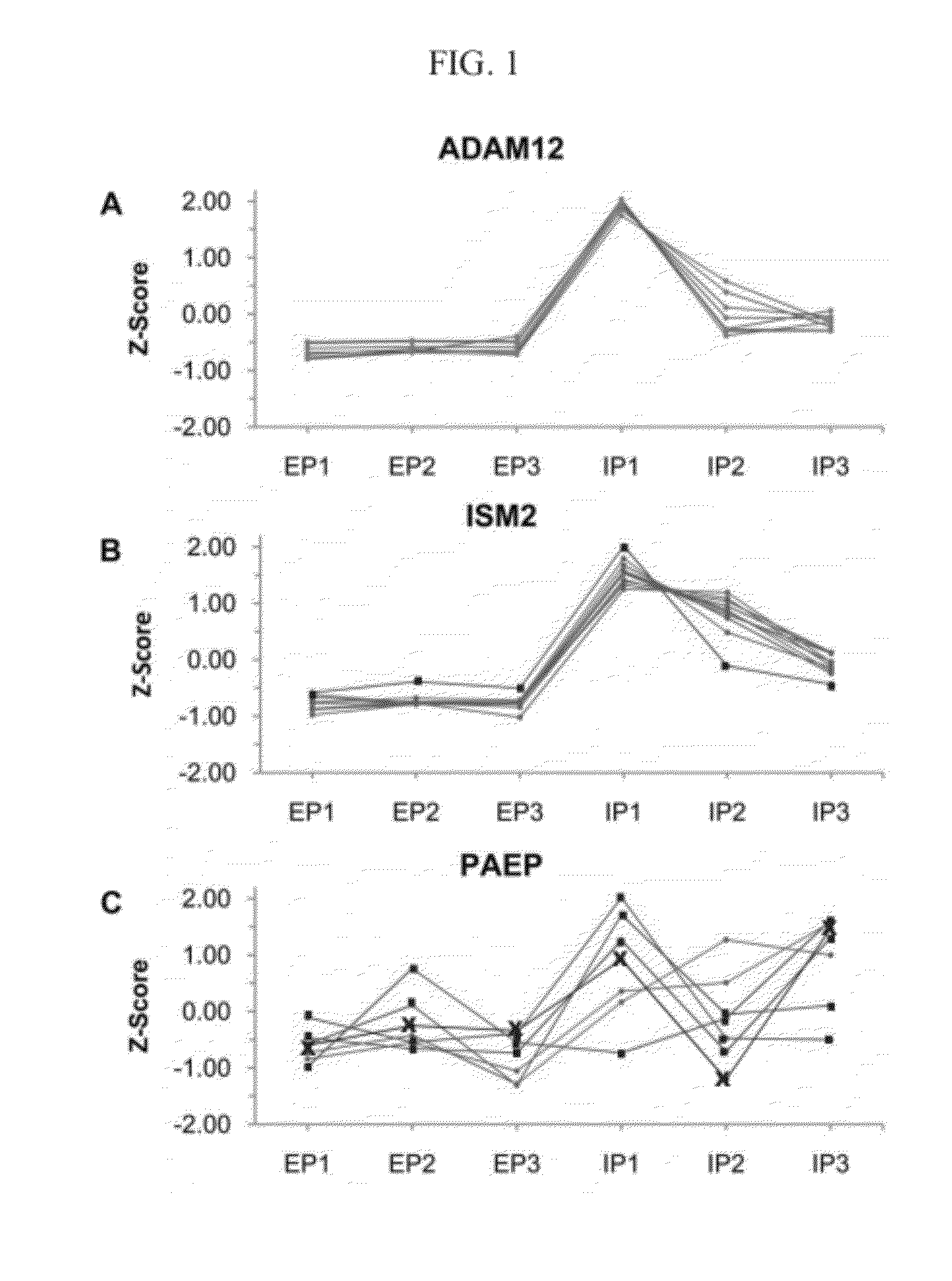
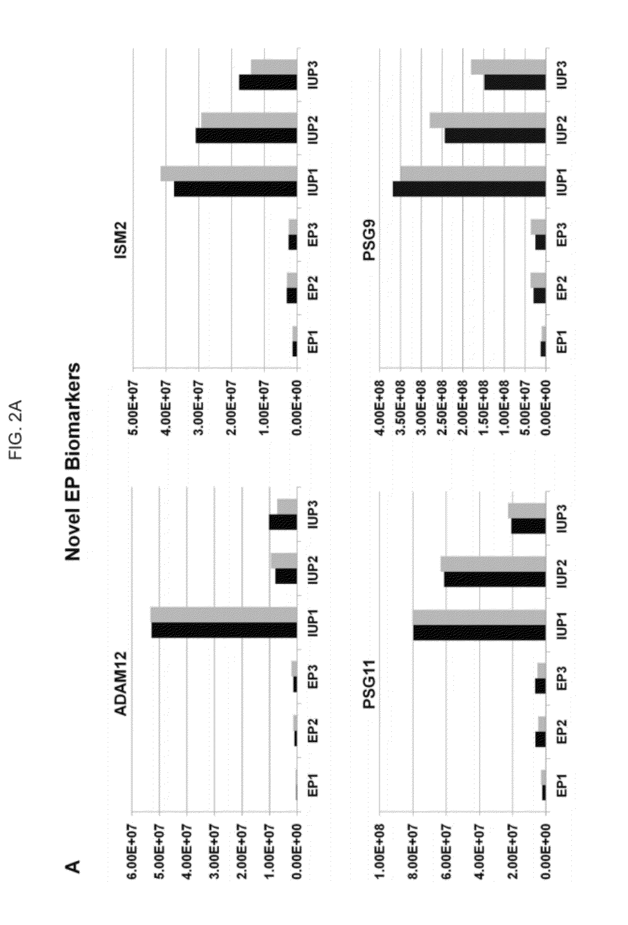
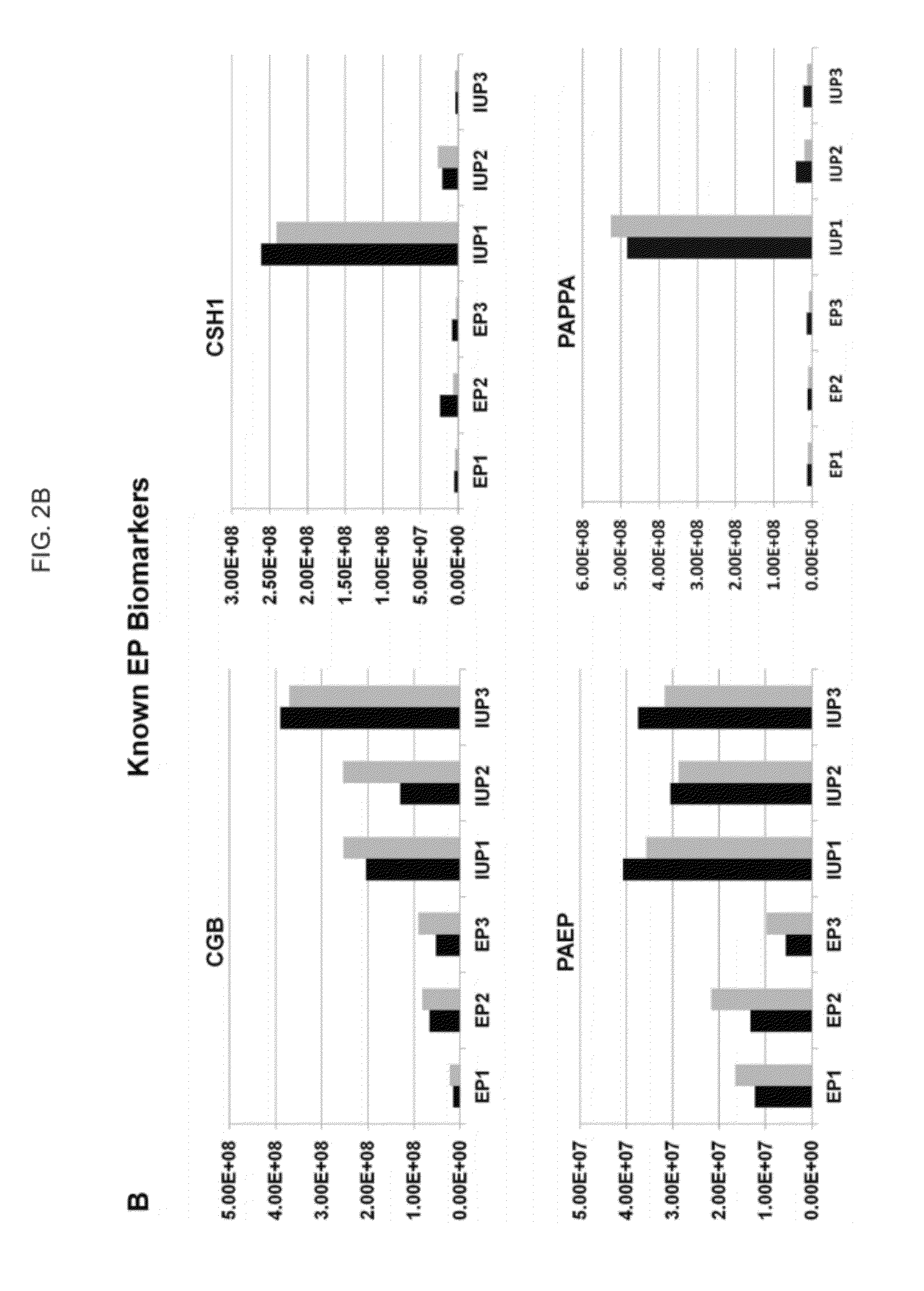
[0123]This analysis identified 70 candidate biomarkers with greater than 2.5-fold difference between the EP and IUP groups, and a high-priority biomarker subset was selected based upon the statistical probability that annotated peptides could properly classify samples into the EP or IUP group. Pilot validation of several biomar...

example 2

Strategy for Discovery of EP Serum Biomarkers Using Label-Free GeLC-MS / MS

[0147]A flow diagram summarizing the 3-D method for quantitative comparisons of serum from EP and IUP patients can be found at Beer et al, J. Proteome Res., 10(3):1126-38 (2011) at FIG. 1, incorporated by reference herein. Major protein depletion followed by GeLC-MS / MS is an efficient approach to identify a wide range of proteins in complex biological fluids such as serum.6,23,34 In this study, the SDS gel separation was performed until the tracking dye migrated 2.0 cm. While performing longer gel separations and using a greater number of gel slices would further increase depth of proteome coverage, the major trade-offs are that throughput proportionally decreases and the complexity of the data set can exceed the capacity of existing software to perform quantitative comparisons.

example 3

GeLC-MS / MS Comparison of EP and IUP Serum Pools

[0148]Depleted sera from nine EP and nine IUP patients were quantitatively compared by label-free LC-MS / MS analysis of pooled tryptic digests. Table 1 summarizes the scope of the experiment, which included a total of 252 LC-MS / MS runs for the discovery phase. Isotope groups (note 1) are the multiple features (discrete m / z signals) that comprise a peptide's isotopic envelope. The isoltope groups were filtered on: z>1, z<5, Peak time score=0.7; Peak m / z score=0.8 prior to DTA creation.

TABLE 1Summary of GeLC-MS / MS Comparison of EP and IUP SeraSamples6 pools × duplicatesFractions / Pool21Total LC-MS / MS Runs252High Quality Features1,095,293High Quality Isotope Groups1251,889Filtered Isotope Groups for DTAs2227,663

[0149]All runs for a given gel slice were performed in a group starting at the top of gel to minimize variations in HPLC and mass spectrometer performance, although the order of performing analyses was randomized within gel slice grou...


PUM

PropertyMeasurementUnit
Gene expression profileaaaaaaaaaa
Levelaaaaaaaaaa

Abstract

Methods and compositions are provided for diagnosing ectopic pregnancy in a mammalian subject by detecting changes in expression of ISM2, ADAM12, PST1, PSG7, PST11, PSG9, PSG2 and other genes identified therein, including combinations thereof. A selected gene, gene transcript or protein / peptide expression product, or profiles or signatures formed by combinations of same, detected in a biological fluid of a subject, enables comparison of the corresponding genes, proteins or profiles from that of a reference or control having a normal intrauterine pregnancy. Detection of characteristic changes in the gene profile or protein expression signature of the subject is correlated with a diagnosis of ectopic pregnancy. Various compositions for use in such diagnosis include PCR primer-probe sets or ligands, labeled or immobilized, which are capable of detecting the changes in expression or translation of these targets.

Description

CROSS REFERENCE TO RELATED APPLICATIONS[0001]This application claims the benefit of the priority of U.S. Provisional Patent Application No. 61 / 443,026, filed Feb. 15, 2011, which is incorporated herein by reference.STATEMENT REGARDING FEDERALLY SPONSORED RESEARCH OR DEVELOPMENT[0002]This invention was made with government support under Grant No. 5R01HD036455 and NCI Cancer Core Grant CA10815 awarded by the National Institutes of Health. The government has certain rights in this invention.BACKGROUND OF THE INVENTION[0003]Ectopic Pregnancy (EP) is a clinical condition that occurs when the embryo implants at a site other than in the uterus, typically the fallopian tube. As the fetus grows, this condition becomes life-threatening due to potential tubal rupture and internal hemorrhage. The incidence of EP is increasing due to a number of factors, and it is now the second-most-common cause of maternal death in the first trimester of pregnancy. Nearly a third of all cases do not exhibit an...

Claims


Application Information

Patent Timeline
IPC IPC(8): C40B30/04C40B40/06C40B40/10C07K2/00G01N33/573C07K16/18G01N33/566C12Q1/68C40B30/10C07K16/40C07H21/00C07K14/00
CPCG01N33/6893G01N33/76C07K14/705G01N2800/60G01N2333/70503G01N2800/368G01N2333/471G01N2560/00
Inventor SPEICHER, DAVID W.BARNHART, KURT T.BEER, LYNN A.
Owner THE TRUSTEES OF THE UNIV OF PENNSYLVANIA
Learn More