

Display of disulfide linked dimeric proteins in filamentous phage
a technology of dimeric proteins and filamentous phages, which is applied in the field of display libraries of disulfide linked dimeric proteins in filamentous phages, can solve the problems of not being able to demonstrate the correct assembly and display of complete antibody heavy chains in filamentous phages
- Summary
- Abstract
- Description
- Claims
- Application Information
AI Technical Summary
Benefits of technology
Problems solved by technology
Method used
Image

Examples
example 1
Display of an Fc-Fusion Protein on pIX
A. Phagemid Vector Construction
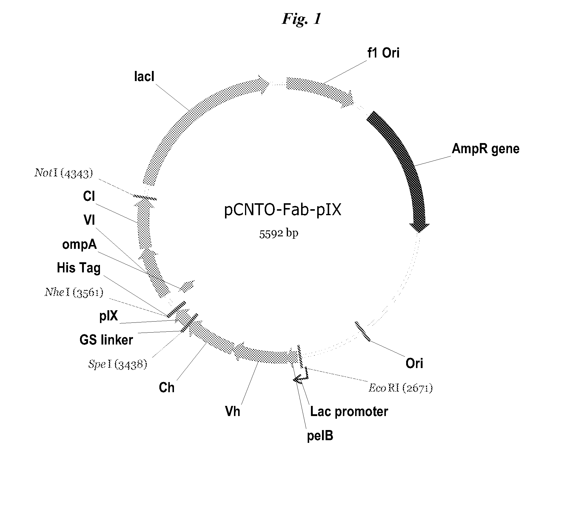
[0055]Phagemid vector, pCGMT9 (Gao et al., Proc. Natl. Acad. Sci. 96:6025-6030, 1999, U.S. Pat. No. 6,472,147) served as the backbone for the development of a phagemid pIX display vector capable of inserting heavy chain constant domains for phage display via pIX fusion. In this phagemid, origins of replication for E. coli (colE1) and filamentous phage (f1) are present, along with a beta-lactamase gene conferring resistance to ampicillin.
[0056]The pIX phagemid vectors for displaying Fc-containing proteins, including MIMETIBODY™ molecules, were constructed based on the Gao vector which had been adapted for bicistronic expression, pCNTO-Fab-pIX, as disclosed in WO2009 / 085462 and FIG. 1. Unlike the strategy used for Fab phage display in which a soluble light chain is expressed in the same cells and associates with the tethered polypeptide, no soluble Fc was expressed (FIG. 2A).
[0057]The Fab light chain sequence in the ...
example 2
[0068]To generate a Peptide-Fc fusion library, a template phagemid, which contains a hairpin loop at the site of random amino acid sequences, was generated. The hairpin was designed in such a way that a unique restriction site, XbaI, was placed where the hairpin formed double-stranded DNA. This would later be used to remove template DNA via restriction digest with XbaI, thereby reducing phage packed with the template phagemid in the final constructed library. Double-stranded template plasmids were transformed into a dut-lung-E. coli host strain, CJ236, as passage through this cell line causes incorporation of uracil into the ssDNA. The uracil containing ssDNA template is then degraded by enzymes of the final library host cell. A single colony harboring the plasmid was grown in a liquid culture that was subsequently infected with VCS-M13 helper phage. The phage was precipitated with PEG plus saline and used for purification of single strand DNA.
[0069]DNA libr...
example 3
Full IgG Display on Phage Particles
A. Vector Design.
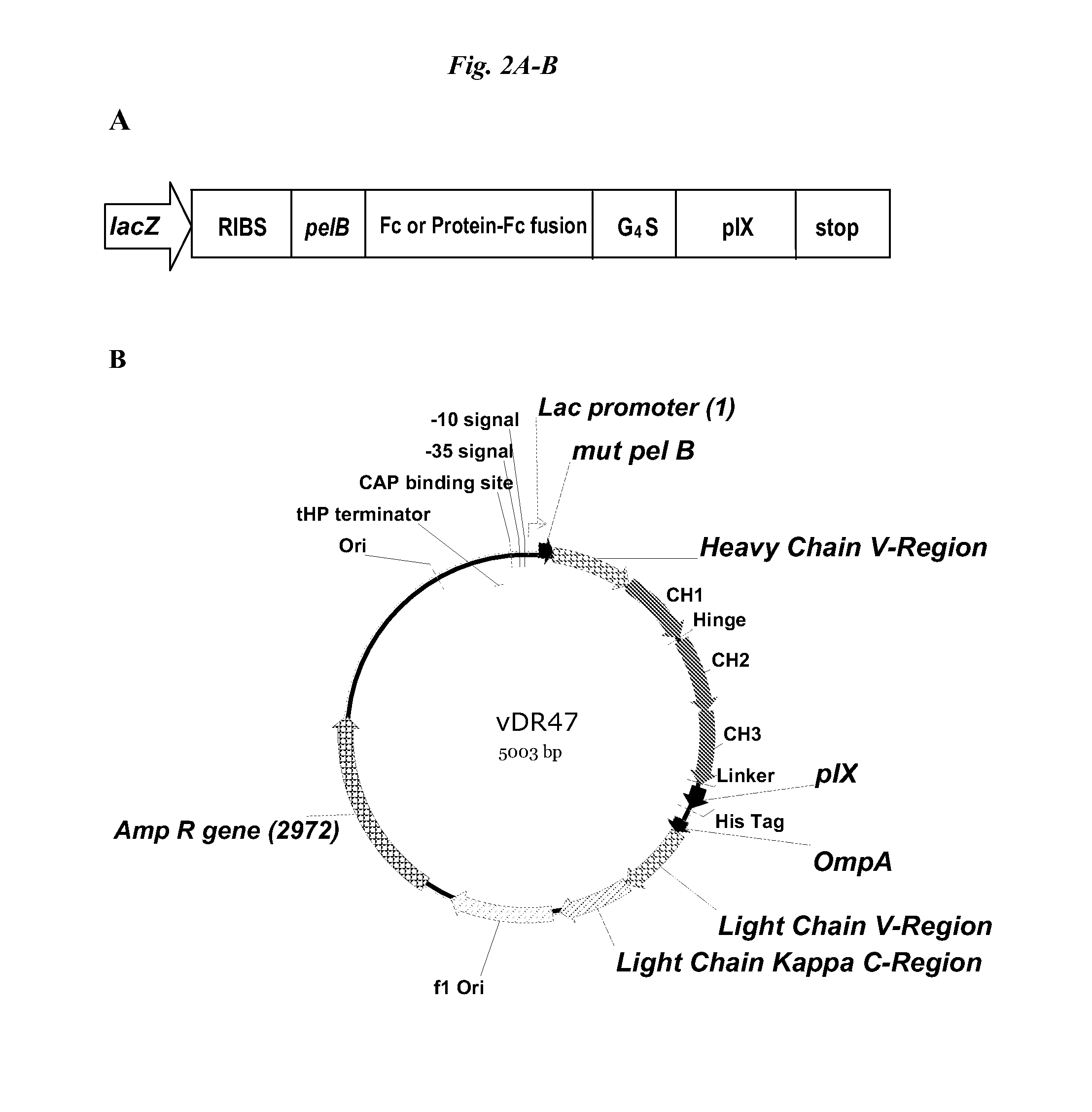
[0078]The full IgG display phagemid (vDR47, FIG. 2B) was construct using the pCNTO Fab IX construct shown in FIG. 1, and as described in WO2009 / 085462, which comprised a Vh and CH1 (SEQ ID NO: 19) domain of the heavy chain. Sequences encoding the hinge, CH2 and CH3 domains of a human IgG1 (SEQ ID NO: 20) were added as well as a variant pelB signal sequence, with a single mutation from the wild-type sequence, P6S (SEQ ID NO: 14), causing a significant improvement in peptide display on pVII minor coat protein and protein secretion (applicants co-pending application) and the vector does not have a lad gene but does have a lac promoter.
B. Characterization of Constructs Used for Full IgG Display.
[0079]A panel of test constructs was made to assess the display of full IgG on pIX. Antibodies to IL13, designated 6-2 and 16-7, and an anti-cytokine antibody 9-4 were chosen as prototypes for constructing the new full IgG molecules. To determin...
PUM
| Property | Measurement | Unit |
|---|---|---|
| Composition | aaaaa | aaaaa |
| Structure | aaaaa | aaaaa |
| Biological properties | aaaaa | aaaaa |
Abstract
Description
Claims
Application Information