Method of collecting specimen and method of diagnosing subject to detect upper digestive system disease

a technology of upper digestive system and specimen collection, which is applied in the direction of instruments, diagnostics, vaccination/ovulation diagnostics, etc., can solve the problems of not all doctors being able to easily perform the examination method, the method is invasive to patients, and the outcome is extremely poor. , to achieve the effect of improving the examination method and simplifying the examination method

Active Publication Date: 2012-10-11
OLYMPUS CORP
View PDF4 Cites 4 Cited by
  • Summary
  • Abstract
  • Description
  • Claims
  • Application Information

AI Technical Summary

Benefits of technology

[0013]The present invention has been achieved in light of the issues of the conventional method of examination described above

Problems solved by technology

The prognosis is extremely poor particularly for pancreatic cancer and biliary tract cancer among the upper digestive system diseases.
In addition, catheter insertion may cause pancreatitis as an accidental symptom.
Therefore, not all doctors can easily perform the method of examination using pancreatic juice and bile.
Therefore, the methods are invasive to patients.
In addition, the cost of examination is high due to the cost of the requi

Method used


Image

  • Method of collecting specimen and method of diagnosing subject to detect upper digestive system disease
  • Method of collecting specimen and method of diagnosing subject to detect upper digestive system disease
  • Method of collecting specimen and method of diagnosing subject to detect upper digestive system disease

Examples


example 1

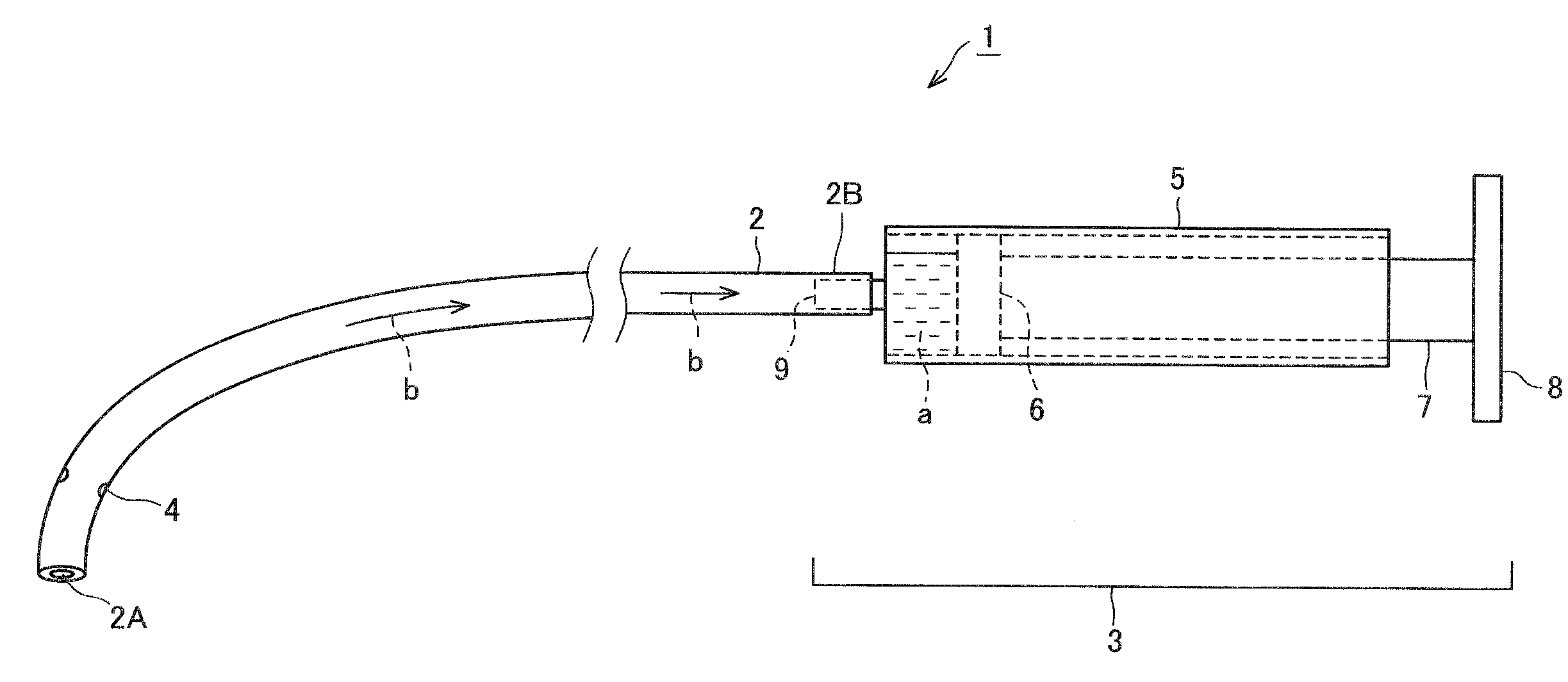
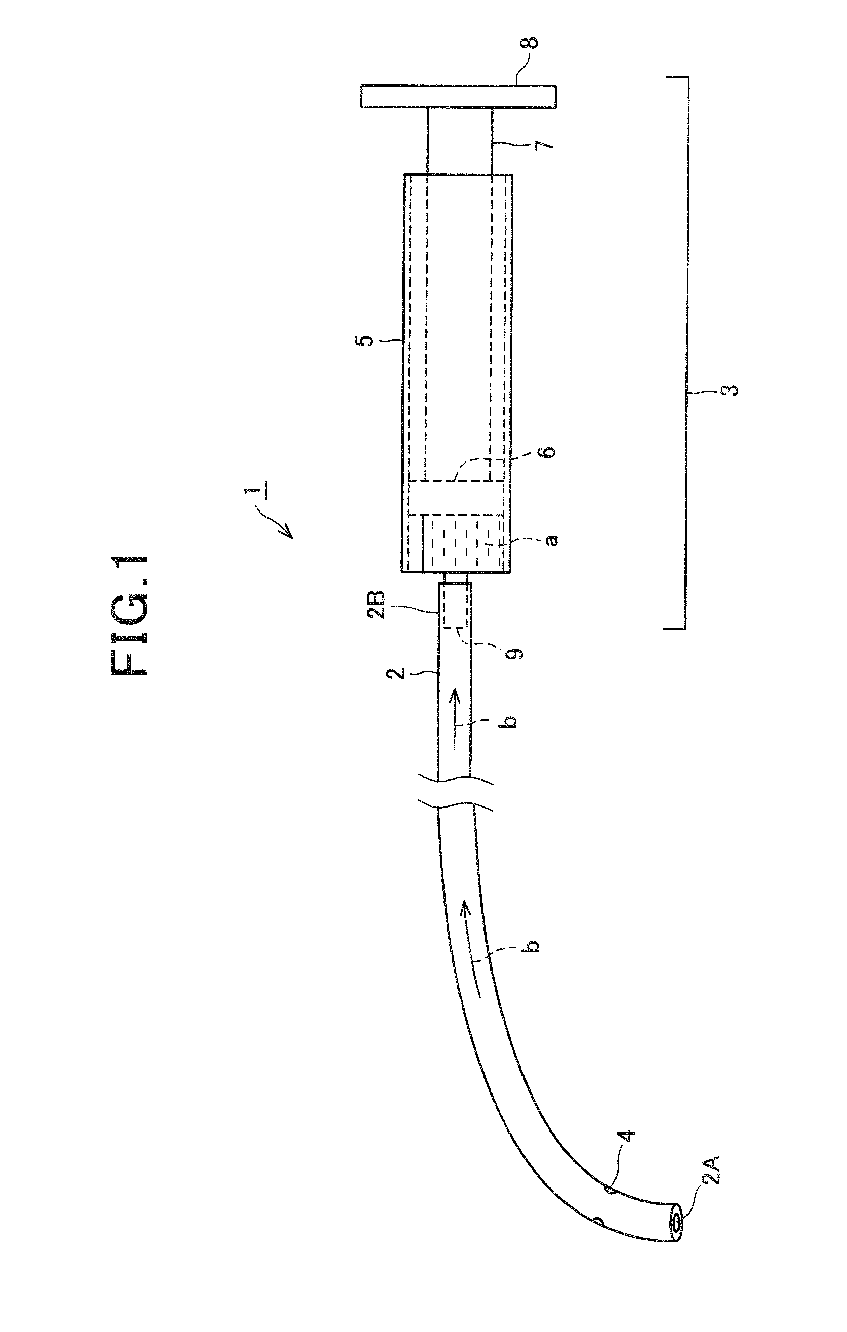
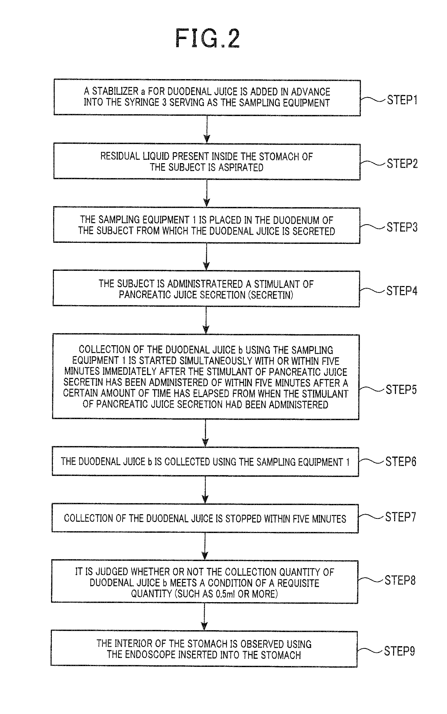
[0117]An upper gastrointestinal endoscope was inserted into 38 upper digestive system disease patients (32 pancreatic cancer patients (malignant) and 6 control group patients (benign) [2 IPMN patients, 1 pancreatic endocrine tumor (NET) patient, 1 MCN patient, 1 SCN patient, and 1 retention cyst patient]: diagnostic results based on postoperative pathological diagnostic results) after fasting overnight. Gastric juice c was aspirated from each patient. When the endoscope reached the duodenum 14, a catheter 2 for aspiration (PR-130Q manufactured by Olympus Medial Systems Corporation) was inserted into the forceps insertion opening of the endoscope and projected outward from the tip section of the endoscope. The duodenal juice b was present in a plurality of sites in the second position 14X and the third position 14Y of the duodenum 14. The duodenal juice b was collected from the tip side of the catheter 2. At this time, a 20 ml syringe 3 was connected to the base side of the catheter ...

example 2

[0128]In the present example, 32 cases of pancreatic disease patients (27 cases of pancreatic cancer, 5 cases in a control group [IPMN, NET, MCN, SCN, and retention cyst]) were subjected to examination. The effects of addition of a protease inhibitor were studied.

[0129]An upper gastrointestinal endoscope was inserted into the patients of the 32 cases of upper digestive system disease after fasting overnight. Gastric juice c was aspirated from the patients. When the endoscope reached the duodenum 14, a catheter 2 for collection (PR-130Q manufactured by Olympus Medial Systems Corporation) was inserted into the forceps insertion opening of the endoscope and projected outward from the tip section of the endoscope. The duodenal juice b was present in a plurality of sites in the second position 14X and the third position 14Y of the duodenum 14. The duodenal juice b was collected from the tip side of the catheter 2.

[0130]Here, the base side of the catheter 2 was bifurcated. The catheter 2 ...

example 3

[0136]In the present example, the IL-8 concentration in the duodenal juice b was measured using an ELISA kit (manufactured by R&D Systems Inc.). Here, the duodenal juice b was collected using the same method as that described in Example 2. The collected duodenal juice b was used as the specimen.

[0137]The results are shown in FIG. 9A and FIG. 9B as a box-plot. In FIGS. 9A and 9B, malignant indicates pancreatic cancer. Benign indicates the control group. The sensitivity (%) of IL-8 to pancreatic cancer is shown in Table 3.

TABLE 3Elapsed TimeFrom The StartOf CollectionOf DuodenalCut-OffJuiceValueSensitivitySpecificity(minutes)Inhibitor(pg / ml)(%)(n)(%)(n)2.5~5.0(−)1.53711 / 30804 / 57.5~10.0228 9 / 32835 / 62.5~5.0(+)25916 / 271005 / 57.5~10.016216 / 26674 / 6

[0138]As shown in FIG. 9A and FIG. 9B, it is has been found that the IL-8 concentration in the duodenal juice b significantly increases in the duodenal juice b after administration of secretin when the protease inhibitor is added, compared to when...


PUM


Abstract

A method of collecting a specimen of the present invention detects upper digestive system disease. The method of collecting a specimen includes: a step of administering a stimulant of pancreatic juice secretion or a stimulant of bile secretion to a subject; a step of positioning sampling equipment in the duodenum of the subject into which duodenal juice is secreted, the sampling equipment being used to collect and store the duodenal juice; a step of starting collection of the duodenal juice by the sampling equipment within five minutes from the administration of the stimulant of pancreatic juice secretion or the stimulant of bile secretion; and a step of stopping collection of the duodenal juice such that collection time for the duodenal juice is within five minutes from the start of collection.

Description

CROSS REFERENCE TO RELATED APPLICATION[0001]This application is based upon and claims the benefit of priority from the prior provisional U.S. patent application Ser. No. 61 / 471,868, filed Apr. 5, 2011, the entire content of which is incorporated herein by reference.BACKGROUND OF THE INVENTION[0002]1. Field of the Invention[0003]The present invention relates to a method of collecting a specimen and a method of diagnosing a subject to detect upper digestive system disease. In particular, the present invention relates to a method of collecting a specimen and a method of diagnosing a subject suitable for detection of early cancers, such as pancreatic cancer, biliary tract cancer, and liver cancer.[0004]2. Description of the Related Art[0005]Rapid advancements are being made in methods of examination for upper digestive system diseases (for example, pancreatic cancer, biliary tract cancer, gallstones, stomach cancer, cholecystitis, pancreatitis, liver cancer, and cirrhosis). The prognosi...

Claims


Application Information

Patent Timeline
IPC IPC(8): A61M1/00C12Q1/04G01N33/566
CPCG01N33/57438G01N33/57446G01N2333/5421A61B2010/0061G01N2800/08G01N2800/14A61B10/0045G01N2800/062
Inventor SANUKI, HIROMIMORIYA, NAOKATAOKA, RIETAKEYAMA, TETSUHIDEYAMAMOTO, KYOKO
Owner OLYMPUS CORP
Why Eureka
Try Eureka