Fasteners, Deployment Systems, and Methods for Ophthalmic Tissue Closure and Fixation of Ophthalmic Prostheses and Other Uses

a technology of ophthalmic prostheses and fasteners, applied in the field of medical devices, systems and methods, can solve the problems of negative cash flow for a particularly complex suture case, time-consuming suturing process, and disadvantages for both surgeons and patients, and achieve the effects of reducing the cost of suturing, and improving the safety of patients

Inactive Publication Date: 2013-01-03
O3 OPTIX LLC
View PDF8 Cites 21 Cited by
  • Summary
  • Abstract
  • Description
  • Claims
  • Application Information

AI Technical Summary

Benefits of technology

[0006]The invention generally provides improved medical devices, systems, and methods. Many embodiments of the invention employ tissue fasteners that can be inserted into (and optionally, though not necessarily, through) tissue structures underlying a tissue surface, often without having to access opposed surfaces behind the tissue structures. Exemplary embodiments of the fasteners are particularly well suited for apposition and closure of tissue edges bordering incisions and other wounds of ophthalmic tissues, to affix prosthetic structures to an ophthalmic tissue structure, and the like. First and second legs of the fastener may be configured to

Problems solved by technology

Depending on the procedure, the suturing process can be very time consuming relative to the total length of a procedure.
Additionally, the overhead expense of the surgical facility (which can be incurred not only during the underlying therapeutic procedure but also throughout the time dedicated to suturing of the access site and the like) will often result in a negative cash flow for a particularly complex suture case.
However, adhesives have been associated with disadvantages for both the surgeon and patient.
For th

Method used


Image

  • Fasteners, Deployment Systems, and Methods for Ophthalmic Tissue Closure and Fixation of Ophthalmic Prostheses and Other Uses
  • Fasteners, Deployment Systems, and Methods for Ophthalmic Tissue Closure and Fixation of Ophthalmic Prostheses and Other Uses
  • Fasteners, Deployment Systems, and Methods for Ophthalmic Tissue Closure and Fixation of Ophthalmic Prostheses and Other Uses

Examples


exemplary embodiment 10

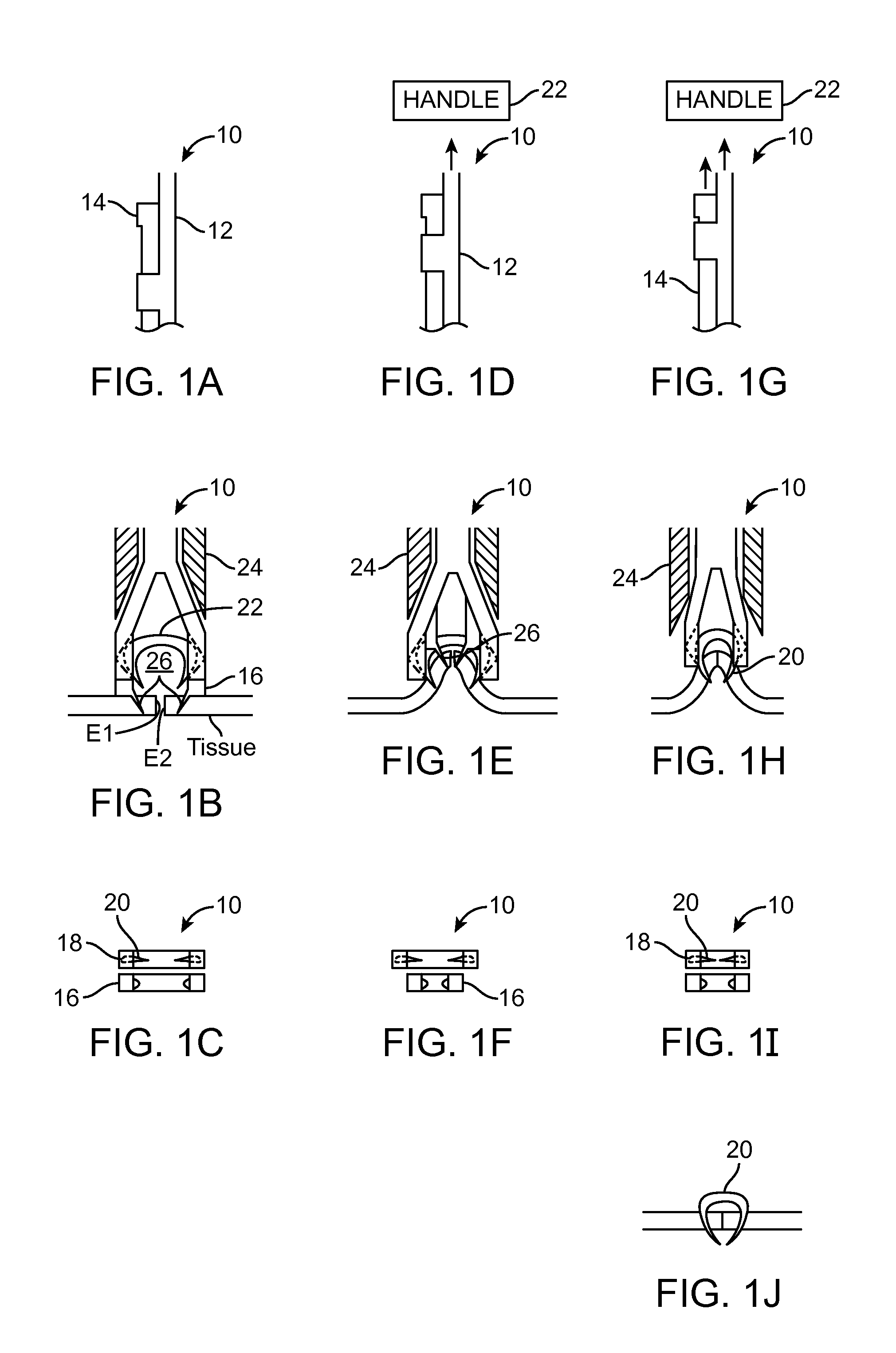
[0067]FIGS. 1A-1J illustrates an exemplary embodiment 10 of an apparatus (mechanism) for simultaneously grasping and clipping together the edges E1, E2 of tissue that have been wounded or incised. The apparatus 10 may include two sets of stacked shafts 12, 14, each with a distal jaw 16, 18. One shaft 12 and jaw 16 are designed to grasp and pull together the edges of the tissue E1, E2. The second shaft 14 and jaw 18 are designed to carry a normally open malleable clip 20 that may be compressed by the jaws 18 to form a closed clip 20 to secure the two edges E1, E2 of tissue together. The stacked shafts 12, 14 may be connected to a handle that provides fore and aft axial movement of each of the jaws against an anvil 24 that surrounds the shafts 12, 14.

[0068]In operation of the exemplary embodiment in FIGS. 1A-1C, as the grasping shaft 12 is pulled by the handle, the jaws 16 of the grasping shaft interfere with the anvil 24 and are compressed. Hooks or protrusions 26 at the distal edge ...

exemplary embodiment 30

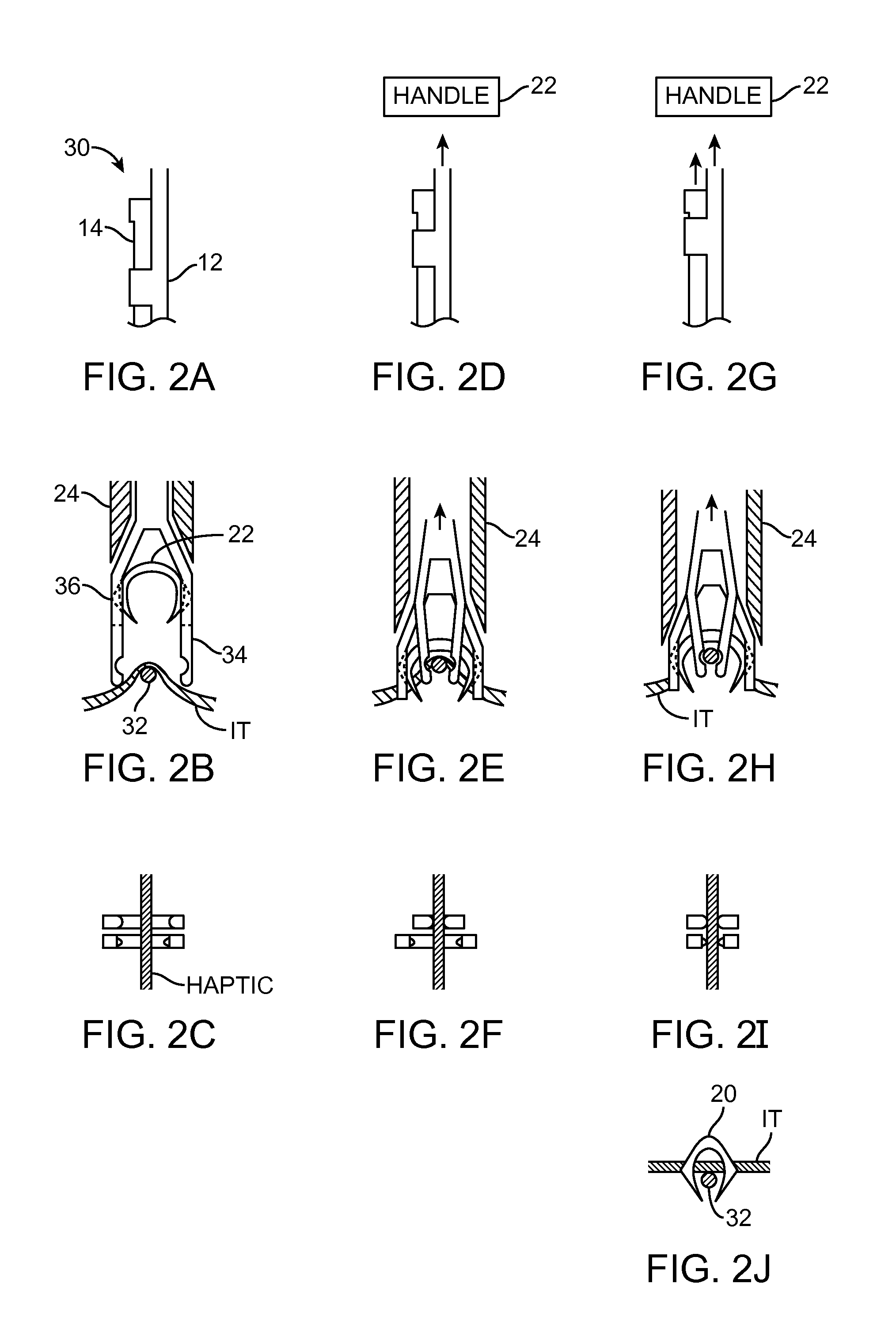
[0069]FIGS. 2A-2J illustrates an exemplary embodiment 30 of an apparatus for simultaneously grasping and clipping together prosthesis 32 to ophthalmic tissue. The embodiment illustrates, by example, the fixation of an intraocular lens haptic 32 (prosthesis) to iris tissue IT. The apparatus in FIGS. 2A-2J may include two sets of stacked shafts 34, 36, each with a distal jaw. One shaft and jaw is designed to grasp and draw the tissue and prosthetic toward the distal instrument. The second shaft and jaw is designed to carry a normally open malleable clip 22 that can be compressed by the jaws to form a closed clip to secure the intraocular lens haptic to the iris. The stacked shafts 34, 36 may be connected to a handle 22 that provides fore and aft axial movement of the jaws against an anvil that surrounds the shafts.

[0070]In operation of the exemplary embodiment in FIGS. 2A-2C, as the grasping shaft is pulled by the handle in operation, the jaws 34 of the grasping shaft 12 interfere wit...

exemplary embodiment 40

[0071]FIGS. 3A and 3B illustrate an exemplary embodiment 40 of the apparatus in which the forceps to apply the ophthalmic clip 20 may be positioned at an angle, on a plane, approximately tangent to the surface of the eye E and the clip may be positioned approximately perpendicular to the tissue to be closed or fixated. The apparatus 40 includes forceps 42 may include jaws for securing a normally open malleable clip. In exemplary operation, when the handles 44 of the forceps are squeezed together, the hinged forceps jaws 46 are drawn together, which close the malleable clip 20. A leaf spring 48 or other spring may be coupled to the handles of the forceps to keep the jaws in a normally open position until the surgeon desires to deploy the clip. Once the clip is deployed, the surgeon may release pressure on the handles 44 such that the spring returns the forceps to the open position leaving the clip in place on the tissue and allowing for removal of the forceps.

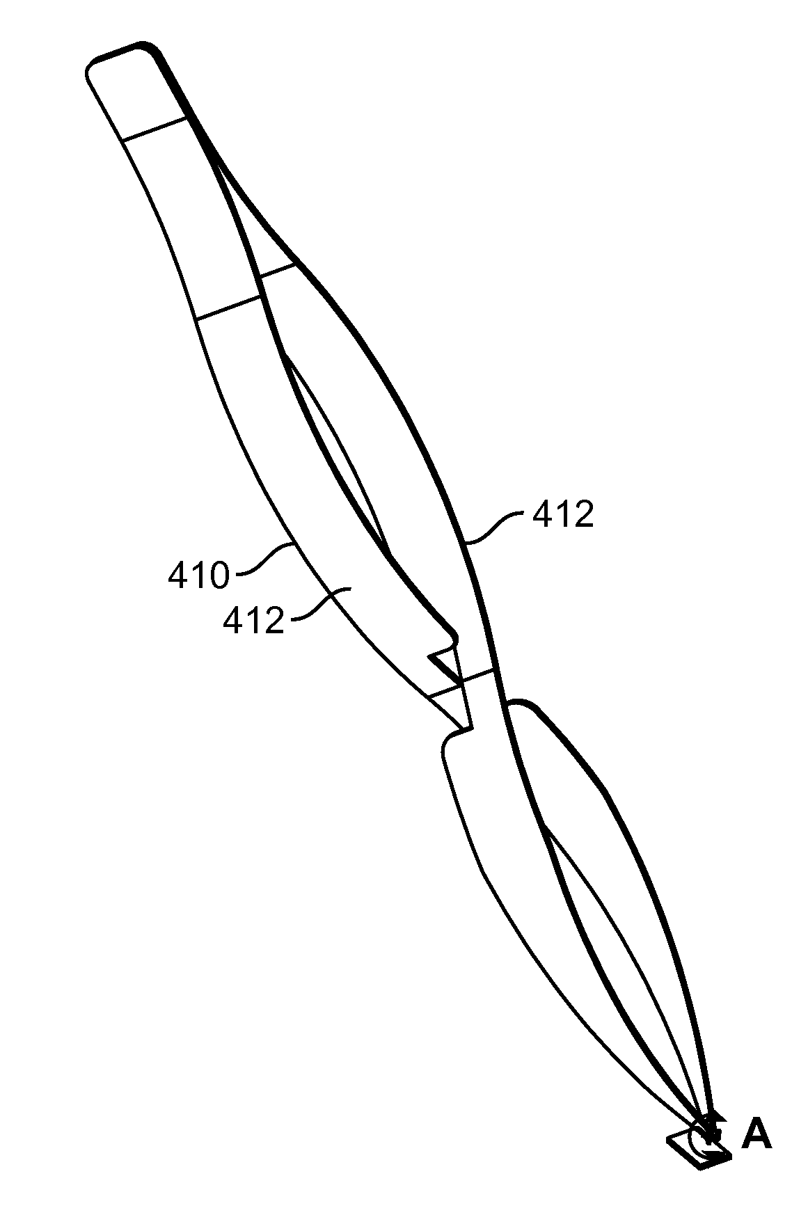
[0072]FIGS. 4A-4C illust...


PUM

PropertyMeasurementUnit
Coloraaaaaaaaaa
Shape memory effectaaaaaaaaaa
Adhesivityaaaaaaaaaa

Abstract

Methods and devices for ophthalmic tissue closure and fixation of ophthalmic prostheses are provided. In accordance with some embodiments, devices for both grasping and clipping a plurality of ocular tissue and ocular prostheses are provided. Various device embodiments are provided for both malleable clips and delivery of normally closed clips (i.e. shape memory). The device may accommodate a plurality of clips which include, but are not limited to: malleable metals, absorbable, shape memory, drug-eluting, and adhesive dispensing. The clips may be pigmented to match the colors of associated tissue (cornea, iris, conjunctiva, sclera, retina) to serve to camouflage fixation clips for healing duration or permanently. According to one aspect, shallow angle access to anatomy may be provided by specialized angulation of device shaft and closure jaws that are intended to access the eye through a small self-healing cornea incision and/or any ocular tissue.

Description

CROSS-REFERENCES TO RELATED APPLICATIONS[0001]The present application claims the benefit under 35 USC 119(e) of U.S. Provisional Application No. 61 / 468,827 filed Mar. 29, 2011; the full disclosure of which is incorporated herein by reference in its entirety for all purposes.FIELD OF THE INVENTION[0002]The present invention relates generally to medical devices, systems and methods, with many of the embodiments described herein providing fasteners such as clips, staples, or the like, optionally for ophthalmic surgery and, more particularly, to repair of wounds, closure of incisions, and fixation of prosthetic structures in ophthalmic surgery.BACKGROUND OF THE INVENTION[0003]In the field of ophthalmology, there exist distinct clinical subspecialties (e.g., cataract, retina, cornea, etc.) organized around disease classifications of the eye. Within each subspecialty, there exist distinct surgical therapies that involve specialized wound closures and prosthesis fixation requiring substant...

Claims


Application Information

Patent Timeline
IPC IPC(8): A61B17/10A61B17/03
CPCA61F9/007A61F2220/0016A61B17/064A61B17/0682A61B2017/2918A61B2017/00004A61B2017/00867A61B2017/0649A61B17/0684A61F2210/0004A61B17/068
Inventor VOLD, STEVEN D.PEARTREE, KENNETH A.BUCKLEY, TIMOTHY D.FEUSTEL, AARON
Owner O3 OPTIX LLC
Why Eureka
Try Eureka