
Methods of detecting nucleic acid sequences with high specificity

a nucleic acid sequence and high specificity technology, applied in the field of nucleic acid chemistry and biochemical assays, can solve the problems of false positive signals, insufficient binding strength of each capture probe to capture sgp stably, false positive signals, etc., and achieve significant affecting assay performance, high background noise, and discrimination. good

Inactive Publication Date: 2013-01-24
ADVANCED CELL DIAGNOSTICS INC
View PDF3 Cites 73 Cited by
  • Summary
  • Abstract
  • Description
  • Claims
  • Application Information

AI Technical Summary

Benefits of technology

This patent describes methods for detecting nucleic acid targets in single cells, particularly rare cells from large populations. The methods involve using multiple capture probes and label probes to capture the targets and detect them using various techniques such as fluorescence detection. The patent also describes methods for improving the accuracy and sensitivity of these detection assays. The technical effects of this patent include improved accuracy and sensitivity for detecting nucleic acid targets in single cells, as well as methods for reducing false positive signals and improving the signal-to-background ratio.

Problems solved by technology

This non-specific sequence could share the same sequence as the target or it could carry small number of mis-matches that are insufficient to be prevented from binding to CP nonspecifically.
This will result in a false positive signal because the label is mistakenly captured to non-targets.
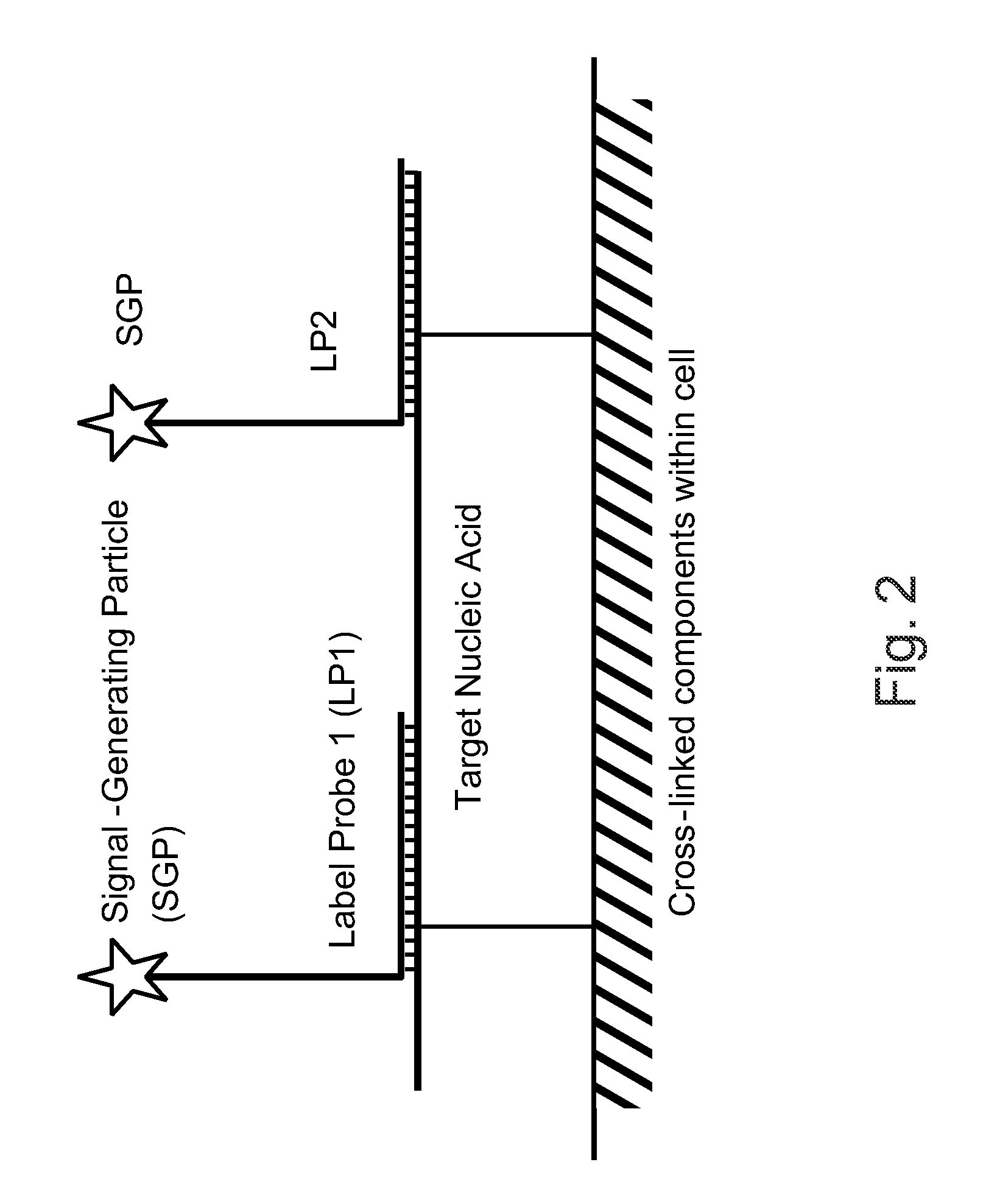
So each capture probe does not have sufficient binding strength to capture the SGP stably.
Therefore, if one of the capture probes hybridizes non-specifically to a non-target sequence, it does not have sufficient binding strength to capture the SGP to the target through out the assay, thus preventing the generation of false positive signals and reducing the background signal.
It may get “stuck” or trapped non-specifically in a void in solid surface in a solution-based assay or within cellular matrix in an in situ detection assay, which will also result in false positive signals and reduce signal-to-background ratio.
If the SGP structure is large enough to contain many label molecules, the false positive or background signals can be significant, making it hard to be distinguished from the real signal.
In addition, in in situ detection applications, the large structure may have difficulty to gain access to the target molecule inside cellular matrix, which may result in reduction in signal level.
For example, the rearrangement of DNA through a translocation can lead to the fusion of two genes, potentially disrupting importing protein coding regions.
In addition to gene fusion events leading to chimeric transcripts, mutations affecting RNA splicing can also create mis-joined RNA sequences that lead to disease.
For nucleic acid detection assays, low specificity not only may produce false positive results, but also increases background noise, leading to reduced detection sensitivity.
However, when the probe becomes long, its binding stability becomes not very sensitive to mis-matches in a small number of bases, which leads directly to increased possibility on non-specific binding.
The problem is particularly severe in assays designed to detect single nucleotide polymorphisms (SNPs), where the target sequence is different from other genotypes by only a single base.
There are two issues in the current genotyping technologies related to probes used in genotyping.
First, because the probes could bind non-specific locations of the genome, all current genotyping technologies with only a few exceptions require the PCR amplification step.
However, PCR amplification presents a major obstacle in genotyping throughput.
Secondly, the genotyping probe that hybridizes to a SNP region may not offer sufficient difference in thermal stability between perfect match and mismatch sequences, thus they may not be able to achieve reliable allelic discrimination.
The second is for in situ detection of nucleic acids, where low specificity will produce a high level of background noise, severely restricting detecting sensitivity.

Method used


Image

  • Methods of detecting nucleic acid sequences with high specificity
  • Methods of detecting nucleic acid sequences with high specificity
  • Methods of detecting nucleic acid sequences with high specificity

Examples


example 1

Detection of Nucleic Acids in Individual Cells

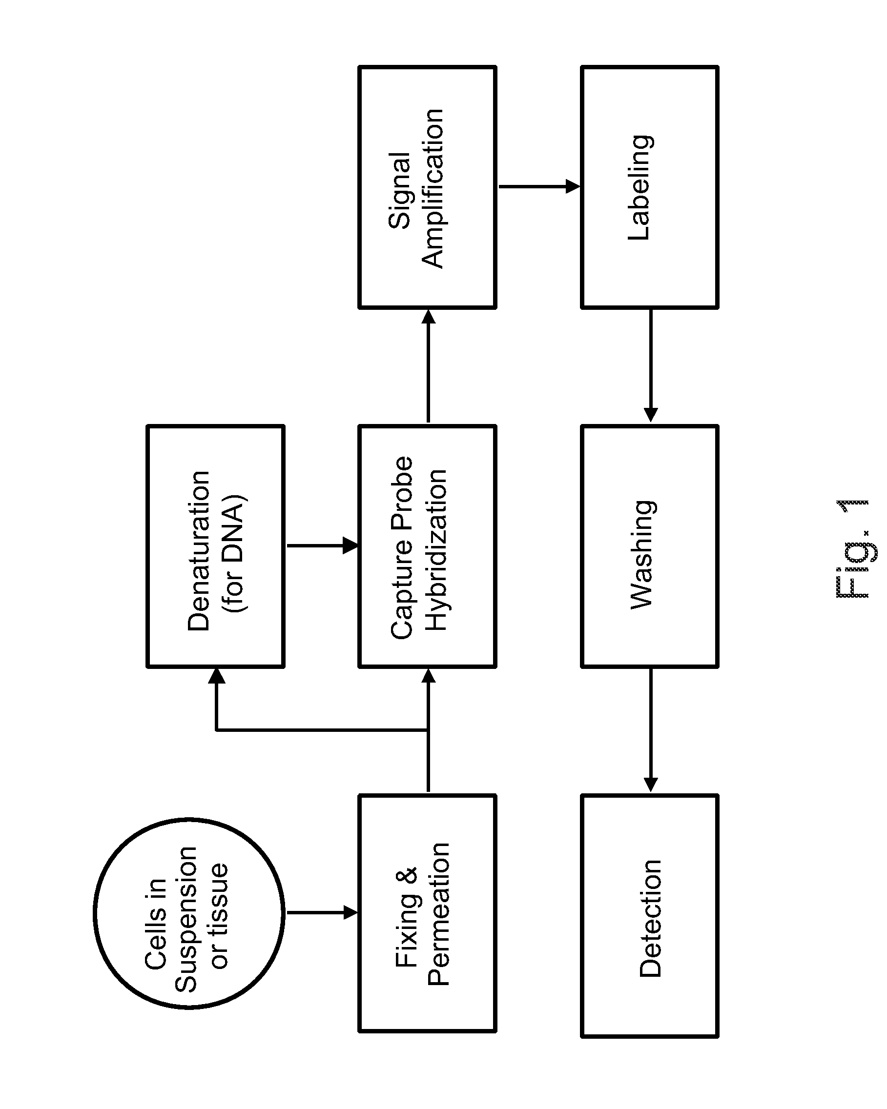
[1033]The following sets forth a series of experiments that demonstrate in-cell detection of nucleic acid. The results demonstrate, for example, that when staining cells on a glass substrate with QMAGEX, we can obtain a highly specific signal with a sensitivity of detecting a single mRNA molecule. Moreover, we can achieve staining of multiple mRNAs at the same time using a combination of different target probes and amplifiers. These results further demonstrate the feasibility of detecting cancer cells exhibiting transcriptional upregulation within a population of cells with normal gene expression. The results also demonstrate staining of cells in suspension and identification of them using flow cytometry, eliminating need for a solid support for the cells and allowing for rapid detection of stained cells. These results further demonstrate the ability to detect cells exhibiting transcriptional upregulation from those with low basal levels...

example 2

In Situ Detection of BCR-ABL Gene Fusion

[1079]To demonstrate the feasibility of detecting RNA fusion transcripts using our assay, we simultaneously hybridized cultured K562 and Jurkat cells with probe sets to BCR and ABL. K562 cells are known to carry the BCR-ABL gene fusion, while Jurkat cell do not. The BCR and ABL probe sets were simultaneously detected with a signal amplification system labeled with a green fluorescent dye and a signal amplification system with a red fluorescent dye, respectively. As shown in FIG. 56, Jurkat cells stained in this manner showed individual green or red dots, indicating the presence of wild type BCR and ABL transcripts. However, as expected K562 cells showed a large number of yellow dots due to the juxtaposition of the BCR and ABL probe sets on the same transcript, indicating that a fusion gene was present. To our knowledge this is the first demonstration of in situ visualization of a fusion transcript.

example 3

Capture Probe (Label Extender) Design

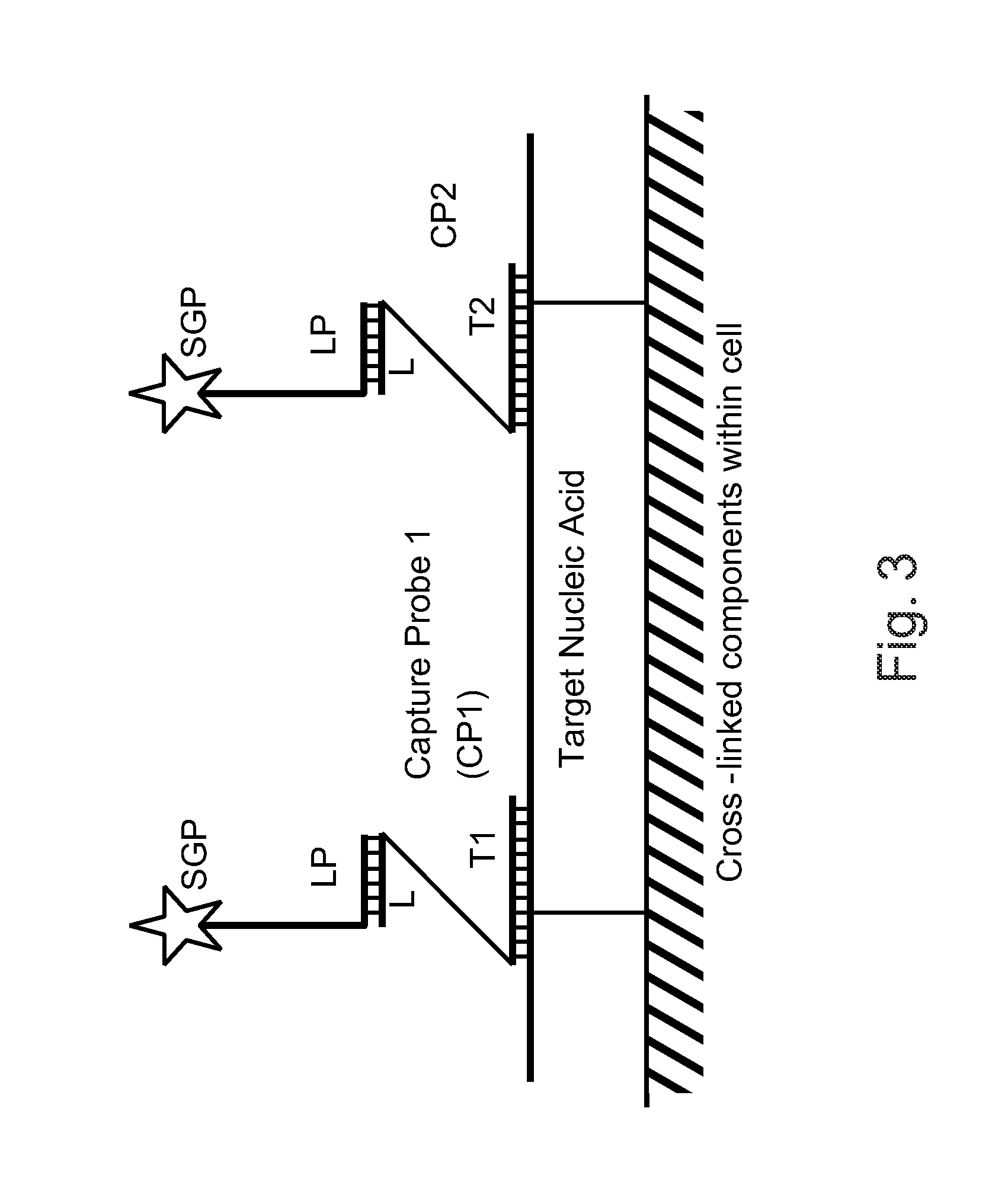
[1080]The following sets forth a series of experiments that illustrate label extender design and that demonstrate that a configuration in which the 5′ ends of the label extenders hybridize to a nucleic acid of interest while the 3′ ends of the label extenders hybridize to a preamplifier results in stronger binding of the preamplifier to the nucleic acid than does a cruciform arrangement of the label extenders.

[1081]Two subsets of label extenders were designed to bind to a human GAPD nucleic acid target and to a preamplifier, as schematically illustrated in FIG. 60. Two label extenders bind each copy of the preamplifier. As shown in Panel A, in one subset of label extenders, the two label extenders in each pair bind the preamplifier through the same end (the 5′ end, in this example) and bind the target nucleic acid through the other end (double Z configuration). As shown in Panel B, in the other subset of label extenders, the two label extenders i...


PUM

PropertyMeasurementUnit
Tmaaaaaaaaaa
Tmaaaaaaaaaa
Tmaaaaaaaaaa

Abstract

The invention relates to methods of detecting nucleic acids, including methods of detecting one or more target nucleic acid sequences in multiplex branched-chain DNA assays, are provided. Nucleic acids captured on a solid support or suspending cells are detected, for example, through cooperative hybridization events that result in specific association of a label with the nucleic acids. The invention further relates to methods to improve probe hybridization specificity and their application in genotyping. The invention also relates to in situ detection of mis-joined nucleic acid sequences. The invention relates to reducing false positive signals and improve signal-to-background ratio in hybridization-based nucleic acid detection assay. The invention further relates to method to improve specificity in hybridization based nucleic acid using co-location probes. Compositions, tissue slides, sample of suspended cells, kits, and systems related to the methods are also described.

Description

CROSS-REFERENCE TO RELATED APPLICATIONS[0001]This application also claims priority to and benefit of U.S. Provisional Application No. 61 / 277,563, filed Sep. 28, 2009, entitled “METHODS TO IMPROVE PROBE HYBRIDIZATION SPECIFICITY AND THEIR APPLICATION IN GENOTYPING”; U.S. Provisional Application No. 61 / 355,244, filed Jun. 16, 2010, entitled “1N SITU DETECTION OF MIS-JOINED NUCLEIC ACID SEQUENCES”; U.S. Provisional Application No. 61 / 355,246, filed Jun. 16, 2010, entitled “METHODS TO REDUCE FALSE POSITIVE SIGNALS AND IMPROVE SIGNAL-TO-BACKGROUND RATIO IN HYBRIDIZATION-BASED NUCLEIC ACID DETECTION ASSAY”; and U.S. Provisional Application No. 61 / 283,503, filed Dec. 7, 2009, entitled “METHOD TO IMPROVE SPECIFICITY IN HYBRIDIZATION BASED NUCLEIC ACID DETECTION”.[0002]Each of the aforementioned applications and patents is incorporated herein by reference in its entirety for all purposes.FIELD OF THE INVENTION[0003]The invention relates generally to nucleic acid chemistry and biochemical ass...

Claims


Application Information

Patent Timeline
Patent Type & Authority Applications(United States)
IPC IPC(8): C12Q1/68C40B30/04G01N33/53
CPCC12Q1/6841C12Q2565/543C12Q2525/313
Inventor LUO, YULINGFLANAGAN, JOHN JAMESSU, NANWANG, HUEI-YU FAY
Owner ADVANCED CELL DIAGNOSTICS INC
Learn More