Method and system for imaging using nuclear medicine imaging apparatus, nuclear medicine imaging system, and radiation therapy control system

Active Publication Date: 2013-02-14
NAT INST FOR QUANTUM & RADIOLOGICAL SCI & TECH
View PDF2 Cites 6 Cited by
  • Summary
  • Abstract
  • Description
  • Claims
  • Application Information

AI Technical Summary

Benefits of technology

[0012]The present invention was developed to solve the conventional problems. It is therefore an object of the invention to

Problems solved by technology

However, there is no denying the risk that a tumor would vary in shape in several weeks from the creation of a therapy plan to the practicing of the therapy, and there is no way for checking whether irradiations have been performed as planned, under current circumstances except for prognostic diagnosis after several weeks.
However, reviewing the processing from the measurement to imaging of radiation, it took several minutes to compute a reconstructed image.
Thus, in any conventional methods, it was impossible to modify the therapy plan in synchronization with the therapy on the basis o

Method used


Image

  • Method and system for imaging using nuclear medicine imaging apparatus, nuclear medicine imaging system, and radiation therapy control system
  • Method and system for imaging using nuclear medicine imaging apparatus, nuclear medicine imaging system, and radiation therapy control system
  • Method and system for imaging using nuclear medicine imaging apparatus, nuclear medicine imaging system, and radiation therapy control system

Examples


Example

EXAMPLE

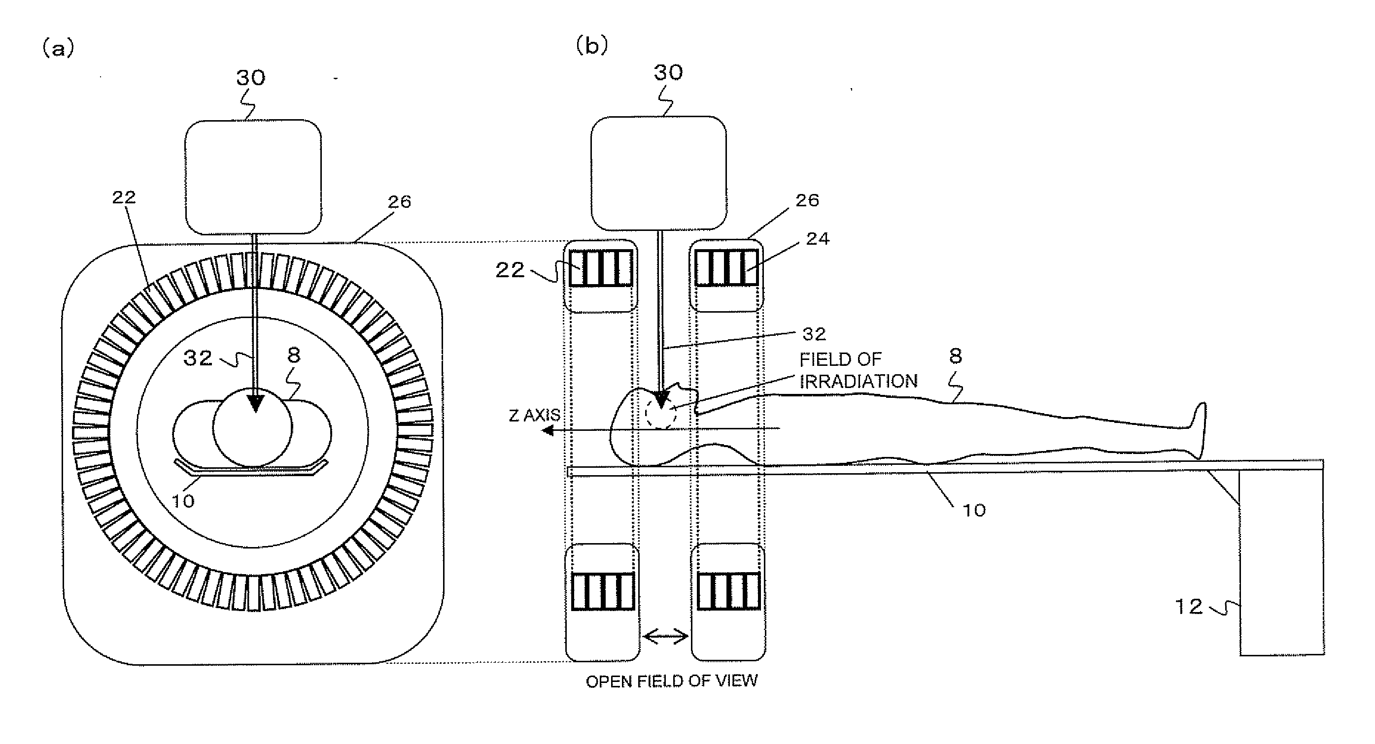
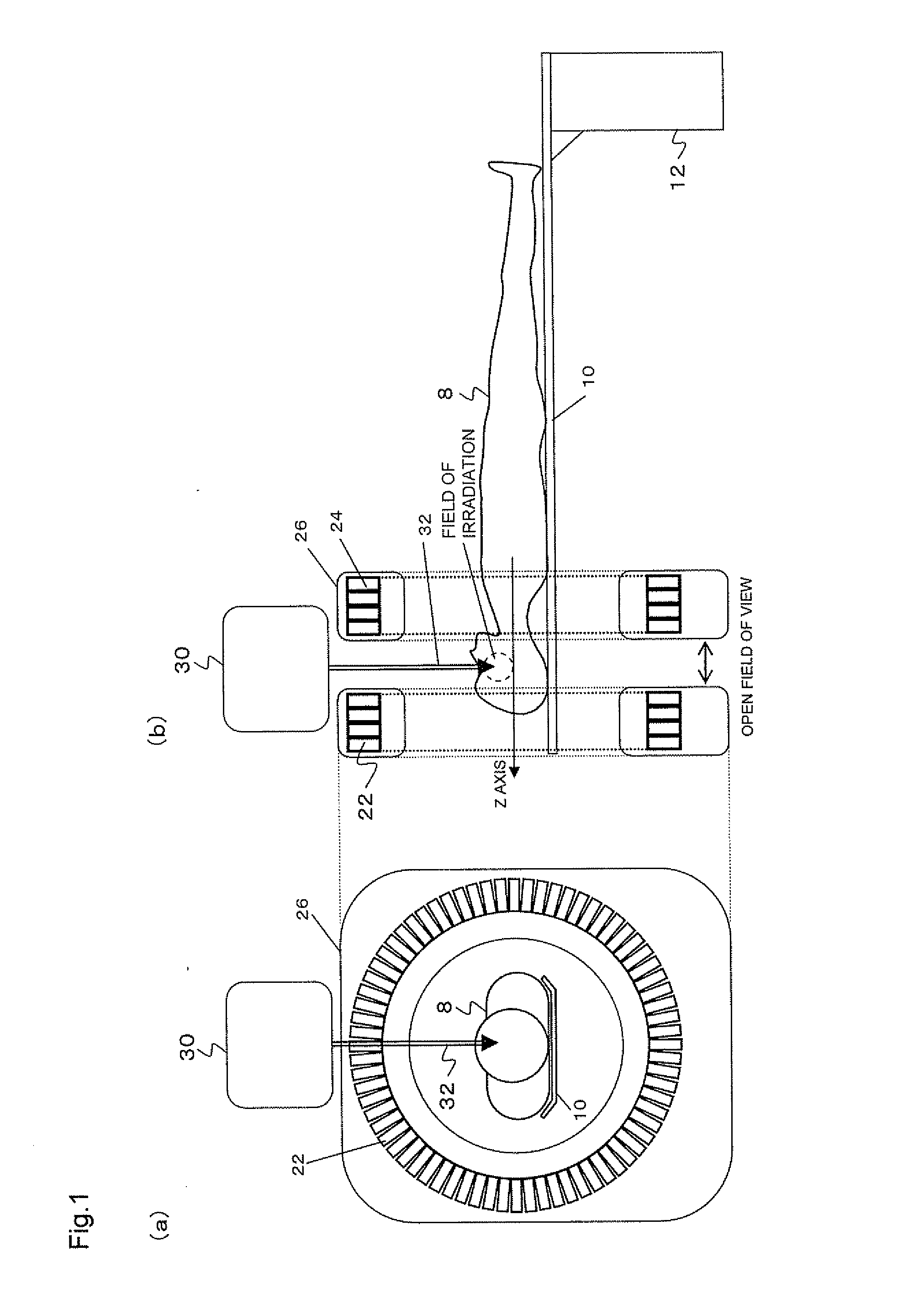
[0076]The present invention is best available when radiation cancer therapy is to be provided under the guidance of PET images.

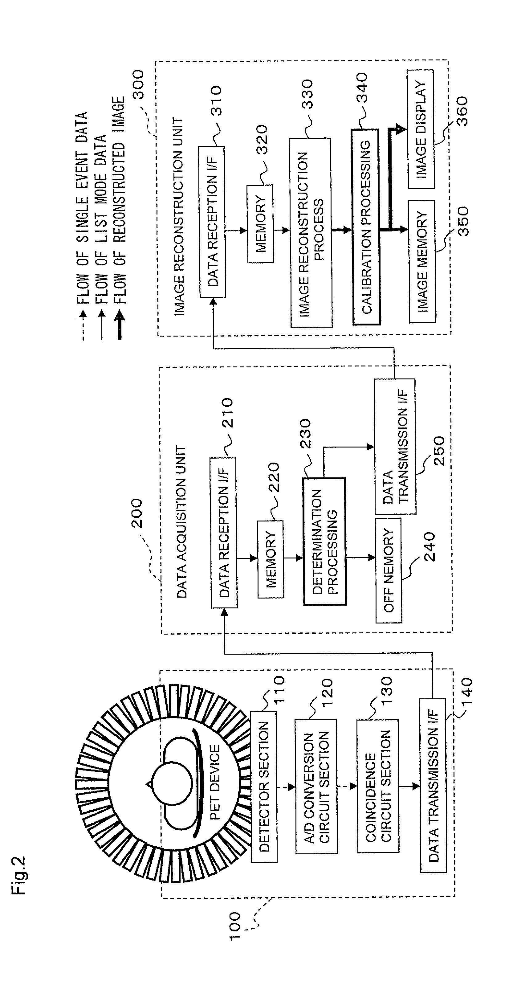
[0077]FIG. 10 shows an example in which a radiation (cancer) therapy apparatus 400 is combined with the open PET device 100 to implement a real time image reconstruction system according to the present invention. The figure shows a patient 8, a bed 10, a base 12 of the bed 10, detector rings 22 and 24, a PET controller 150, an image reconstruction apparatus 500 which includes the data acquisition unit 200 and the image reconstruction unit 300, a therapy planning device 600, and a therapy apparatus controller 700.

[0078]Using a marker such as fludeoxyglucose (FDG) representative of a PET probe that collects in cancer, only a target such as lung cancer moving within the body can be accurately irradiated with radiation while tracking the target in real time on the PET images.

[0079]FIGS. 11 and 12 show the specific procedures.

[0080]FIG. 11 shows a method ...


PUM


Abstract

In imaging on the basis of list mode data of a list of radioactive count data detected by a nuclear medicine imaging apparatus for measuring radiation in a pulse mode, the processing from the measurement to imaging of radiation is accelerated substantially to the real time level by selecting the number of count data to be used for online imaging computations on the basis of the counting rate of radiation.

Description

TECHNICAL FIELD[0001]The present invention relates to methods and systems for imaging using nuclear medicine imaging apparatus which conduct radiation measurements in a pulse mode, nuclear medicine imaging systems, and radiation therapy control systems. More particularly, the invention relates to a method and a system for imaging using a nuclear medicine imaging apparatus, a nuclear medicine imaging system, and a radiation therapy control system, which are preferred for use with gamma cameras, positron cameras, single photon emission computed tomography (SPECT) device, positron emission tomography (PET) device or the like, and which are capable of accelerating the processing from the measurement to imaging of radiation substantially to a real time level so as to display or analyze images in real time.BACKGROUND ART[0002]The positron emission tomography (PET) has received attention as being effective for early diagnosis of cancer. The PET or an examination method has been used for ex...

Claims


Application Information

Patent Timeline
IPC IPC(8): G01T1/164
CPCA61B6/5205A61B6/037
Inventor YAMAYA, TAIGAYOSHIDA, EIJI
Owner NAT INST FOR QUANTUM & RADIOLOGICAL SCI & TECH
Why Eureka
Try Eureka