
Method for Automated Autoantibody Detection and Identification

a technology of autoantibody detection and identification, applied in the direction of fluorescence/phosphorescence, instruments, material analysis, etc., can solve the problems of large amount of autoantibody production, limited number of known autoantibodies, and malfunction of the immune system of the body

Inactive Publication Date: 2013-02-28
RUTGERS THE STATE UNIV
View PDF3 Cites 4 Cited by
  • Summary
  • Abstract
  • Description
  • Claims
  • Application Information

AI Technical Summary

Benefits of technology

The invention is a method for detecting and identifying autoantibodies in a clinical sample. This involves fixing and permeabilizing cells, incubating them with a patient sample and a fluorescently labeled anti-human antibody, and then staining them with a reagent for staining a cellular compartment or marker. The cells are then subjected to imaging flow cytometry to capture cellular images, which are then compared to pre-defined templates with automated pattern recognition. The invention can detect various autoantibodies and can be used with different types of cells. A kit containing all the necessary components is also provided.

Problems solved by technology

However, when a person has an autoimmune disease, the body's immune system malfunctions, producing large amounts of autoantibodies.
However, one of the problems with the solid phase immunoassay is that only a limited number of known autoantigens can be measured.
In this respect, the primary disadvantage of the ANA-HEp-2 test is the subjective evaluation of HEp-2 slides that complicates standardization and reproducibility.
Interpretation of immunofluorescence patterns is dependent on the individual's knowledge and experience and therefore high intra- and inter-laboratory variability is common and represents a major diagnostic problem, especially in non-specialized laboratories.
Moreover, this method is labor intensive and time consuming for the large quantity of samples that are processed annually.
However, these systems are not widely used.

Method used


Image

  • Method for Automated Autoantibody Detection and Identification
  • Method for Automated Autoantibody Detection and Identification
  • Method for Automated Autoantibody Detection and Identification

Examples


example 1

ANA Protocol

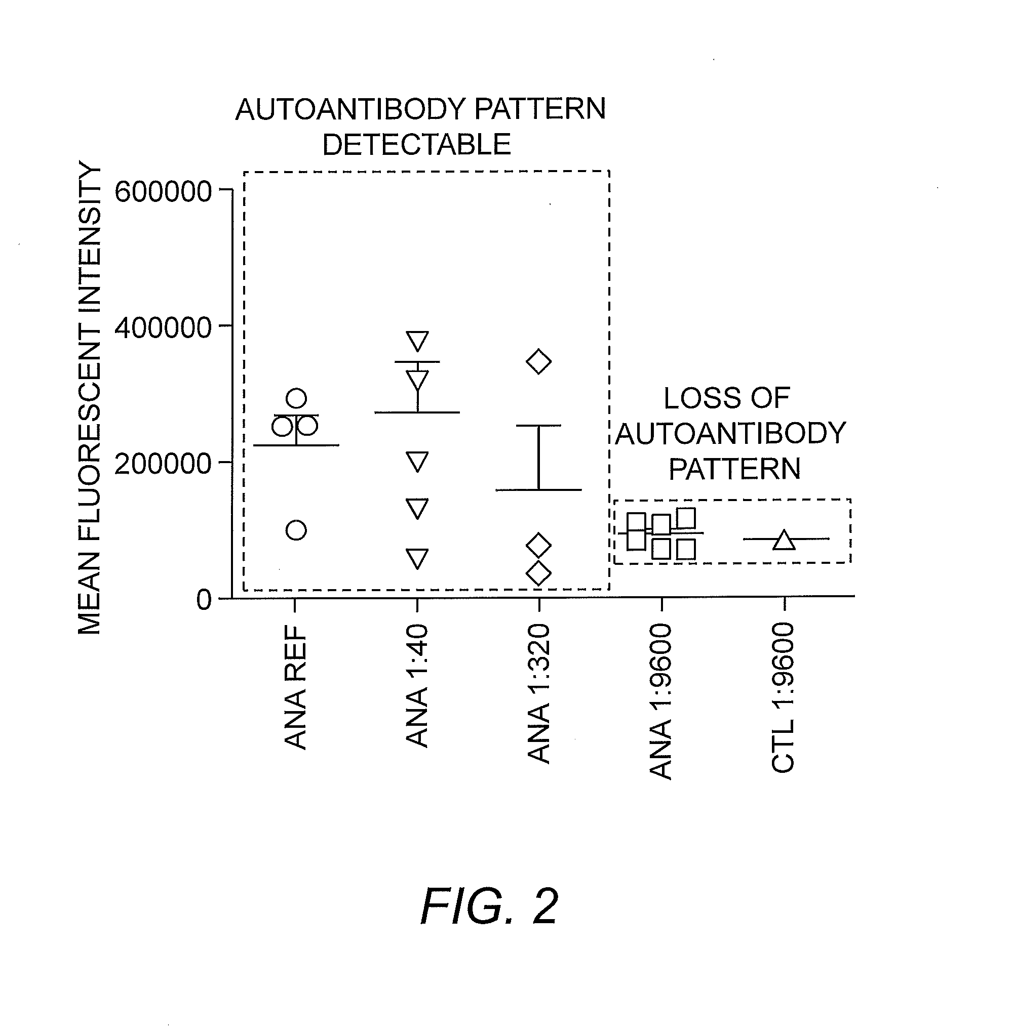
[0038]Patient samples are prepared at an appropriate dilution in phosphate buffered sample (PBS; e.g., 10 μL of sample plus 390 μL of PBS). Subsequently, the patient sample is incubated with a fixed and permeabilized suspension of HEp-2 cells. After an appropriate amount of time for the autoantibodies to bind autoantigens, the HEp-2 cells are incubated with fluorescent markers for cellular compartments and multiple serial dilutions can be prepared to determine titer. Generally, titers of 1:40 or less (e.g., in the range of 1:40 to 1:320) are particularly useful, and, as shown in FIG. 2, provide a sufficient level of intensity for autoantibody pattern detection.

[0039]HEp-2 cells are subsequently incubated with fluorescent anti-IgG secondary antibody and localization of autoantibodies to cell compartments is carried out with imaging flow cytometry. Cellular images are captured and analyzed with pre-defined templates and software algorithms. A statistical report indicates p...

example 2

Classification of Patterns

[0040]Various patterns of autoantibody binding and the basis thereof are as follow:

Nuclear Patterns

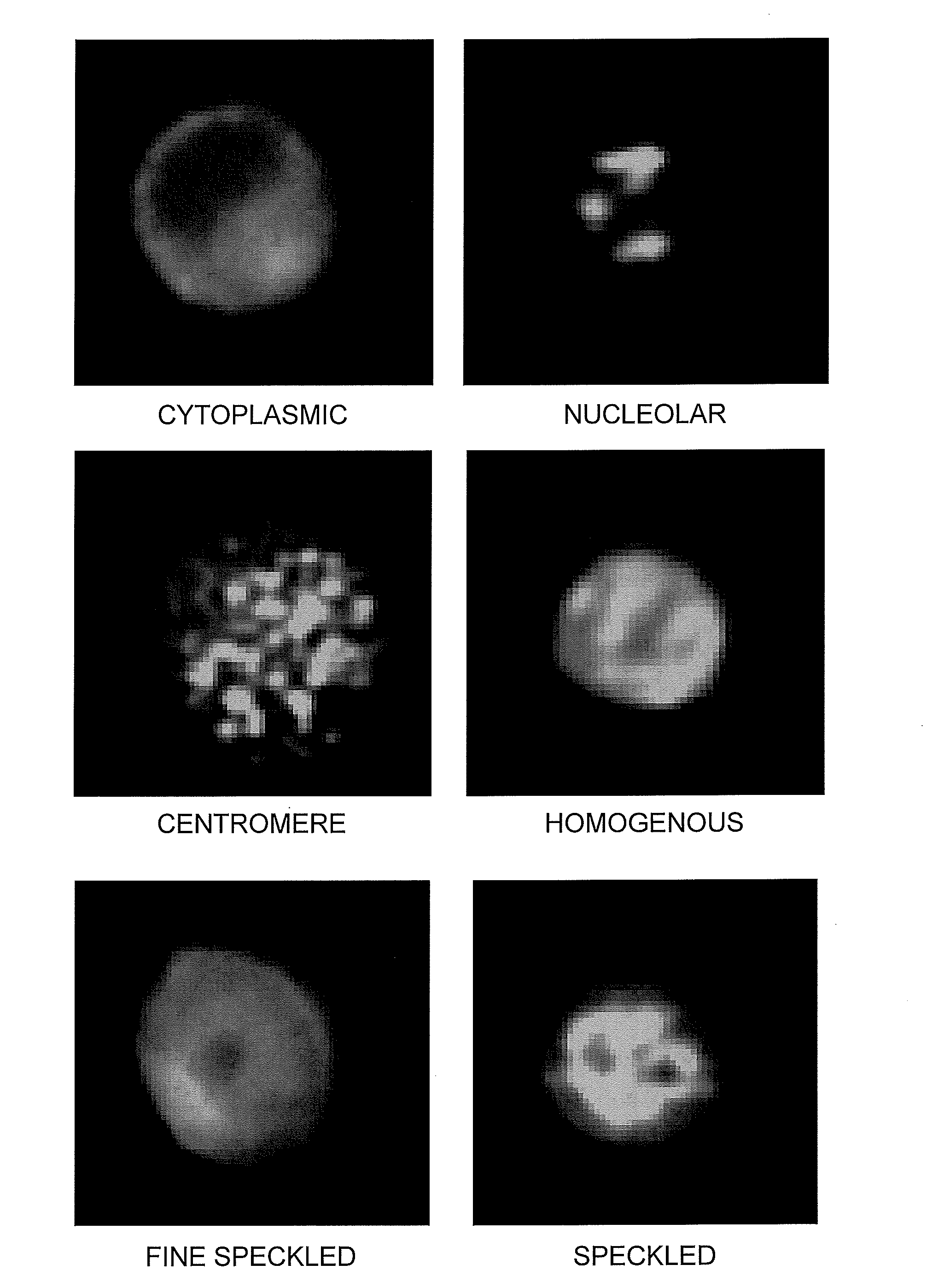
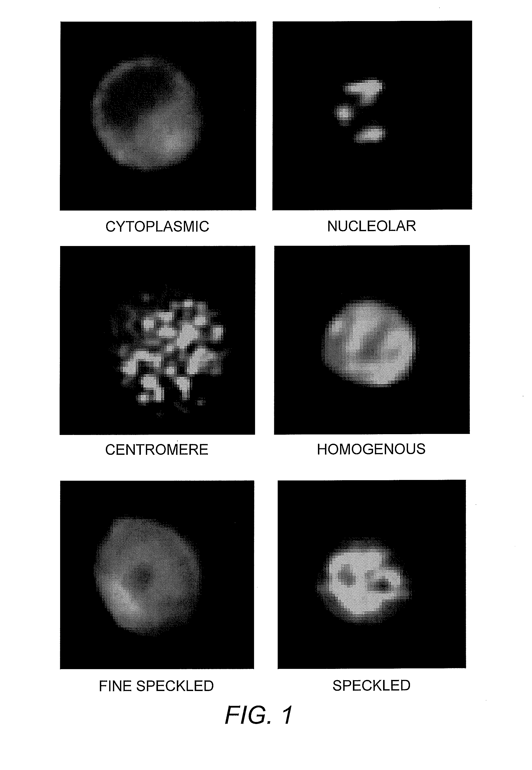
[0041]1. Homogeneous. A homogeneous or diffuse staining pattern of the nucleus is consistent with autoantibodies to native DNA (nDNA) histones and / or deoxyribonucleoprotein (DNP) (Lachman & Kunkel (1961) Lancet 2:436; Friou (1964) Arthritis Rheum. 7:161).

[0042]2. Speckled Patterns. A speckled pattern is the most commonly observed ANA pattern and can be distinguished from a homogenous pattern by, e.g., dark spot areas or areas of increased contrast. A uniform, true speckled pattern may be seen with centromere antibodies in cells not in division. A clumpy speckled patterns may be seen with antibodies to n-RNP, Sm, and SSB / La.[0043]i. Fine speckled pattern, chromosome-negative: Numerous small uniform points of fluorescence uniformly scattered throughout the nucleus. The nucleoli generally appear unstained. The mitotic cells may demonstrate a few speckles in their...


PUM

PropertyMeasurementUnit
wavelengthaaaaaaaaaa

Abstract

The present invention is a kit and method for detecting and identifying autoantibodies. The invention employs the use of indirect immunofluorescence, imaging flow cytometry and pattern recognition software to automatically identify autoantibodies associated with autoimmune disorders.

Description

INTRODUCTION[0001]This application claims priority from U.S. Provisional Application No. 61 / 526,447, filed Aug. 23, 2011, the content of which is incorporated herein by reference in its entirety.BACKGROUND OF THE INVENTION[0002]Antibodies are proteins that are made as part of an immune response. Normally the immune system responds to infection by producing large numbers of antibodies to fight bacteria or viruses. However, when a person has an autoimmune disease, the body's immune system malfunctions, producing large amounts of autoantibodies. Autoantibodies, unlike normal antibodies that fight bacteria, viruses, parasites, and fungi, attack the body's own tissues and cells. Autoantibody-mediated inflammation and cell destruction can affect blood cells, skin, joints, kidneys, lungs, nervous system, and other organs of the body resulting in autoimmune or connective tissue disorders such as systemic lupus erythematosus (SLE), autoimmune hepatitis, rheumatoid arthritis, polymyositis / der...

Claims


Application Information

Patent Timeline
Patent Type & Authority Applications(United States)
IPC IPC(8): G01N21/64
CPCG01N21/6458G01N21/6428
Inventor BARNES, BETSY J.FENG, DISTONE, RIVKA C.
Owner RUTGERS THE STATE UNIV
Learn More