Method and magnetic resonance system for imaging a partial region of an examination subject

a magnetic resonance system and partial region technology, applied in the field of partial region imaging of an examination subject, can solve the problem of not being able to compensate for the distortion in the subsequent exposure of magnetic resonance, and achieve the effect of precise attenuation correction, low distortion, and high quality

Active Publication Date: 2013-03-07
SIEMENS HEALTHCARE GMBH
View PDF14 Cites 15 Cited by
  • Summary
  • Abstract
  • Description
  • Claims
  • Application Information

AI Technical Summary

Benefits of technology

[0024]Due to the low distortion, the determined image is of high quality even in the boundary region of the field of view, which is why examination regions of adipose patients (for example) that cannot be arranged closer to the center of the tunnel of the magnetic resonance system can also be examined by means of MR. Furthermore, the determined image can be used, for example, in the positioning of stereotactic devices or implementation of stereotactic procedures, and / or the attitude of the partial region can be reliably determined in the determined image of the examination subject.
[0025]According to a further embodiment, an attenuation correction for a positron emission tomography (PET) scanner is determined depending on an attitude of the partial region of the examination subject. Due to the low distortion, the attitude of the partial region (an arm, for example) can be determined reliably from the image of the examination subject. In positron emission tomography, consideration of attenuation of the received radiation (photons) by the structure or anatomy of the examination subject in the beam direction is of decisive importance. Because the attitude of the partial region of the examination subject can be determined even at the edge of the field of view of the magnetic resonance system, a complete determination of the attitude and structure of the examination subject or patient in the magnetic resonance system is possible, and thus a precise attenuation correction for positron emission tomography is achievable. Since the attenuation correction is in this case based solely on information from the magnetic resonance imaging, positron emission tomography can also be implemented with less strongly enriched PET tracers (rubidium, for example).

Problems solved by technology

Given severe distortions in these boundary regions in which the B0 field inhomogeneities and the gradient field have nonlinearities, a subsequent compensation of the distortion in the magnetic resonance exposure is frequently not possible since the distorted regions overlap in the magnetic resonance exposure.

Method used


Image

  • Method and magnetic resonance system for imaging a partial region of an examination subject
  • Method and magnetic resonance system for imaging a partial region of an examination subject
  • Method and magnetic resonance system for imaging a partial region of an examination subject

Examples


Embodiment Construction

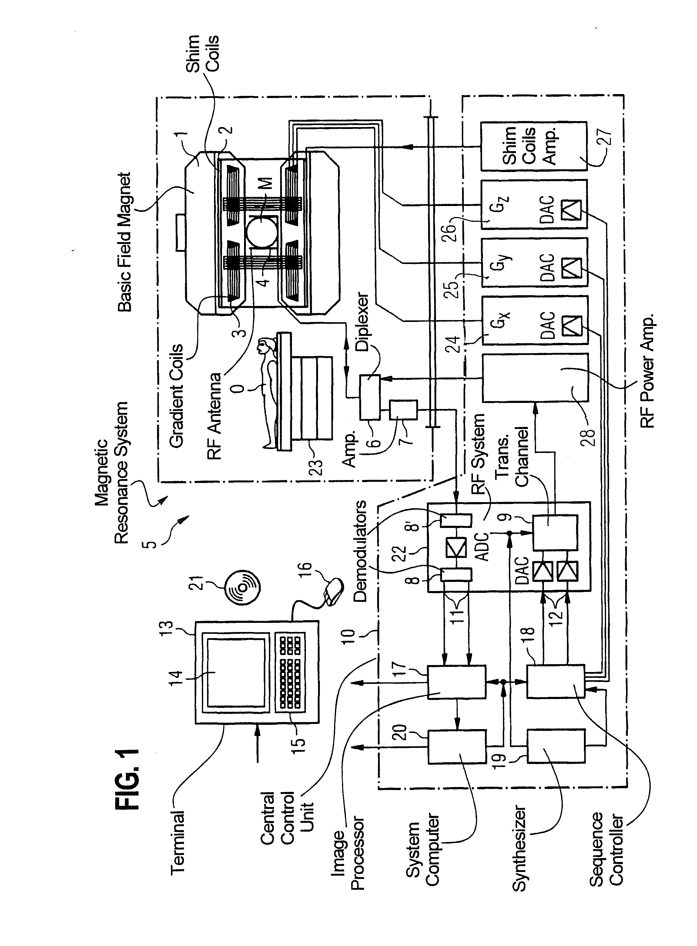
[0036]FIG. 1 is a schematic representation of a magnetic resonance system 5 (of a magnetic resonance imaging or magnetic resonance tomography apparatus). A basic magnet 1 generates a temporally constant, strong magnetic field for polarization or alignment of the nuclear spins in an examination region of an examination subject U (for example a part of a human body that is to be examined) that lies on a table 23 and is moved into the magnetic resonance system 5. The high homogeneity of the basic magnetic field that is required for the magnetic resonance measurement (data acquisition) is defined in a typically spherical measurement volume M in which the parts of the human body that are to be examined are introduced. Shim plates made of ferromagnetic material (which may be selectively adaptable) are mounted at a suitable location to support the homogeneity requirements, and in particular to eliminate temporally invariable influences. Temporally variable influences are eliminated by shim...


PUM


Abstract

In a method and magnetic resonance (MR) apparatus to image a partial region of an examination subject by means of a multislice measurement, which partial region includes at least two measurement slices, and is located at least in part at the edge of a field of view of the magnetic resonance apparatus, for each voxel to be optimized that is located at the edge of the field of view, a gradient field is configured for each measurement slice of the partial region that is to be measured and is used to acquire magnetic resonance data in the multislice measurement. The gradient field is configured so as to cause a nonlinearity of the gradient field and a B0 field inhomogeneity to cancel at each of the aforementioned voxel to be optimized at the partial region at the edge of the field of view. An image of the partial region of the examination subject is determined from the magnetic resonance data acquired in this manner.

Description

BACKGROUND OF THE INVENTION[0001]1. Field of the Invention[0002]The present invention concerns a method for imaging a partial region of an examination subject in a magnetic resonance system, and a magnetic resonance system designed to implement such a method.[0003]2. Description of the Prior Art[0004]In a magnetic resonance system, the volume in which magnetic resonance data are acquired is limited in all three spatial directions due to physical and technical conditions (for example a limited magnetic field homogeneity and the nonlinearity of the gradient field). The acquisition volume—known as a “field of view” (FoV)—is therefore limited to a volume in which the aforementioned physical features lie within a predetermined tolerance range, and thus in which an image of the subject to be examined that is true to scale is possible with typical measurement sequences. The FoV is limited in the x-direction and y-direction, i.e. perpendicular to a longitudinal axis of a tunnel of the magne...

Claims


Application Information

Patent Timeline
Patent Type & Authority Applications(United States)
IPC IPC(8): G01R33/565G01R33/341
CPCG01R33/481A61B5/055G01R33/56572G01R33/56563
Inventor BLUMHAGEN, JAN OLEFENCHEL, MATTHIASLADEBECK, RALF
Owner SIEMENS HEALTHCARE GMBH
Why Eureka
Try Eureka