
Detection of neighboring variants

a neighboring variant and detection technology, applied in the field of detection of neighboring variants, can solve the problems of not being able to distinguish, unable to analyze one sample at a time, and prior techniques for cystic fibrosis (cf) testing using high-resolution thermal melt analysis lacked the ability to discern between benign and disease-causing variants in close proximity, etc., to achieve high-resolution thermal melting curves

Active Publication Date: 2013-05-16
CANON US LIFE SCIENCES INC
View PDF1 Cites 7 Cited by
  • Summary
  • Abstract
  • Description
  • Claims
  • Application Information

AI Technical Summary

Benefits of technology

The present invention provides a method for distinguishing between different variants of a target nucleic acid molecule. This can be useful in identifying or determining polymorphisms associated with a disease or disorder. The method involves amplifying the target nucleic acid molecule and detecting specific variants using a first and second unlabeled probe. The method can also be used to detect a disease in a patient based on their genotype and knowledge of benign and disease-causing variant gene sequences associated with the disease. The technical effect of this invention is the ability to distinguish between different variants of a target nucleic acid molecule, which can be useful in identifying or determining polymorphisms associated with a disease or disorder.

Problems solved by technology

However, it suffers from a limitation that it can only analyze one sample at a time, and the sample container must be replaced manually.
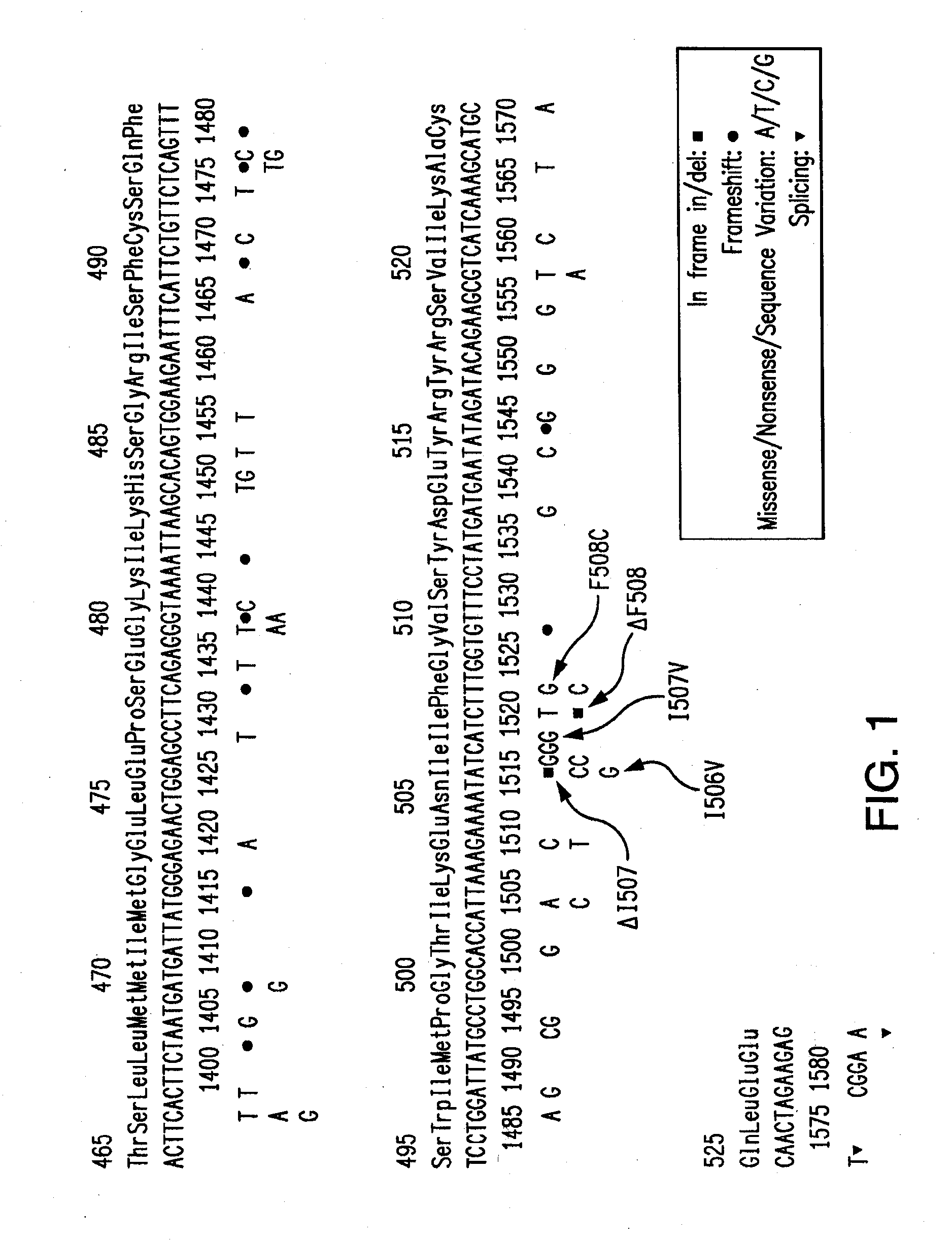
For example, prior techniques for Cystic Fibrosis (CF) testing using high resolution thermal melt analysis lacked the ability to discern between benign and disease-causing variants in close proximity in Exon 10 of the CFTR (cystic fibrosis transmembrane conductance regulator) gene.
Although commercial kits are available to detect CF mutations, these kits do not make it possible to distinguish between all benign and disease-causing mutations in Exon 10 in a rapid detection system (Johnson et. al., J. Mol. Diagn. 2007.

Method used


Image

  • Detection of neighboring variants
  • Detection of neighboring variants
  • Detection of neighboring variants

Examples


example 1

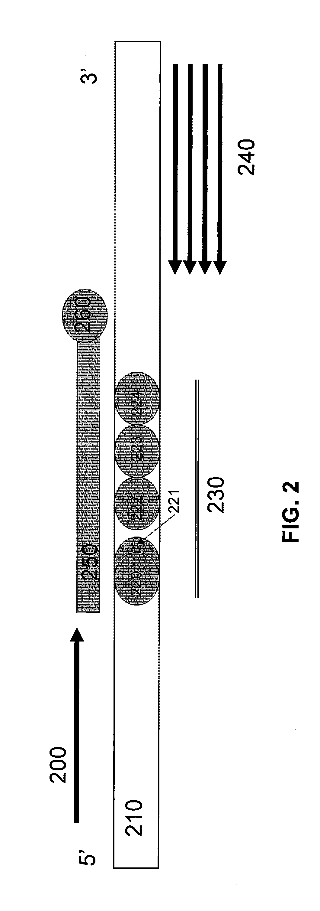
[0091]FIG. 4 illustrates a schematic diagram of primers, probes, and CFTR Exon 10. A forward primer (400), which acts as the limiting primer, extends in the 5′ to 3′ direction of the target nucleic acid having a locus of interest (410) having mutations of interest (420-424) at a locus of interest (430). The mutations of interest (420-424) include benign variants I506V (420), I507V (422), and F508C (424), as well as disease-causing variants ΔI507 (421) and ΔF508 (423). A reverse primer (440), which acts as the excess primer, extends in the 3′ to 5′ direction of the target nucleic acid having a locus of interest (410). Unlabeled probe (450) having a blocker (460) at its 3′ end, has been designed to hybridize to the reverse strand of the target nucleic acid having a locus of interest (410) at the locus of interest (430).

[0092]In one non-limiting embodiment, a primer pair (F2 / R4) that produces a specific amplicon that includes the mutations of ΔI507 / ΔF508 and neighboring benign variants...

example 2

[0095]Biological samples having a locus of interest (see details in Table 1) were incubated with limiting primer (F2), excess primer (R4), and an unlabeled probe (UP3). The two primers were provided in different concentrations along with buffer, dNTPs, MgCl2, LC Green Plus, and polymerase. Parallel mixtures using the same primers and reagents, but a different unlabeled probe (UP4), were also prepared. Each of the mixtures was loaded onto a 96-well plate on a LC 480 and subjected to asymmetric PCR. Initially, the PCRs preceded using the limiting and excess primers to produce small amplicons. In each mixture subjected to PCR, once the limiting primer was exhausted, the unlabeled probe then attached to the locus of interest on a small amplicon in conjunction with the excess primer and PCR continued. It is noted that the unlabeled probes each had a blocker in place so as to not extend during the PCRs.

TABLE 1NA01531CoriellExon 10deltaF508homNA07552CoriellExon 10 / Exon 11deltaF508 / R553XCom...

example 3

[0102]Primers and probes were designed for use in a Cystic Fibrosis American College of Obstetricians and Gynecologists (ACOG) panel. The ACOG panel included the primers and probes described in Examples 1 and 2, above. The sequences of these primers and probes are listed in Table 3 below and details pertaining thereto are listed in Table 4. Forward primers are denominated with the suffix “F#,” reverse primers are denominated with the suffix “R#,” and probes are denominated with the suffix “UP#.”

TABLE 3ExonPrimer or Probe NameSequence 5′-3′ 3Exon 3 G85E F1GCCCTTCGGCGATGTTTTExon 3 G85E R1gatccttacCCCTAAATATAAAAAG 4Exon 4 R117H F3ATGACCCGGATAACAAGGAGExon 4 R117H R2CATAAGCCTATGCCTAGATAAATCGIntronExon 4 621 + 1G > T F2GAGAATAGCTATGTTTAGTTTGATTT 4Exon 4 621 + 1G > T R3gcctgtgcaaggaagtattaIntronExon 5 711 + 1G > T F2GTCTCCTTTCCAACAACCTGAA 5Exon 5 711 + 1G > T R1agtgcctaaaagattaaatcaa 7Exon 7 R334W F2GCACTAATCAAAGGAATCATCCTCExon 7 R334W R2CAGAATGAGATGGTGGTGAAT 7Exon 7 R347P F3CCACCATCTCATTC...


PUM

PropertyMeasurementUnit
Tmaaaaaaaaaa
Tmaaaaaaaaaa
timeaaaaaaaaaa

Abstract

The present invention relates to methods, kits, probes, and systems for distinguishing between nucleotide variants that are close in proximity on a gene. The methods, kits, probes, and systems can include the use of a small amplicon assay in combination with two unlabeled probes in a high resolution thermal melting analysis of a biological sample containing a locus of interest in order to discern between disease-causing and benign variants that are close in proximity on a gene within the biological sample. The present invention also relates to method of detecting a disease in a patient based on the patient's genotype by determining whether the patient has a disease-causing variant at a locus of interest. The signature melt curves produced by the unlabeled probe tests can be analyzed using HRMA software to distinguish between disease-causing and benign variants that are close in proximity on a gene within the biological sample.

Description

FIELD OF THE INVENTION[0001]The present invention relates to methods, kits, primers, probes, and systems for distinguishing between nucleotide variants that are close in proximity on a gene. More particularly, aspects of the present invention relate to methods, kits, primers, probes, and systems for using a small amplicon assay in combination with unlabeled probes in conducting a high resolution thermal melting analysis of a biological sample containing a locus of interest in order to discern between disease-causing and benign variants that are close in proximity on a gene. The present invention also relates to methods of detecting a disease in a patient based on the patient's genotype by determining whether the patient has a disease-causing variant at a locus of interest on the patient's genome.DESCRIPTION OF THE BACKGROUND[0002]Melt curve analysis or high resolution thermal melting is an important technique for analyzing nucleic acids. In accordance with some methods, a double str...

Claims


Application Information

Patent Timeline
Patent Type & Authority Applications(United States)
IPC IPC(8): C12Q1/68C12M1/34C07H21/00
CPCC12Q2600/156C12Q1/6827C12Q1/6883C12Q2525/204C12Q2527/107C12Q2531/107
Inventor XU, LINGHOWELL, RENEE
Owner CANON US LIFE SCIENCES INC
Learn More