

Method for recording and displaying at least two 3D subtraction image data records and c-arm x-ray apparatus
- Summary
- Abstract
- Description
- Claims
- Application Information
AI Technical Summary
Benefits of technology
Problems solved by technology
Method used
Image

Examples
Embodiment Construction
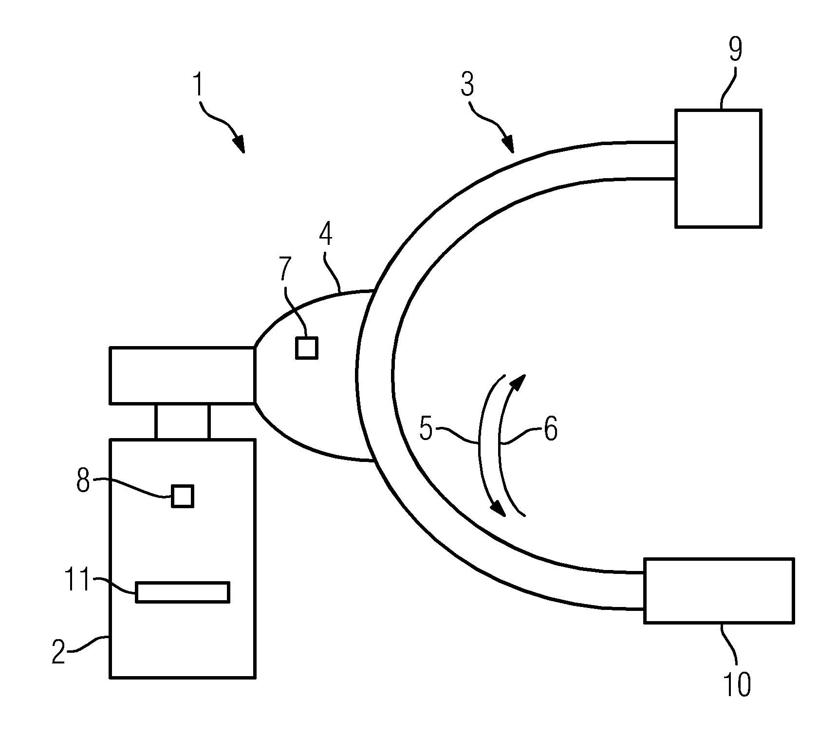
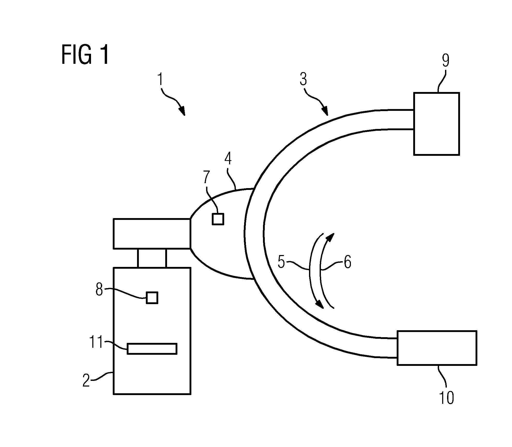
[0037]FIG. 1 shows a C-arm x-ray apparatus 1 with a supporting apparatus 2 and a C-arm 3. The C-arm 3 is connected to the supporting apparatus 2 via the suspension 4. To record data, the C-arm, 3 can be moved in the direction of the arrows 5 and 6 by means of a motor 7 found in the suspension 4. A control apparatus 8 for activating the motor 7 in the suspension 4 and for controlling the x-ray source 9 and the detector 10, which are arranged on the ends of the C-arm 3, is disposed in the supporting apparatus 2. The generator 11 for high voltage is further accommodated in the supporting apparatus 2.
[0038]The control apparatus 8 naturally also includes a storage unit for storing the recorded data. It may also comprise a computing facility for further processing the recorded data and a display apparatus. The recorded data may however also be further processed on an external computing facility.
[0039]Aside from the rotation directions shown in the direction of the arrows 5 and 6, the C-ar...
PUM
 Abstract
Description
Claims
Application Information