Probe for ultrasonic diagnostic apparatus and method of manufacturing the same

Inactive Publication Date: 2013-07-25
SAMSUNG MEDISON
View PDF5 Cites 4 Cited by
  • Summary
  • Abstract
  • Description
  • Claims
  • Application Information

AI Technical Summary

Benefits of technology

[0010]Therefore, it is an object of the present disclosure to provide a probe for an ultrasonic diagnostic apparatus which may increase attenuation

Problems solved by technology

However, in such a configuration, the powder may not be regularly distributed, and it may be difficult to produce powder with a uniform size.
Further, pores that are unexpectedly produ

Method used


Image

  • Probe for ultrasonic diagnostic apparatus and method of manufacturing the same
  • Probe for ultrasonic diagnostic apparatus and method of manufacturing the same
  • Probe for ultrasonic diagnostic apparatus and method of manufacturing the same

Examples


Embodiment Construction

[0039]Reference will now be made in detail to exemplary embodiments of the present disclosure, examples of which are illustrated in the accompanying drawings, wherein like reference numerals refer to like elements throughout.

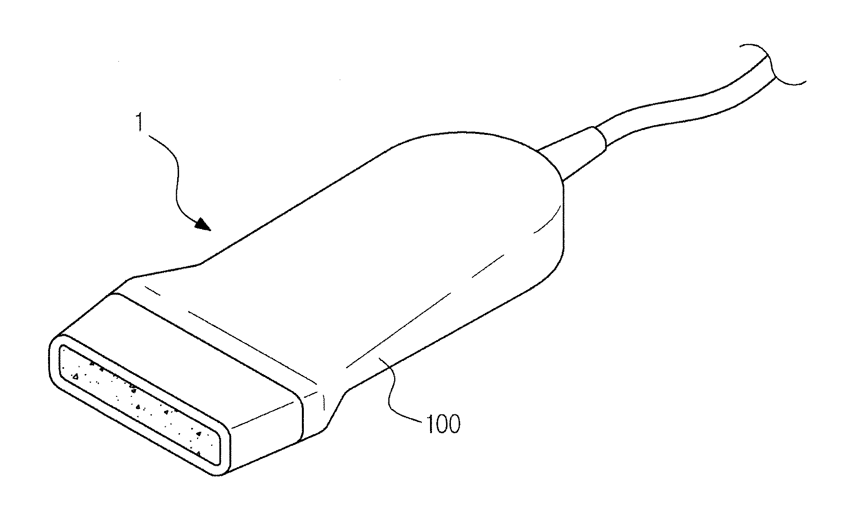
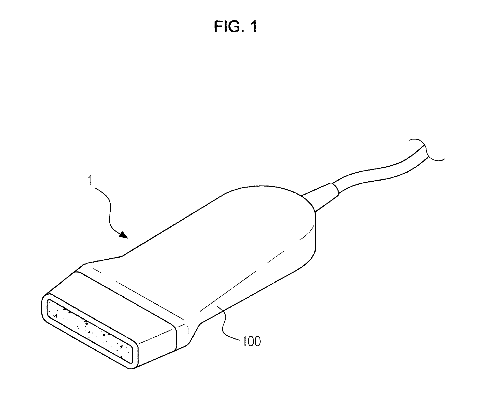
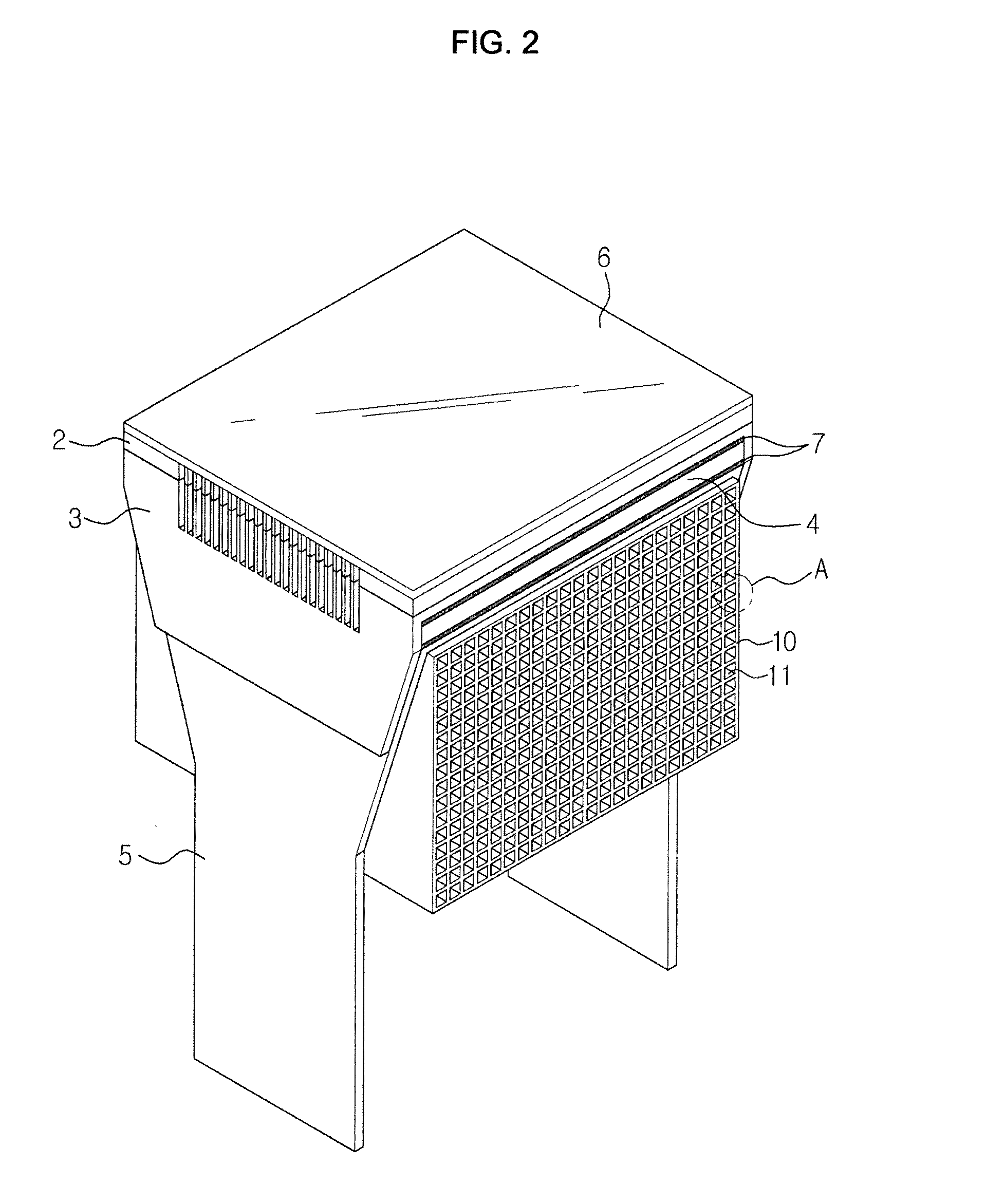
[0040]FIG. 1 is a view illustrating exterior of the probe for an ultrasonic diagnostic apparatus according to an exemplary embodiment of the present disclosure, FIG. 2 is a perspective view illustrating a probe for an ultrasonic diagnostic apparatus according to an exemplary embodiment of the present disclosure, and FIG. 3 is an exploded view illustrating the probe of FIG. 1.

[0041]As shown in FIGS. 1, 2 and 3, a transducer includes a piezoelectric member 4, acoustic matching layers 2 and 3, an acoustic lens (6) and a backing member 10. The illustrated transducer may be a constituent of a probe 1 for an ultrasonic diagnostic apparatus according to an exemplary embodiment of the present disclosure, and in practice may be positioned inside a case (100).

[0042]The ac...


PUM

PropertyMeasurementUnit
Temperatureaaaaaaaaaa
Densityaaaaaaaaaa

Abstract

A probe for an ultrasonic diagnostic apparatus includes a case, a piezoelectric member provided inside the case, at least one acoustic matching layer disposed at a front of the piezoelectric member, an acoustic lens disposed at a front of the acoustic matching layer, and a backing member disposed at a rear of the piezoelectric member and provided with a plurality of pores to reduce sound waves transmitted to the rear of the piezoelectric member and to allow to adjust an acoustic impedance. A method of manufacturing the probe for an ultrasonic diagnostic apparatus includes the operations of preparing a fluid to form a backing layer, pouring the fluid into a mold, hardening the fluid and removing the mold.

Description

CROSS-REFERENCE TO RELATED APPLICATION(S)[0001]This application claims the benefit of Korean Patent Application No. 10-2012-0006227, filed on Jan. 19, 2012 in the Korean Intellectual Property Office, the entire disclosure of which is incorporated herein by reference.BACKGROUND[0002]1. Field[0003]Exemplary embodiments of the present disclosure relate to a probe for an ultrasonic diagnostic apparatus which may increase sensitivity of the probe, and a method of manufacturing the same.[0004]2. Description of the Related Art[0005]An ultrasonic diagnostic apparatus is a noninvasive apparatus that transmits an ultrasound signal to a body surface of a subject towards a target organ in the body and obtains cross-sectional images of soft tissue and blood flow using information from a reflected ultrasound signal. Compared to other medical imaging systems such as, for example, X-ray imaging systems, computed tomography (CT) scanners, magnetic resonance imaging (MRI) systems and diagnostic syste...

Claims


Application Information

Patent Timeline
IPC IPC(8): A61B8/00B29C39/02
CPCA61B8/4494A61B8/4281B29C39/026B06B1/0674A61B8/4483A61B8/4444A61B8/00G01N29/24
Inventor JUN, SUNG JAELEE, SUNG JAEKIM, JI SEONSEO, MIN SEON
Owner SAMSUNG MEDISON
Why Eureka
Try Eureka