Separation of living untouched neurons

a technology for separating living neurons and untouched cells, applied in the field of cells, can solve the problems of large cell loss, low purities, and lengthy procedures, and achieve the effect of increasing purities

Active Publication Date: 2013-11-28
MILTENYI BIOTEC B V & CO KG
View PDF1 Cites 7 Cited by
  • Summary
  • Abstract
  • Description
  • Claims
  • Application Information

AI Technical Summary

Benefits of technology

[0014]The purity can be further increased if an astrocyte specific cell surface marker is used in addition to CD51 to deplete a subpopulation of CD51 negative remaining astrocytes.

Problems solved by technology

Relatively easy techniques like density gradient centrifugation or isolation of living cells by culture conditions lead to low purities and massive cell loss.
But, besides the need of transgenic mice and expensive equipment this procedure is quite cumbersome and takes several hours.
Nevertheless, this approach does not allow for the separation of living neurons as it requires labeling of an intracellular marker.
But, an one step isolation for neurons using this technique has not been described so far.
However, a general neuron specific cell surface marker is not known so far and the direct magnetic cell sorting of viable neurons is therefore not applicable.
Nevertheless, a negative isolation strategy can be applied to isolate cell populations that cannot be addressed directly.

Method used


Image

  • Separation of living untouched neurons
  • Separation of living untouched neurons
  • Separation of living untouched neurons

Examples


example 2

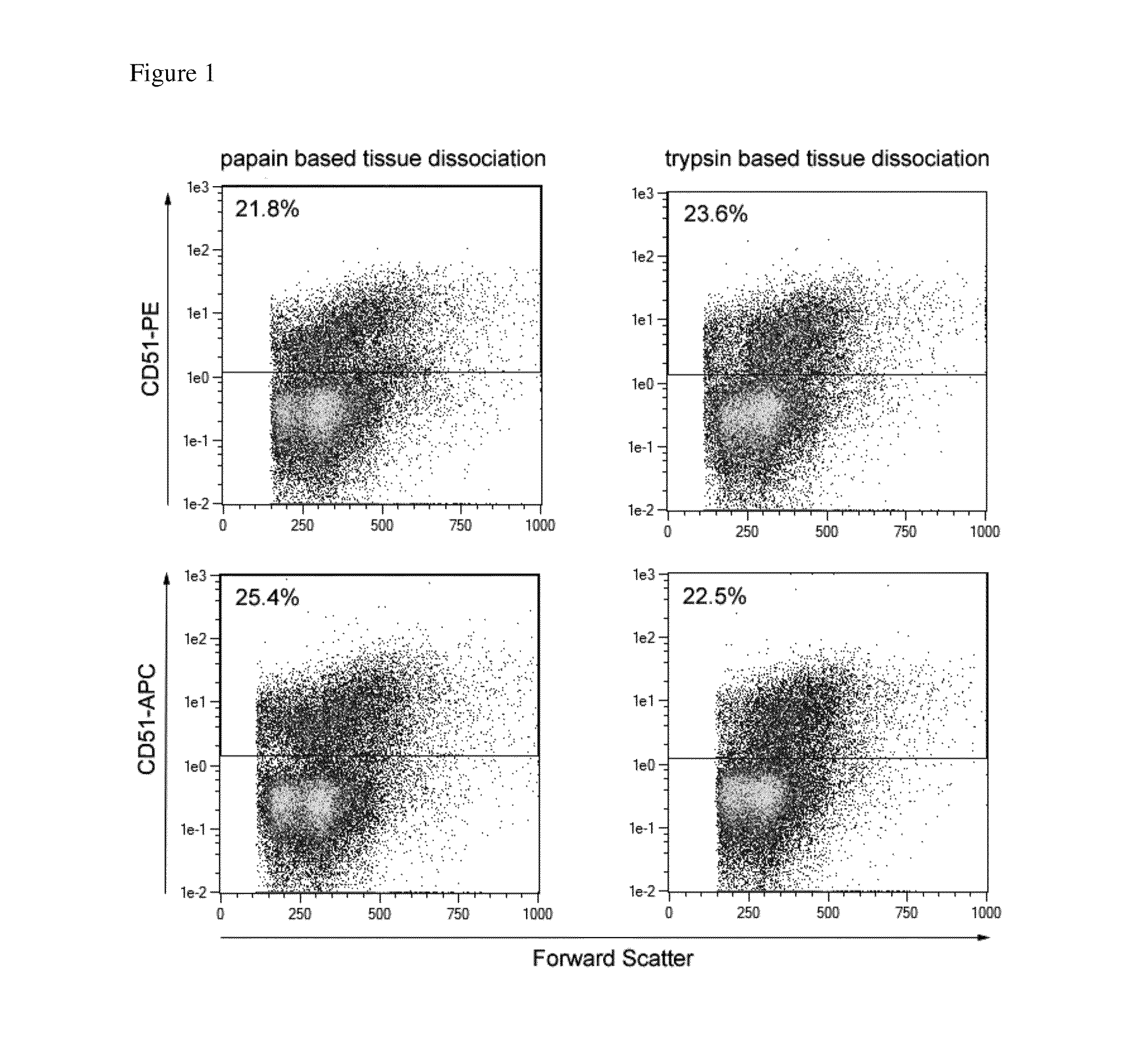
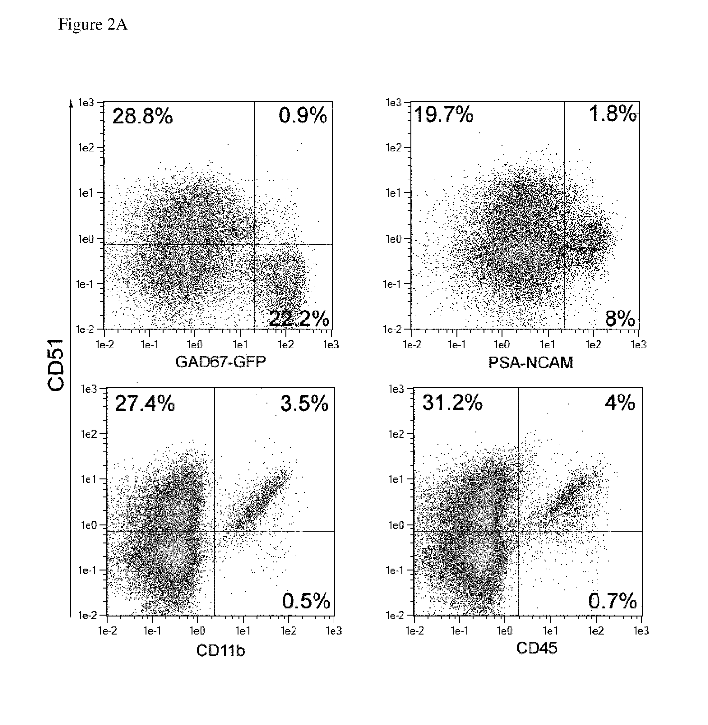
Flow Cytometric Characterization of CD51 Positive Cells in Postnatal Mouse Brain Tissue

[0089]Mouse brain tissue derived from P1, P2, or P3 CD1 mice was dissociated as described before using the NTDK(T) or (P). The resulting single cell suspension was co-stained with CD51 and markers specific for different neural subpopulations and subjected to flow cytometric analysis. For staining of the protease-sensitive epitopes CD31 and AN2, the cell suspension was incubated for 2 h at 37° C. in MACS® Neuro Medium +MACS® Supplement B27 PLUS under continuous rotation to re-express the epitopes. Due to the lack of markers that allow marking of living neuronal cell populations, GAD67-GFP transgenic mice were used to detect GABAergic neurons by their expression of GFP (green fluorescent protein). The analysis showed that GAD67-GFP positive GABAergic neurons lacked CD51 expression. Furthermore, neuronal progenitor cells, identified by PSA-NCAM expression, were identified as CD51 negative (see FIG. 2...

example 3

Depletion of CD51 Positive Cells By Magnetic Cell Separation

[0091]For depletion of CD51 positive non-neuronal cells a direct labeling strategy was compared to an indirect labeling with respect to purity and recovery of the target cells. For the direct labeling a CD51 specific antibody was covalently conjugated to magnetic particles.

[0092]The generation of superparamagnetic particles as used herein is disclosed in U.S. Pat. No. 5,543,289 which is included herewith by reference. Monoclonal antibodies recognizing the CD51 antigen were covalently conjugated to magnetic beads, resulting in 25 μg antibody per mL of bead suspension at a concentration of OD450=10.

[0093]Different concentrations of bead conjugated antibodies (0.75, 1.5, 3, 6 OD450 / ml) were given to 100 μl of a neural cell suspension containing 1×10⁷ cells. Cells were incubated for 15 minutes at 4° C., washed once with 1 ml PBS+0.5% BSA buffer, then resuspended in 1 ml of the same buffer and loaded on an LD column placed in th...

example 4

Efficient Depletion of Non-Neuronal Cells

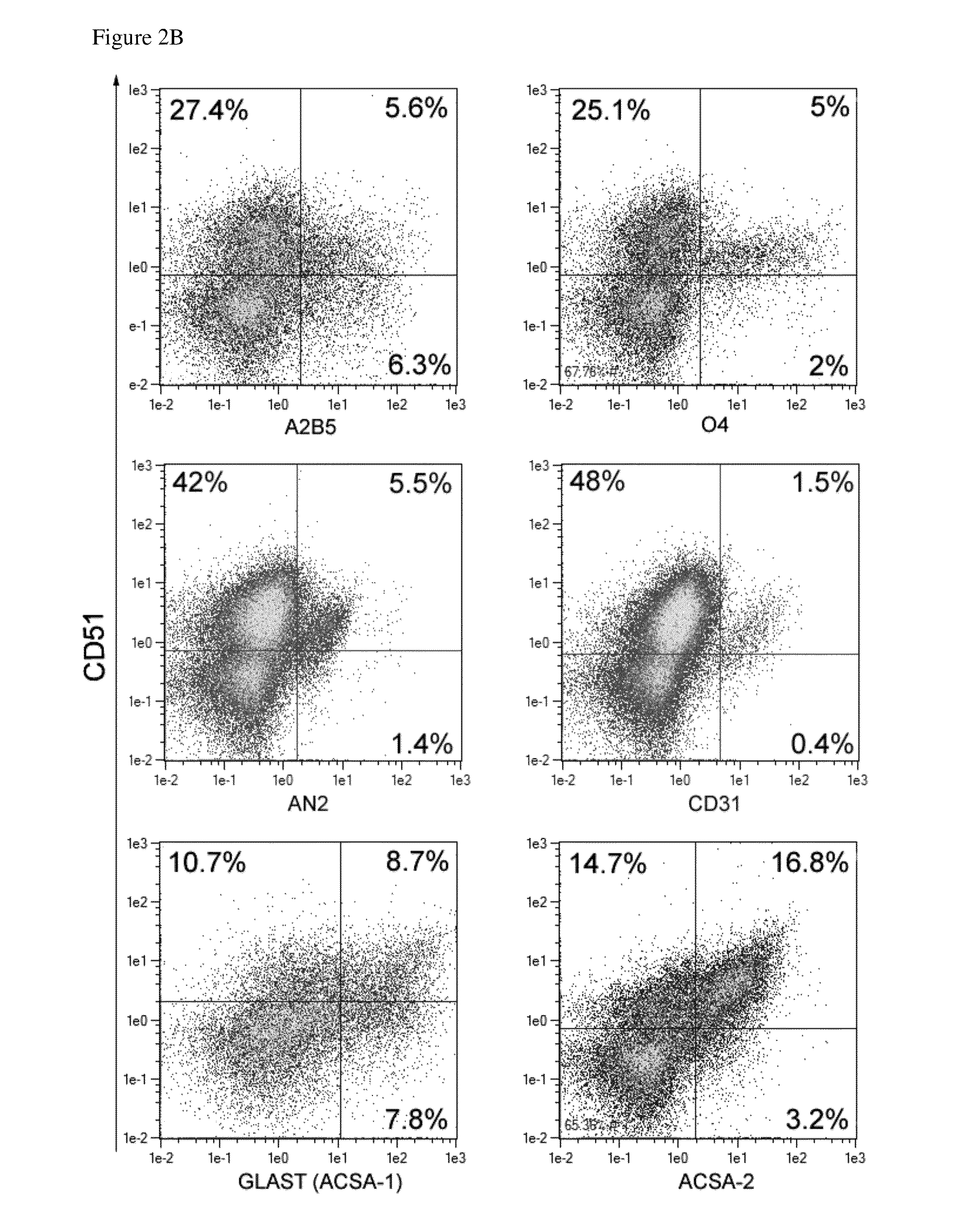
[0097]CD51 positive cells were depleted using whole mouse brain derived from P2 or P3 CD1 mice and an indirect labeling strategy as described before. The original, negative as well as positive cell fraction were co-stained with CD51 and exemplary A2B5, O4, GLAST (ACSA-1), ACSA-2, CD11b specific antibodies to determine the percentage of different neural subtypes in the original, negative as well as positive cell fraction. Use of GAD67-GFP mice allowed detection of GABAergic neurons. FIG. 4A shows that GAD67-GFP positive GABAergic neurons were enriched in the negative target cell fraction, whereas non-neuronal cells, like AN2 positive oligodendrocyte precursors, O4 positive oligodendrocytes, and CD11b positive microglia were depleted and found in the positive fraction (see FIGS. 4A,B). The majority of GLAST (ACSA-1) and ACSA-2 positive astrocytes was also depleted, but approximately 6% of these cells retain within the negative fraction (see FIG...


PUM

PropertyMeasurementUnit
concentrationaaaaaaaaaa
temperatureaaaaaaaaaa
pHaaaaaaaaaa

Abstract

The present invention provides the use of the cell surface antigen CD51 as a negative selection marker for neuronal cells and a method for enrichment, isolation and/or detection of neuronal cells comprising the steps a) contacting a sample containing neuronal cells with an antigen-binding fragment specific for the CD51 antigen coupled to a solid phase, thereby labeling the CD51 positive cells of said sample and b) isolating the non-labeled cells of said sample.

Description

CROSS-REFERENCE TO RELATED APPLICATIONS[0001]This claims priority to European Application No.: EP12157671.4, filed Mar. 1, 2012, the contents of which are incorporated herein by reference in its entirety.FIELD OF THE INVENTION[0002]The present invention relates generally to the field of separating cells, in particular to processes for separation of untouched neuronal cells.BACKGROUND OF THE INVENTION[0003]A cell suspension that is obtained after dissociation of neural tissue comprises a wide variety of different cell types. But, selective targeting and isolation of a special cell type is often the prerequisite for experimental in vitro studies on these cells. Relatively easy techniques like density gradient centrifugation or isolation of living cells by culture conditions lead to low purities and massive cell loss.[0004]A widely used approach in the field of neurobiology is the use of transgenic mice expressing a fluorescent protein controlled by a cell-type specific promoter in com...

Claims


Application Information

Patent Timeline
Patent Type & Authority Applications(United States)
IPC IPC(8): C12N5/0793G01N33/569
CPCC12N5/0619G01N33/56966C07K16/2848C12N5/0622
Inventor BOSIO, ANDREASJUNGBLUT, MELANIE
Owner MILTENYI BIOTEC B V & CO KG
Why Eureka
Try Eureka