Medical image display system

a medical image and display system technology, applied in the field of medical image display system, can solve problems such as unstable medical records, and achieve the effect of enhancing diagnosis accuracy and effective us

Inactive Publication Date: 2014-01-09
KONICA MINOLTA INC
View PDF5 Cites 29 Cited by
  • Summary
  • Abstract
  • Description
  • Claims
  • Application Information

AI Technical Summary

Benefits of technology

[0036]According to the present invention, it is possible to effectively use a reconstructed image created from a moire image g

Problems solved by technology

Moire becomes unstable when something i

Method used


Image

  • Medical image display system
  • Medical image display system
  • Medical image display system

Examples


first embodiment

[0083]Below, the first embodiment of the present invention is described with reference to the drawings.

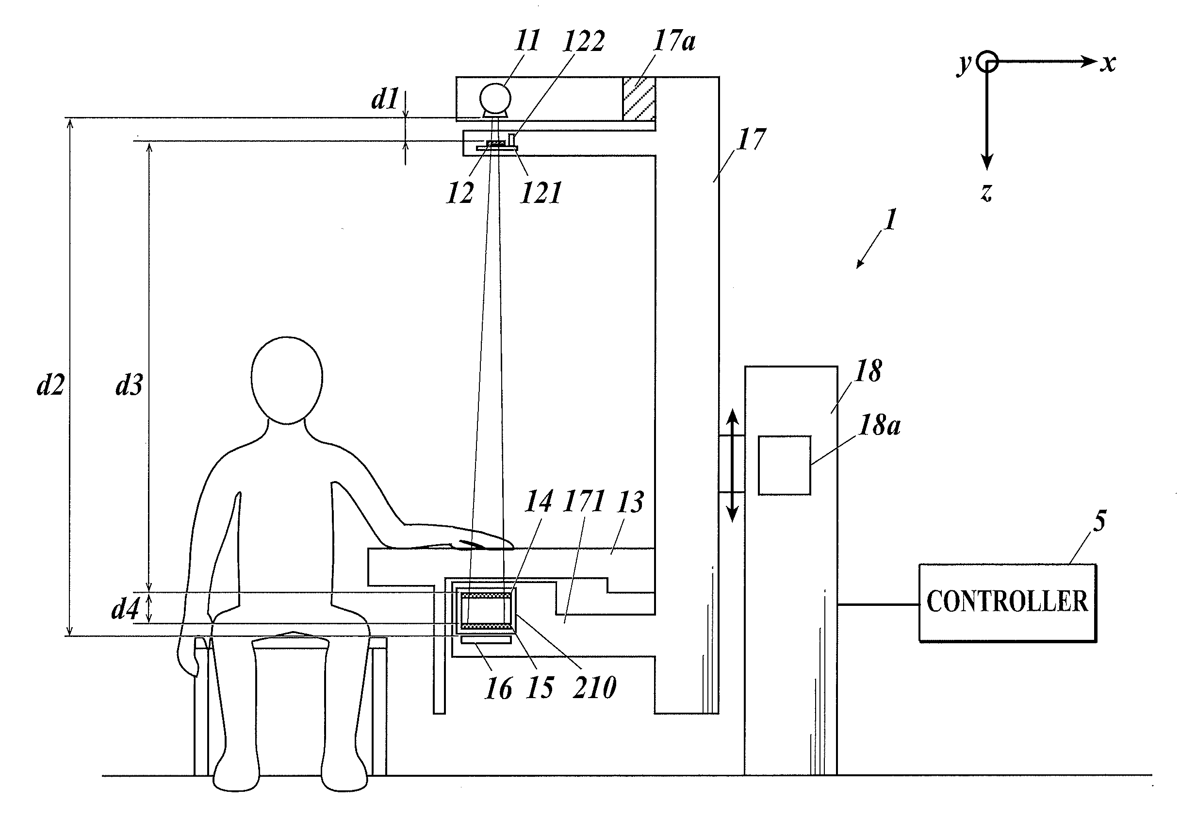
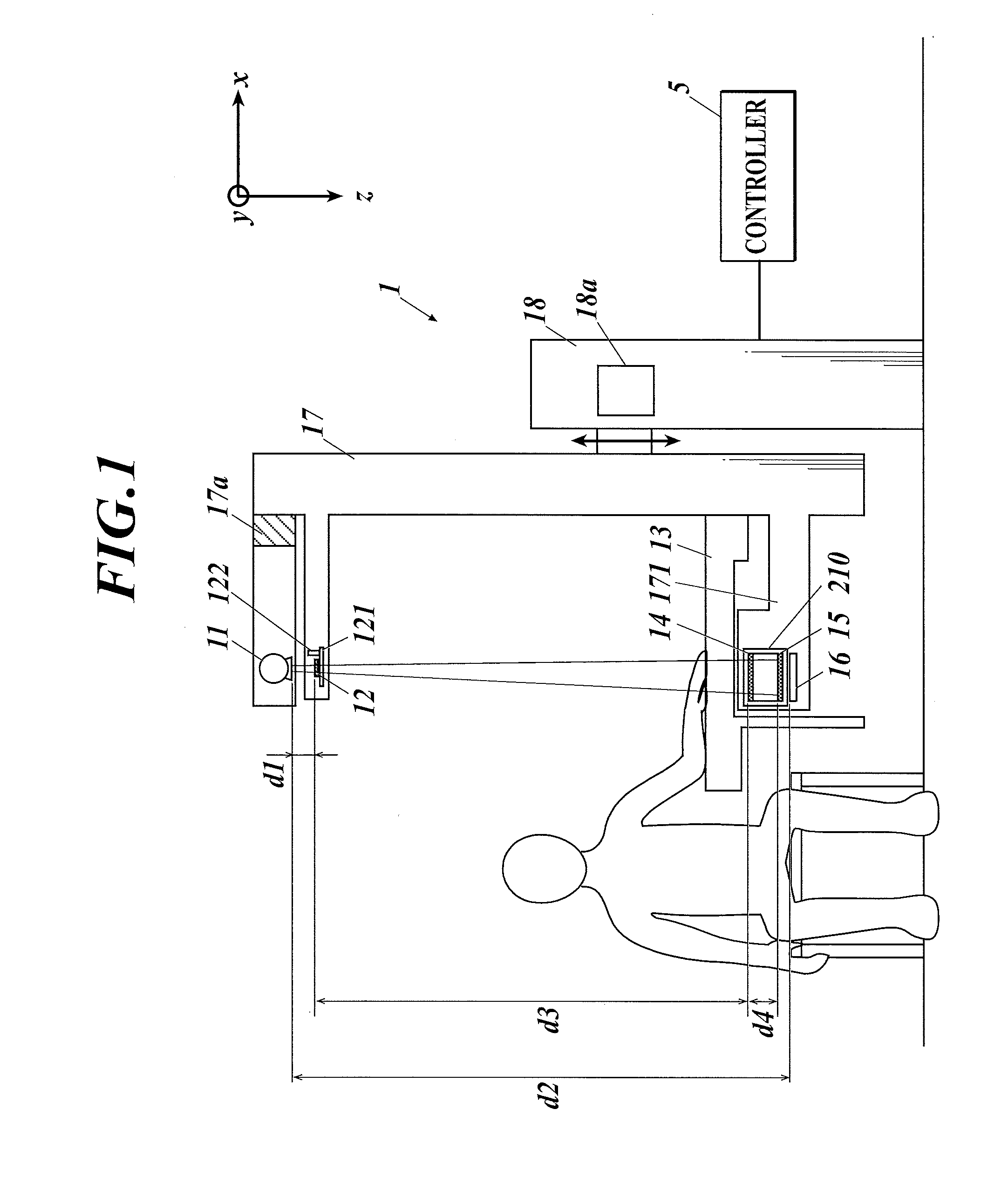
[0084]FIG. 1 shows a medical image display system of a first embodiment. The medical image display system includes an X-ray capturing apparatus 1 and a controller 5. The X-ray capturing apparatus 1 is an apparatus including a first capturing mode to function as a fringe scanning type capturing apparatus and a second capturing mode to function as a Fourier transformation type capturing apparatus. The fringe scanning type capturing apparatus performs capturing of a plurality of steps with a Talbot-Lau interferometer for a reconstructed image by a fringe scanning method to generate a plurality of moire images. The Fourier transformation type capturing apparatus performs capturing in one or two directions for a reconstructed image of a Fourier transformation method to generate one or two moire images.

[0085]The present embodiment describes an example where the configuration of the X-ray...

second embodiment

[0281]Below, the second embodiment of the present invention is described.

[0282]Conventionally, in order to support image diagnosis, an abnormal shadow candidate detecting apparatus (CAD: Computer-Aided Diagnosis) detects an abnormal shadow candidate from a medical image and the result of detecting is displayed with the medical image for diagnosis. Conventionally, as described above, an absorption image is used as the medical image for diagnosis, and only the absorption image is used when the abnormal shadow candidate is detected with the CAD.

[0283]In view of the above, as the second embodiment, described below is an example where, similar to the conventional absorption image based diagnosis system, first the interpreting of the image and the detecting of the abnormal shadow candidate with the CAD is performed with the absorption image among the reconstructed images, and then the small angle scattering image and the differential phase image are used in the secondary diagnosis to judg...

third embodiment

[0306]Below, the third embodiment of the present invention is described.

[0307]In the third embodiment described below, the order of switching display as described in the first embodiment is determined using the result of detecting with the CAD.

[0308]Regarding the medical image display system of the third embodiment, the configuration of the X-ray capturing apparatus 1 and the controller 5, and the operation from capturing to creating the reconstructed image are the same as those described in the first embodiment, therefore the description is incorporated herein.

[0309]In the controller 5, when the creating of the reconstructed image ends, in coordination with the abnormal shadow candidate detecting program stored in the storage section 55, the control section 51 detects the abnormal shadow candidate from each of the absorption image, the small angle scattering image, and the differential phase image. The algorithm of the abnormal shadow candidate detecting program applied to each ima...


PUM


Abstract

A medical image display system is shown. The medical image display system includes, a fringe scanning type capturing apparatus or a Fourier transformation type capturing apparatus; an image processing section; a display section; and a control section. The capturing apparatus includes, an X-ray source; a first grating and a second grating; a subject table; and an X-ray detector. The image processing section generates a plurality of reconstructed images for diagnosis based on an image signal of a subject captured with the capturing apparatus. The display section displays at least two of the plurality of reconstructed images. The control section detects an abnormal candidate on each of the plurality of reconstructed images and controls display order of the plurality of reconstructed images displayed on the display section based on a result of detecting.

Description

TECHNICAL FIELD[0001]The present invention relates to a medical image display system.BACKGROUND ART[0002]Most medical X-ray images used in diagnosis are images by an absorption contrast method. According to the absorption contrast method, a contrast is formed by a difference of attenuation of X-ray intensity when X-rays pass through a subject. Alternatively, a phase contrast method is proposed, where contrast is obtained by phase variation of X-rays instead of absorption of X-rays. For example, phase contrast capturing is performed to obtain an X-ray image with high visibility by edge enhancement using refraction of X-rays in enlarged capturing (for example, see patent documents 1 and 2).[0003]The absorption contrast method is effective for capturing a subject with large X-ray absorption, such as bones, etc. Alternatively, the phase contrast method can image tissue of breasts, articular cartilage, and soft tissue around joints, which have small X-ray absorption difference and which ...

Claims


Application Information

Patent Timeline
IPC IPC(8): A61B6/00
CPCA61B6/52A61B6/502A61B6/06A61B6/483A61B6/484A61B6/5205A61B6/5247G21K2207/005
Inventor NAGATSUKA, SUMIYAKIYOHARA, JUNKOKANEKO, SHIKOUKIDO, KAZUHIRO
Owner KONICA MINOLTA INC
Why Eureka
Try Eureka