In vitro embryo blastocyst prediction methods
a blastocyst and embryo technology, applied in the field of biological and clinical testing, can solve the problems of low birth rate, miscarriage, and well-documented adverse outcomes for both mother and fetus
- Summary
- Abstract
- Description
- Claims
- Application Information
AI Technical Summary
Benefits of technology
Problems solved by technology
Method used
Image
Examples
example 1
[0113]This example describes the development of a blastocyst prediction model and its utility in an IVF clinic.
Methods
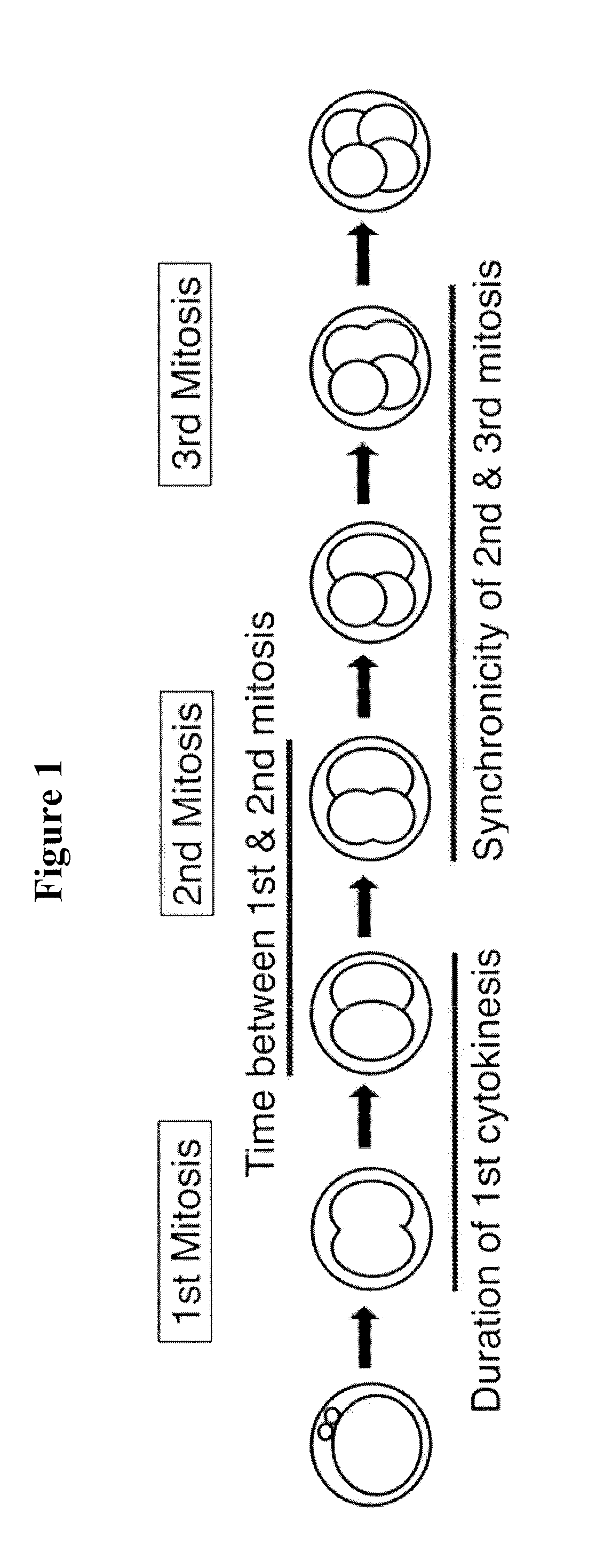
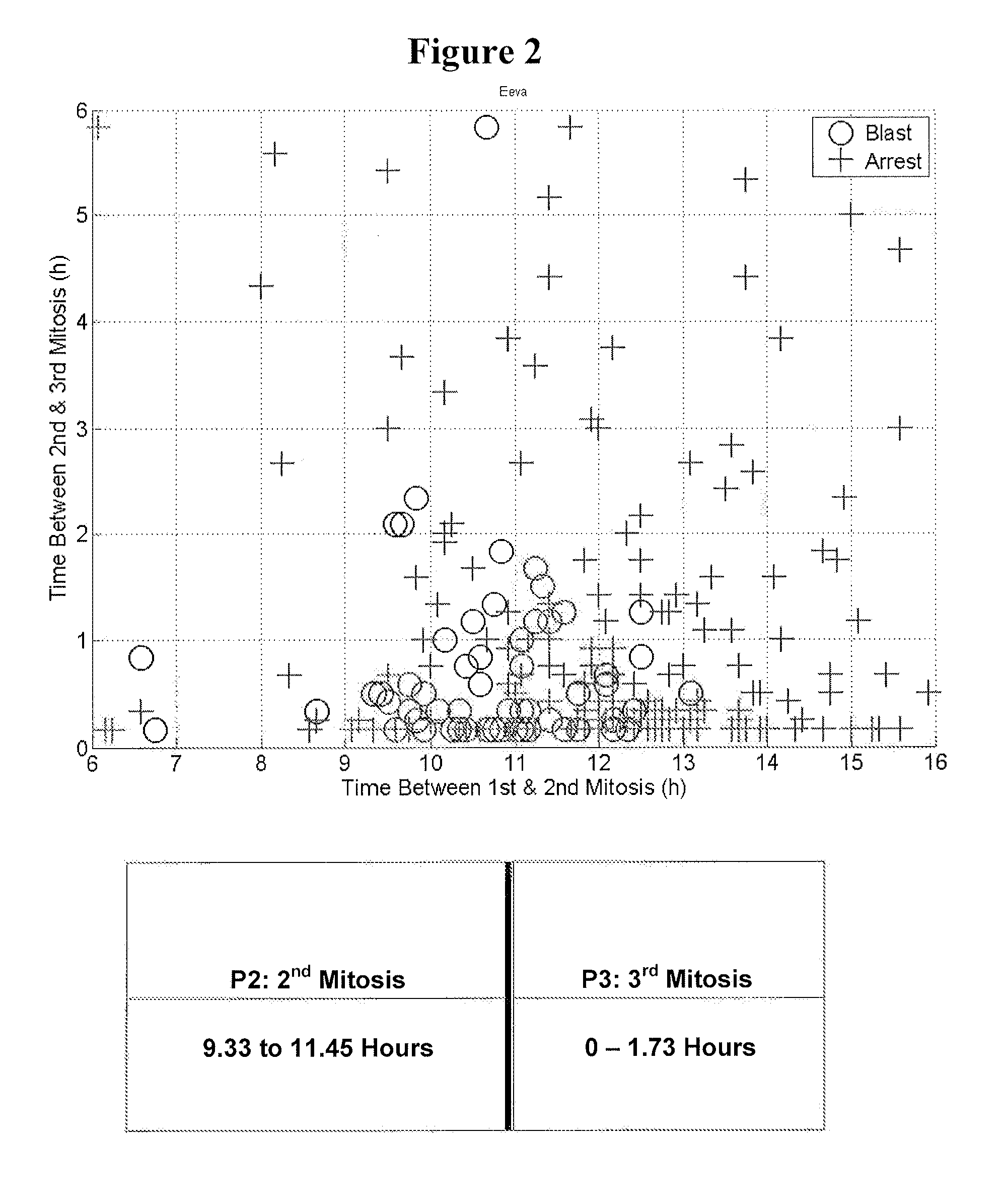
[0114]To develop the blastocyst prediction model, a clinical study was performed to collect data from 3 sites, 54 subjects and 292 embryos. The embryos were cultured using standard procedures in an IVF lab and imaged at 5 minute intervals inside the incubator. By retrospectively analyzing the image data, it was shown that quantification of the timing of early cell division up to approximately 48 hours after fertilization could predict whether an embryo would become a blastocyst on day 5 with a high degree of specificity. During this analysis, it was found that the time between 1st and 2nd mitosis (p2) and the time between 2nd and 3rd mitosis (p3) significantly contributed to the predictive power of the prediction model. Therefore, the blastocyst prediction model was based on the time between 1st and 2nd mitosis (p2) and the time between 2nd and 3rd mitosis (p3).
[011...
example 2
Purpose
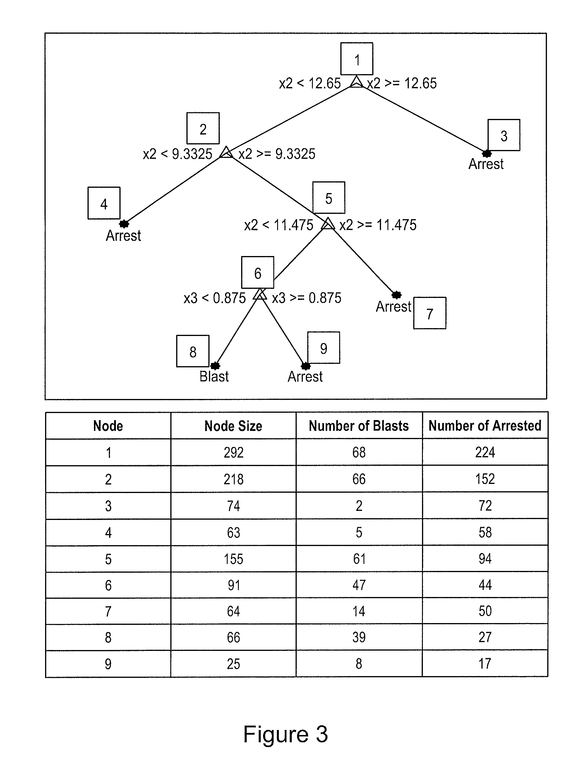
[0121]This example describes the process used to develop statistical classification models for predicting blastocyst formation based on the blastocyst prediction timing parameters.
[0122]The clinical study dataset was collected to help build and evaluate different types of statistical classification models for predicting blastocyst formation. The input parameters to these classifiers were the 3 predictive parameters (based on the paper Wong C C, Loewke K E, Bossert N L, Behr B, De Jonge C J, Baer T M, Reijo Pera R A. Non-Invasive Imaging of Human Embryos Before Embryonic Genome Activation Predicts Development to the Blastocyst Stage. Nat Biotechnol. 2010 October; 28(10):1115-21.): duration of first cytokinesis (P1), time between 1st and 2nd mitosis (P2), and time between 2nd and 3rd mitosis (P3).
[0123]The models were trained on an extensive clinical study dataset The dataset consisted of 292 embryos across 45 patients. The average age of the egg is 33.6±4.8. ...
example 3
[0205]Development and validation of a new test for predicting embryo viability based on time-lapse imaging and automated cell tracking.
Abstract
[0206]The objective of this study was to develop and prospectively validate a new, real-time early embryo viability assessment platform for improving embryo selection in in vitro fertilization (IVF) laboratories.
[0207]The specificity, positive predictive value and overall accuracy of identifying Usable Blastocysts (blastocysts deemed suitable for transfer or freezing) at the cleavage stage are significantly improved when using the new test compared to traditional Day 3 morphology.
[0208]New embryo selection methods are expected to improve IVF success rates and reduce the need for multiple embryo transfer, yet step-by-step approaches to validate new technology for clinical usefulness are lacking. In this study, scientifically-based time-lapse image markers are integrated with cell tracking capabilities to create the first method for quantitati...
PUM
| Property | Measurement | Unit |
|---|---|---|
| time | aaaaa | aaaaa |
| time | aaaaa | aaaaa |
| time | aaaaa | aaaaa |
Abstract
Description
Claims
Application Information
 - R&D
- Intellectual Property
- Life Sciences
- Materials
- Tech Scout
- Unparalleled Data Quality
- Higher Quality Content
- 60% Fewer Hallucinations
Browse by: Latest US Patents, China's latest patents, Technical Efficacy Thesaurus, Application Domain, Technology Topic, Popular Technical Reports.
© 2025 PatSnap. All rights reserved.Legal|Privacy policy|Modern Slavery Act Transparency Statement|Sitemap|About US| Contact US: help@patsnap.com


