

Method of detecting pancreatic disease and pancreas testing kit
- Summary
- Abstract
- Description
- Claims
- Application Information
AI Technical Summary
Benefits of technology
Problems solved by technology
Method used
Image


Examples
example 1
[0054]The test sensitivity of the method of detecting pancreatic disease according to the present invention was assessed by the following experiment.
[0055]Pancreatic juice and body fluid containing pancreatic juice which served as test fluids, were collected by means of catheter suction from test subjects including 31 patients serving as a pancreatic cancer patient group and 7 patients serving as an IPMN patient group. The test fluids collected from the patients were quickly frozen and stored after collection, and, subsequently, the concentrations of S100P contained in the individual test fluids were measured by using an ELISA kit made by CycLex Co., Ltd. By using the same methods, the test fluids were collected and measurements were taken therefrom also for 6 benign pancreatic cyst patients (MCN, SCN), serving as a control group. Diseases of the individual patients were diagnosed by means of pathological diagnosis.
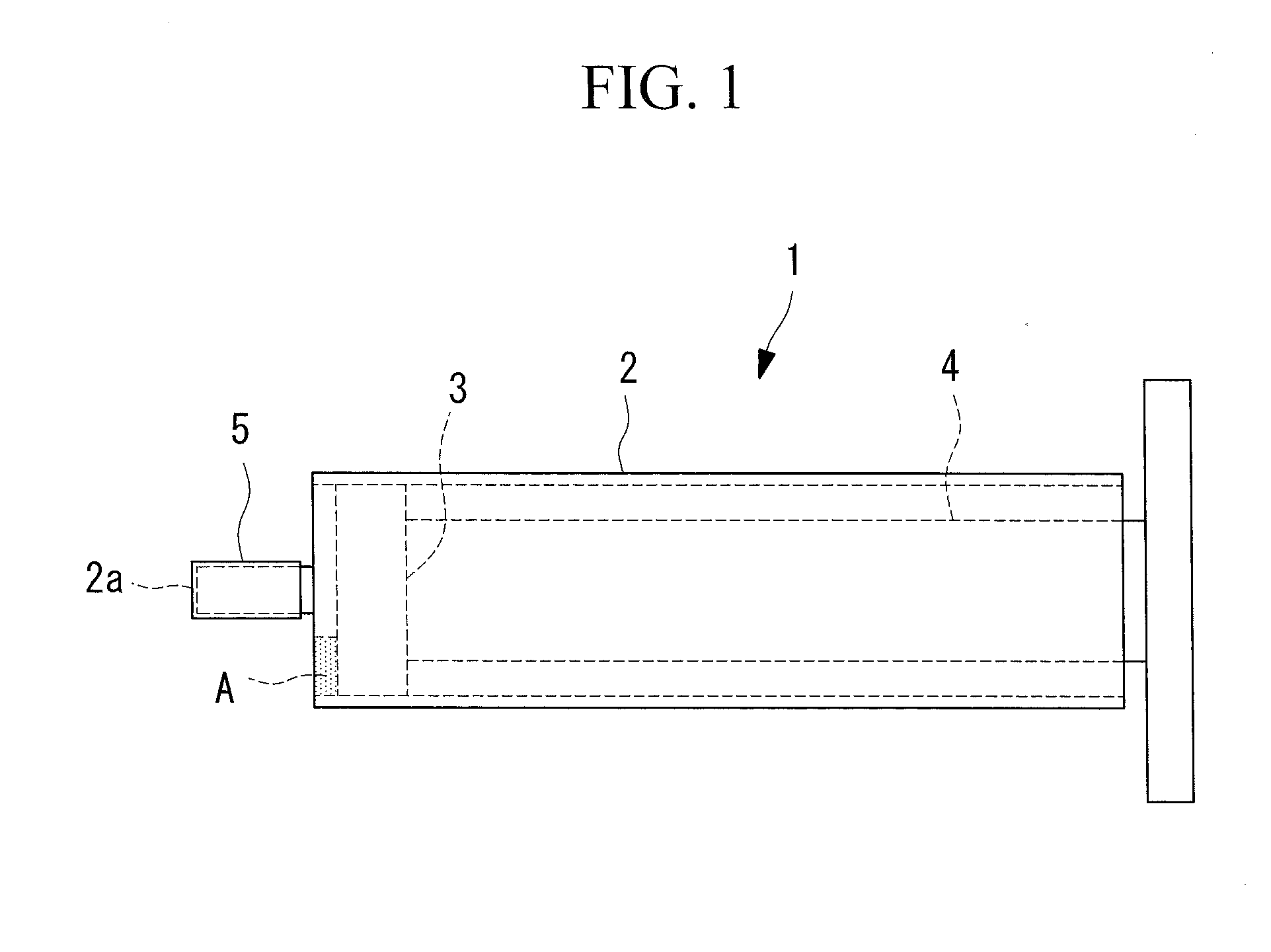
[0056]The pure pancreatic juice collected from the pancreatic duct a...
example 2
[0061]Next, an immunochromatography device according to this Example was fabricated by using the two types of anti-S100P antibodies (made by MBL Co., Ltd.) used for ELISA employed in Example 1 as the first antibody and the second antibody. By using this immunochromatography device to test the test fluids used in Example 1, the correlation between test results respectively obtained by means of the ELISA method and the immunochromatography method was checked.
[0062]The immunochromatography device according to this Example was fabricated by fabricating the support by using a nitrocellulose film, and labeling the first antibody with colloidal gold. Then, the test fluids used in Example 1 were tested by using the fabricated immunochromatography device, and the presence / absence of band color was determined and the shade of the color was measured.
[0063]When the measured shades of the color in the immunochromatography device were divided into three classes, it was confirmed that there was a ...
example 3
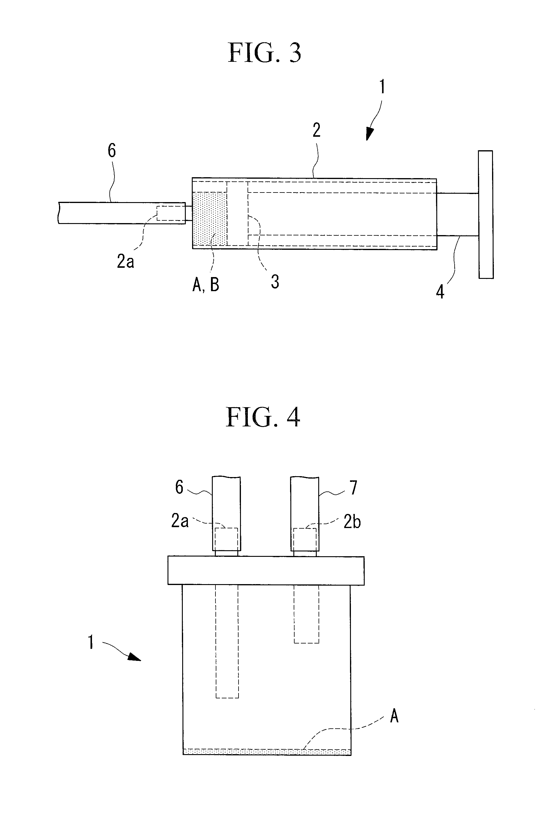
[0064]Based on the results in Example 1, by using the method in which the protease inhibitor is retained in the collection vessel in advance and the secretin administration for promoting secretion is not performed, the pancreatic juice / duodenal juice was collected and the S100P concentrations were measured for 41 pancreatic cancer patients, 17 IPMN patients, 6 benign pancreatic cyst (SCN, MCN) patients, 4 chronic pancreatitis patients, and 3 pancreatic endocrine tumor patients. The collection of the pancreatic juice / duodenal juice and the measurement of the S100P concentration were performed by using the same method as in Example 1. The measurement results are shown in FIG. 8. In FIG. 8, the vertical axis is on a logarithmic scale.
[0065]The highest value among the measured values of the S100P concentrations obtained from the six benign pancreatic cyst patients was set as the cut-off value, and the positive rates were calculated for the respective example diseases. As a result, the p...
PUM
| Property | Measurement | Unit |
|---|---|---|
| Concentration | aaaaa | aaaaa |
| Sensitivity | aaaaa | aaaaa |
Abstract
Description
Claims
Application Information