Apparatus and Method for Aiding Needle Biopsies

a technology of applicability and biopsies, applied in the field of optical coherence tomography, can solve the problems of limited use of oct for imaging interstitial tissue, translation of oct to clinical use, and limited applicability of oct to achieve the effect of increasing the success rate of biopsies

Inactive Publication Date: 2014-01-30
PHYSICAL SCI
View PDF12 Cites 27 Cited by
  • Summary
  • Abstract
  • Description
  • Claims
  • Application Information

AI Technical Summary

Benefits of technology

[0007]What is needed is instrumentation and an approach for aiding transcutaneous and endoscopic biopsies with the goal of increasing the success rate of biopsies. The approach is based on the use of optical coherence tomography (“OCT”), which is a high-resolution imaging technique that can be used to assess tissue cellularity at the tip of a biopsy needle. OCT has proven its capability to image tissue morphology at the micron scale, and thus has enabled the diagnosis of cancer disease in the early stage, at the epithelial level. However, the use of OCT for imaging interstitial tissue is somewhat limited due to the lack of minimally invasive probes that can be deployed alone, or by means of biopsy needles, to image tissue morphology in various organs, for example, lungs, breast, pancreas, liver or kidney, where a natural lumen does not exist or cannot be used to image deeper within the organ. OCT applicability and translation to clinical use, especially for imaging interstitial tissue, has been limited by the lack of adequate probes that can be deployed safely without significantly disrupting tissue morphology and inducing severe bleeding, while providing high-quality images.

Problems solved by technology

However, the use of OCT for imaging interstitial tissue is somewhat limited due to the lack of minimally invasive probes that can be deployed alone, or by means of biopsy needles, to image tissue morphology in various organs, for example, lungs, breast, pancreas, liver or kidney, where a natural lumen does not exist or cannot be used to image deeper within the organ.
OCT applicability and translation to clinical use, especially for imaging interstitial tissue, has been limited by the lack of adequate probes that can be deployed safely without significantly disrupting tissue morphology and inducing severe bleeding, while providing high-quality images.

Method used


Image

  • Apparatus and Method for Aiding Needle Biopsies
  • Apparatus and Method for Aiding Needle Biopsies
  • Apparatus and Method for Aiding Needle Biopsies

Examples


Embodiment Construction

[0039]It should be understood that when an element is referred to as being “on” or “connected” or “coupled” to another element, it can be directly on or above, or connected or coupled to, the other element or intervening elements can be present. In contrast, when an element is referred to as being “directly on” or “directly connected” or “directly coupled” to another element, there are no intervening elements present. Other words used to describe the relationship between elements should be interpreted in a like fashion (e.g., “between” versus “directly between,”“adjacent” versus “directly adjacent,” etc.). When an element is referred to herein as being “over” another element, it can be over or under the other element, and either directly coupled to the other element, or intervening elements may be present, or the elements may be spaced apart by a void or gap.

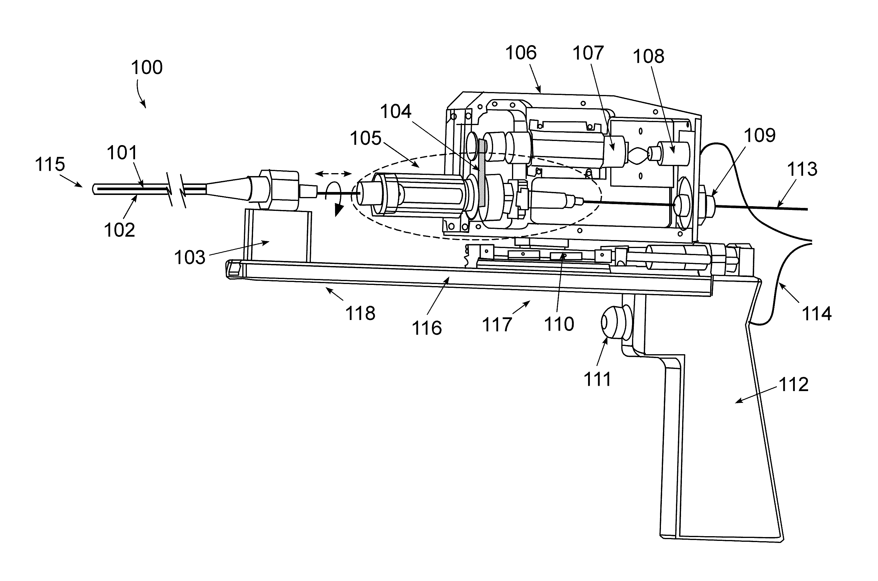
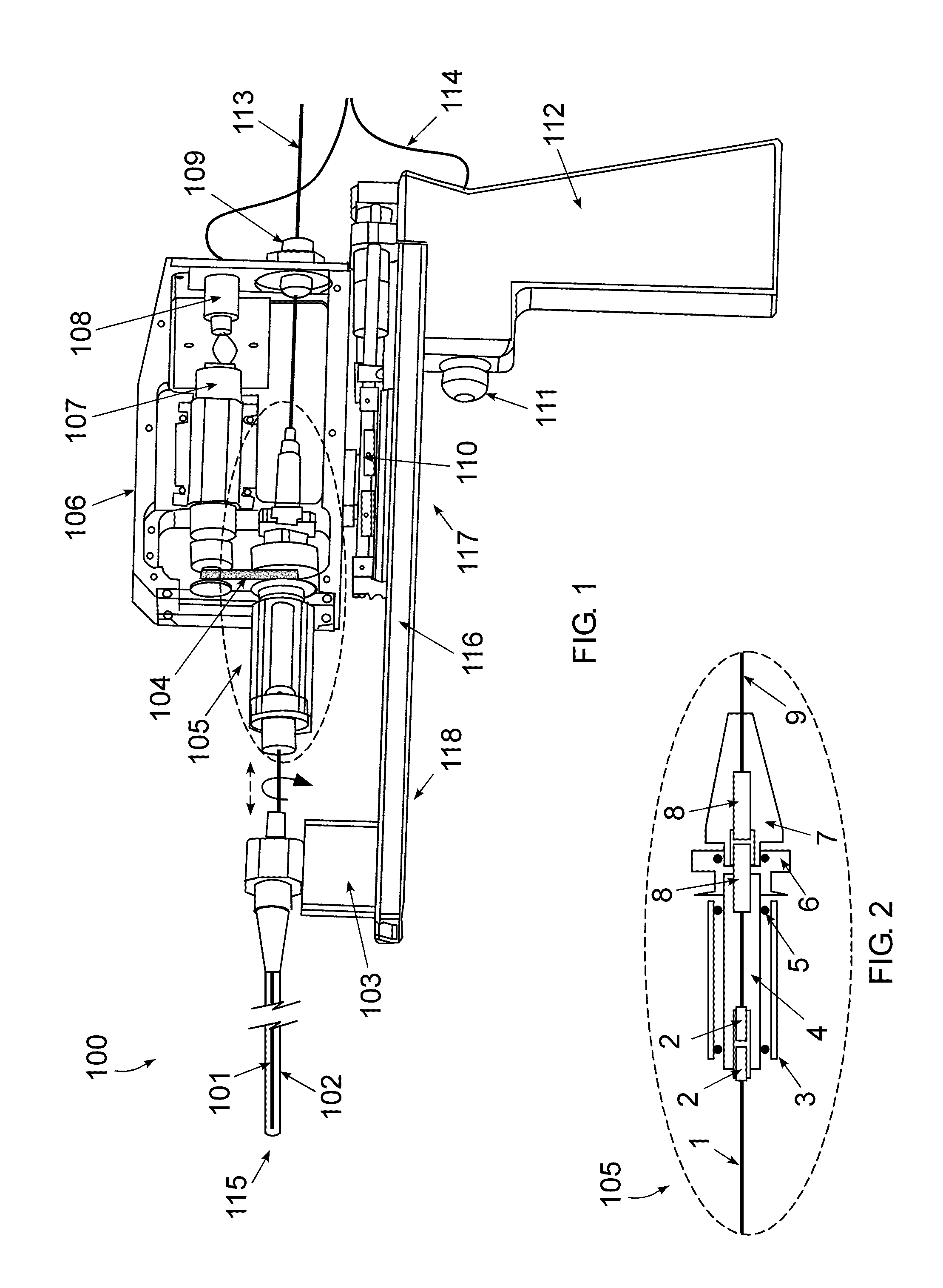
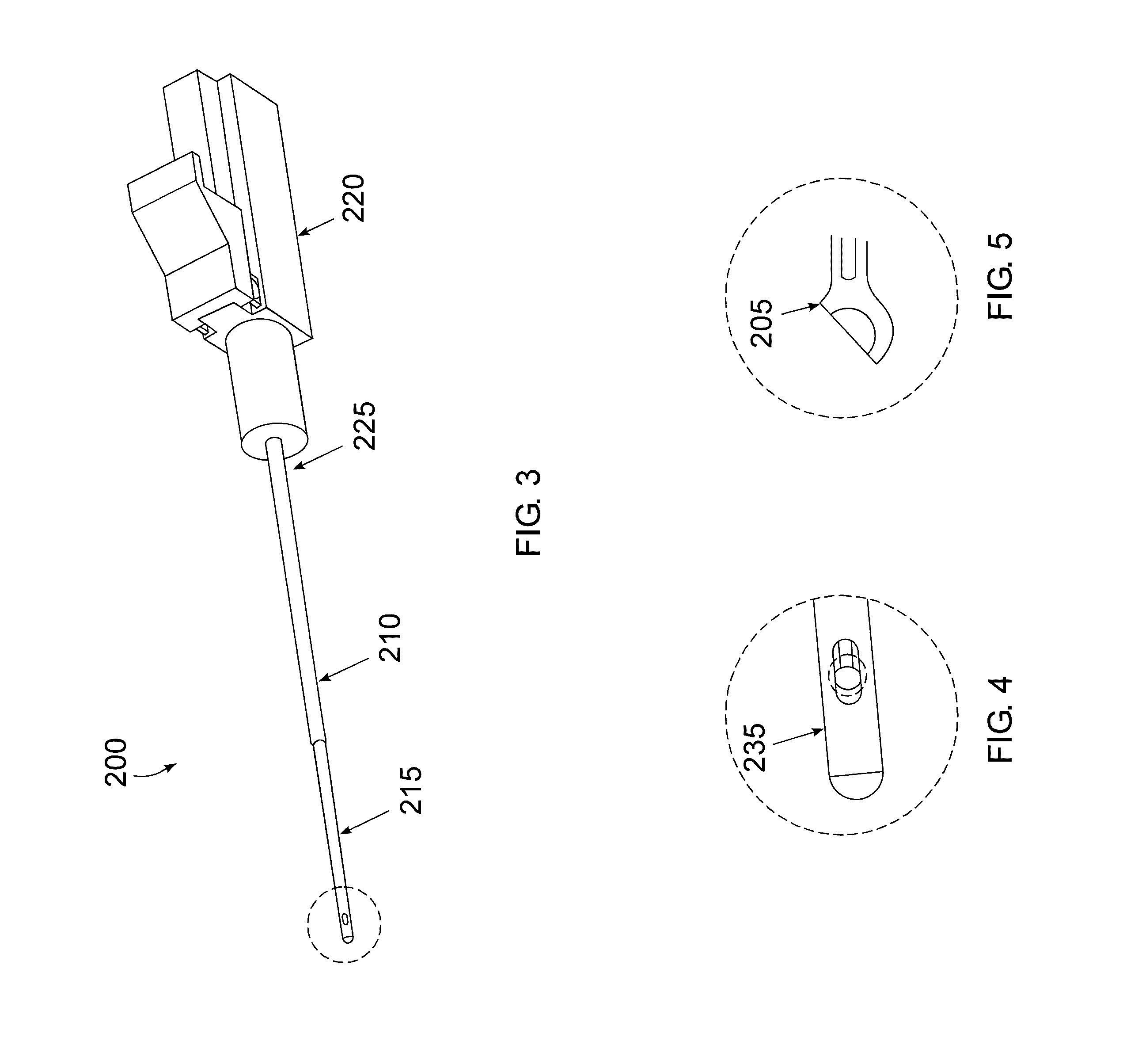
[0040]Embodiments of the present invention relate to the development of a high-resolution optical imaging probe that can be de...


PUM


Abstract

A handheld optical coherence tomography imaging and tissue sampling system and method of imaging and sampling a tissue is disclosed. The method includes inserting a catheter probe into a biopsy needle. The biopsy needle can be attached to a hand-held scanning and sampling device. The biopsy needle is maneuvered to an investigation site. A three-dimensional image of the tissue at the investigation site is captured with the catheter probe.

Description

RELATED APPLICATIONS[0001]This application claims the benefit of and priority to U.S. Provisional Application No. 61 / 589,082, filed Jan. 20, 2012, which is owned by the assignee of the instant application and the disclosure of which is hereby incorporated herein by reference in its entirety.STATEMENT OF GOVERNMENT INTEREST[0002]The subject matter described herein was developed in connection with funding provided by the National Institute of Health under Grant Nos. 2R44CA117218 and 5R42CA114896. The Federal government may have rights in the technology.TECHNICAL FIELD[0003]The invention relates generally to an apparatus and method for aiding needle biopsies, and more particularly, to an optical coherence tomography (“OCT”) imaging and tissue sampling system and method.BACKGROUND[0004]Early diagnosis and a proper stage determination are key factors that contribute to cancer cure success. A cancer's stage is based on the primary tumor's size and whether it has spread to other areas of t...

Claims


Application Information

Patent Timeline
Patent Type & Authority Applications(United States)
IPC IPC(8): A61B5/00A61B10/02
CPCA61B10/02A61B5/0066A61B10/0233
Inventor IFTIMIA, NICUSOR V.MUJAT, MIRCEAFERGUSON, R. DANIELHAMMER, DANIEL X.CHANG, SHING
Owner PHYSICAL SCI
Why Eureka
Try Eureka