Systems and methods for interventional imaging

a technology of interventional imaging and system, applied in the field of interventional imaging, can solve the problems of increasing the ionizing radiation dose and exhaustion of the medical practitioner, challenging the understanding of the relationship of the insertion and navigation of the catheter within the different branches of the vascular system, and limiting the understanding of the medical practitioner

Inactive Publication Date: 2014-02-06
GENERAL ELECTRIC CO
View PDF6 Cites 25 Cited by
  • Summary
  • Abstract
  • Description
  • Claims
  • Application Information

AI Technical Summary

Problems solved by technology

The insertion as well as the navigation of the catheter within the different branches of the vascular system, however, is a challenging procedure.
Conventional fluoroscopic interventional systems, however, may constrain a medical practitioner's understanding of a relation between the catheter and vascular positions primarily based on 2D projection images that provide limited or no information in the depth direction, where the depth direction is parallel to the x-rays that traverse the patient.
Further, certain fluoroscopic procedures may entail frequent switching between aligning the patient along a desired imaging plane and navigating the catheter inside the patient's body.
Consequently, lack of sufficient information in fluoroscopic interventional imaging can result in longer examination time, which may result in increased ionizing radiation dose and exhaustion to the medical practitioner, and increased ionizing radiation dose, contrast dose, anesthesia time, and discomfort to the patient.
Additionally, risk of injury to the patient increases with the scanning duration and / or retention of a catheter in the patient's vasculature.
However, 2D projection imaging data may prove inadequate in certain scenarios due to confounding information (overlaying structures) in the acquired projection information.
However, slower rotation speeds achieved with conventional interventional systems may result in image artifacts in 3D imaging due to voluntary motion, such as due to patient repositioning, and involuntary motion such as peristalsis or heart motion within the patient.
Additionally, certain interventional systems may employ detectors with smaller axial coverage when compared to conventional CT systems, which may cause a truncated imaging field of view (FOV), leading to erroneous measurements and additional scanning time.
The erroneous measurements and the long examination times may prove detrimental to patient health especially in emergencies such as assessment of ischemia in the brain and heart, where immediate and accurate assessment from interactive evaluation of real-time images is critical for improving patient health.
However, conventional C-arm systems rotate too slowly to generate dynamic 3D information during medical procedures such as neurological imaging, during which it is desirable to reconstruct multiple 3D images to characterize passage of a bolus in real-time.
Lack of dynamic 3D data during imaging results in uncertainty, which in turn may lead to incorrect diagnoses, treatment planning, and / or procedure validation.
Thus, neither conventional fluoroscopic interventional systems, nor C-arm systems allow for acquisition of both high-fidelity 2D projection images and 3D cross-section images of the entire organ using the same system to conduct diagnostic evaluation as well as perform interventional procedures.

Method used


Image

  • Systems and methods for interventional imaging
  • Systems and methods for interventional imaging
  • Systems and methods for interventional imaging

Examples


Embodiment Construction

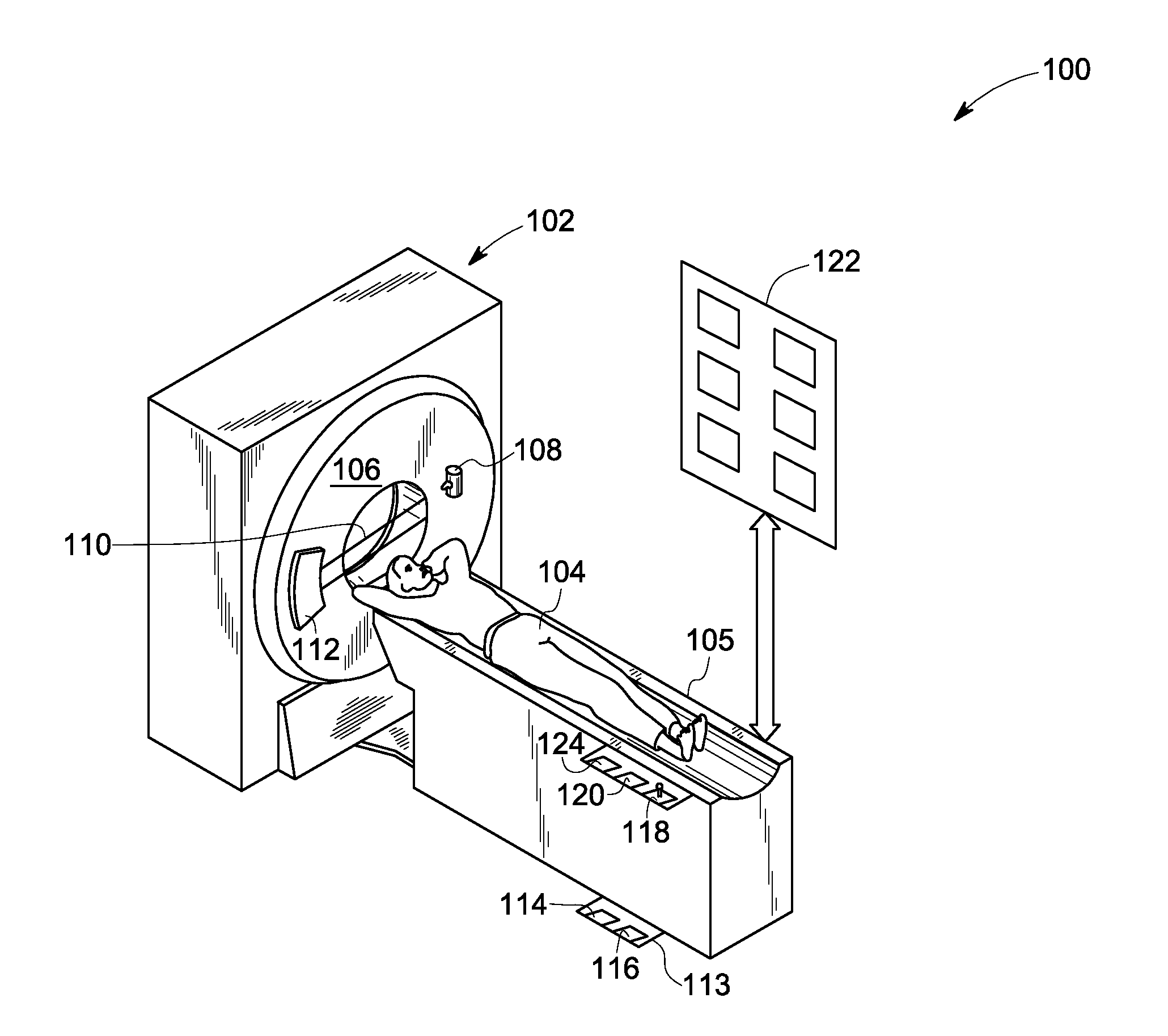
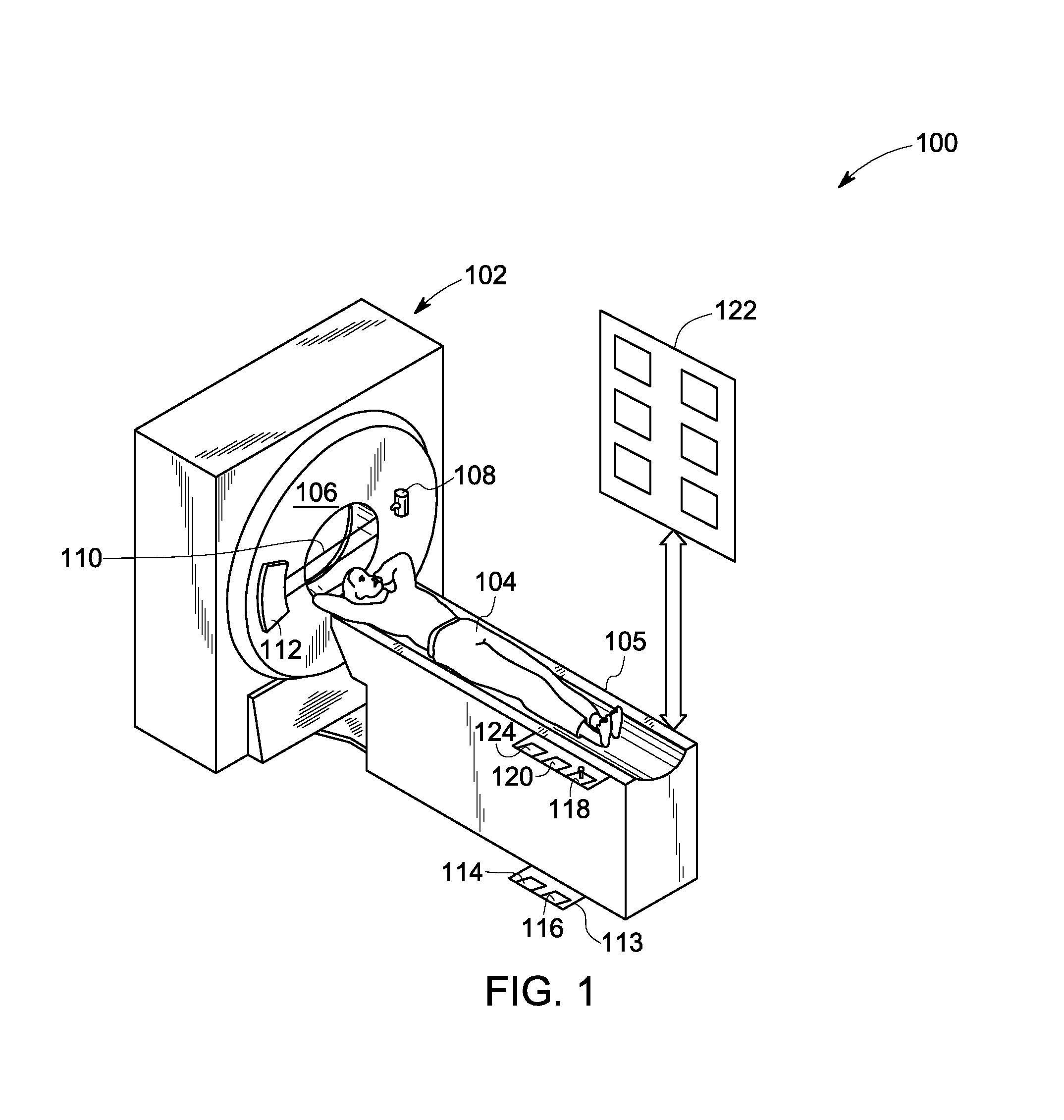
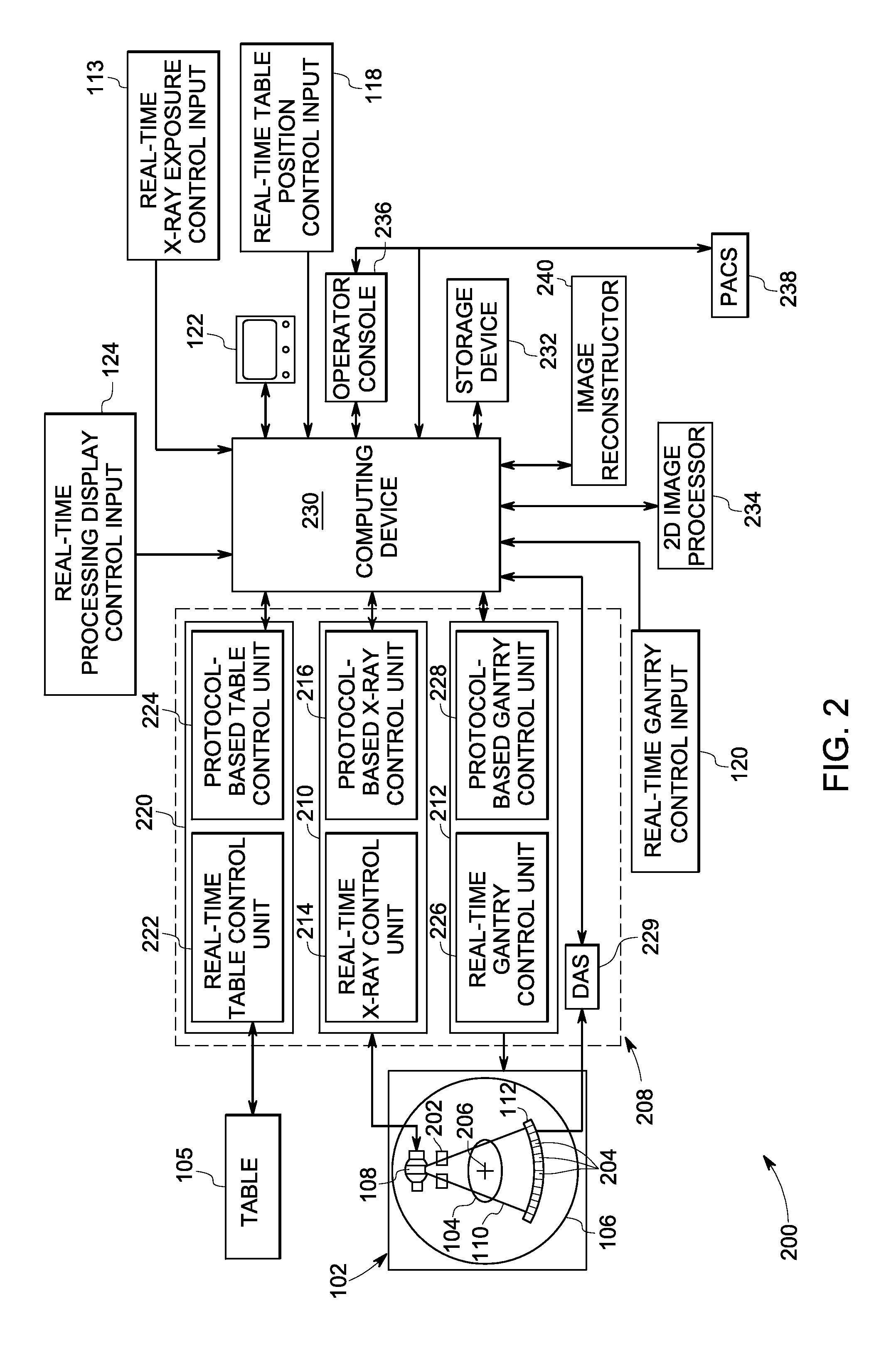
[0018]The following description presents embodiments of imaging systems and methods that provide high-fidelity 2D projection and 3D volumetric images of a scanned organ for use in diagnostic and interventional procedures using reduced radiation dosage and examination time. The interventional procedures, for example, may include angioplasty, stent placement, balloon septostomy, Transcatheter Aortic-Valve Implantation (TAVI), localized thrombolytic drug administration, tumor embolization and / or an electrophysiology study.

[0019]Additionally, the following description presents embodiments of imaging systems and methods that minimize contrast agent dosage, x-ray radiation exposure and scan durations. Certain embodiments of the present systems and methods may also be used for reconstructing high-fidelity 3D cross-sectional images in addition to the 2D projection images for allowing real-time diagnosis and / or therapy delivery, and efficacy assessment. Particularly, certain embodiments illu...


PUM


Abstract

An imaging system including an integral computed tomography and interventional (CT / I) system that includes a large-area detector configured to acquire projection data corresponding to a field of view of the system from one or more view angles is presented. The system includes a computing device operatively coupled to the CT / I system and configured to process the acquired projection data to generate a 2D projection image in real-time, a 3D cross-sectional image of a region of interest in the subject, a combined image using the 2D projection image and the 3D cross-sectional image and / or control selective generation of the 2D projection image, the 3D cross-sectional image and / or the combined image based on one or more imaging specifications. The system also includes a display operatively coupled to the computing device and configured to display the 2D projection image, the 3D cross-sectional image and / or the combined image based on the imaging specifications.

Description

BACKGROUND[0001]Embodiments of the present disclosure relate generally to interventional imaging, and more particularly to integral CT and interventional systems and methods for seamless diagnostic and interventional imaging.[0002]Interventional techniques are widely used for managing a plurality of life-threatening medical conditions. Particularly, certain interventional techniques entail minimally invasive image-guided procedures that provide a cost-effective alternative to invasive surgery. Interventional imaging, for example, may be employed for diagnosing and treating patients who may be suffering from heart disease, coronary artery disease, stroke, osteoporosis, cancer and other medical conditions. In such patients, interventional radiology may facilitate minimally-invasive procedures without the stress of a surgical operation.[0003]Generally, interventional techniques may be employed in various fields of medicine such as neurology, general radiology, cardiology and electrophy...

Claims


Application Information

Patent Timeline
Patent Type & Authority Applications(United States)
IPC IPC(8): A61B6/03
CPCA61B6/032A61B6/0457A61B6/541A61B6/4241A61B6/542A61B6/482A61B6/507A61B6/12A61B6/0487
Inventor LANGAN, DAVID ALLENEDIC, PETER MICHAELCLAUS, BERNHARD ERICH HERMANN
Owner GENERAL ELECTRIC CO
Why Eureka
Try Eureka